. 2023 Aug;11(8):e007031.
doi: 10.1136/jitc-2023-007031.

PPARγ activation modulates the balance of peritoneal macrophage populations to suppress ovarian tumor growth and tumor-induced immunosuppression

Affiliations

PPARγ activation modulates the balance of peritoneal macrophage populations to suppress ovarian tumor growth and tumor-induced immunosuppression

Mélissa Prat et al. J Immunother Cancer. 2023 Aug.

Abstract

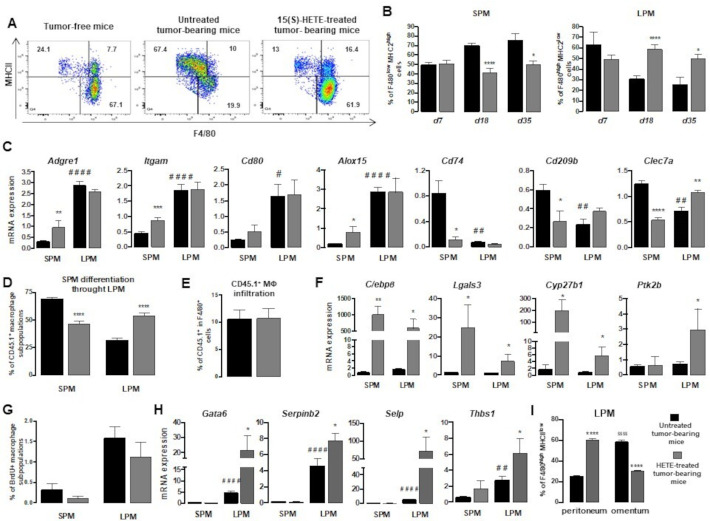
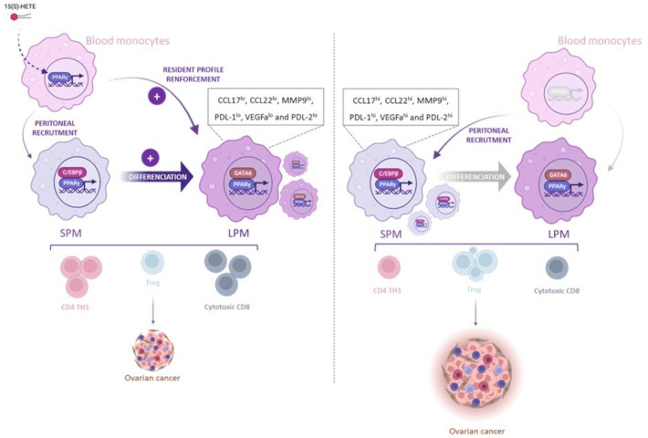
Background: Ovarian adenocarcinoma (OVAD) frequently metastasizes to the peritoneal cavity and manifests by the formation of ascites, which constitutes a tumor-promoting microenvironment. In the peritoneal cavity, two developmentally, phenotypically and functionally distinct macrophage subsets, immunocompetent large peritoneal macrophages (LPM) and immunosuppressive small peritoneal macrophages (SPM), coexist. Because peroxisome proliferator-activated receptor γ (PPARγ) is a critical factor participating in macrophage differentiation and cooperates with CCAAT/enhancer binding protein β (C/EBPβ), a transcription factor essential for SPM-to-LPM differentiation, PPARγ could be also involved in the regulation of SPM/LPM balance and could be a promising therapeutic target.

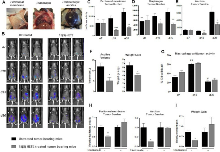
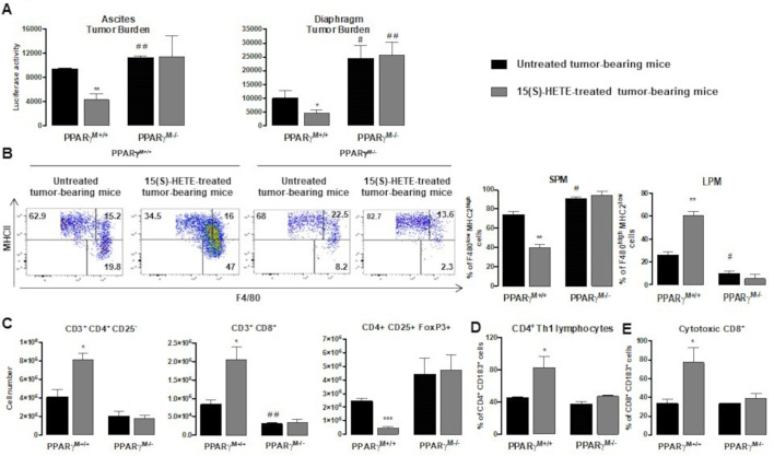
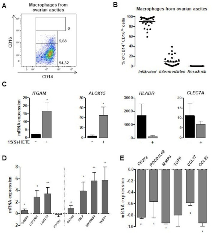
Methods: To evaluate the 15(S)-hydroxyeicosatetraenoic acid (HETE), a PPARγ endogenous ligand, impact on ovarian tumor growth, we intraperitoneally injected 15(S)-HETE into a murine ovarian cancer model. This experimental model consists in the intraperitoneally injection of ID8 cells expressing luciferase into syngeneic C57BL/6 female mice. This ID8 orthotopic mouse model is a well-established experimental model of end-stage epithelial OVAD. Tumor progression was monitored using an in vivo imaging system. Peritoneal immune cells in ascites were analyzed by flow cytometry and cell sorting. To determine whether the impact of 15(S)-HETE in tumor development is mediated through the macrophages, these cells were depleted by injection of liposomal clodronate. To further dissect how 15(S)-HETE mediated its antitumor effect, we assessed the tumor burden in tumor-bearing mice in which the PPARγ gene was selectively disrupted in myeloid-derived cells and in mice deficient of the recombination-activating gene Rag2. Finally, to validate our data in humans, we isolated and treated macrophages from ascites of individuals with OVAD.

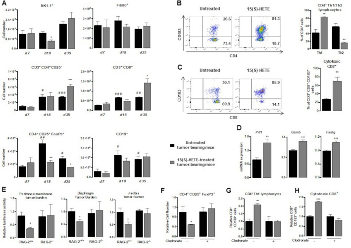
Results: Here we show, in the murine experimental model of OVAD, that 15(S)-HETE treatment significantly suppresses the tumor growth, which is associated with the differentiation of SPM into LPM and the LPM residency in the peritoneal cavity. We demonstrate that C/EBPβ and GATA6 play a central role in SPM-to-LPM differentiation and in LPM peritoneal residence through PPARγ activation during OVAD. Moreover, this SPM-to-LPM switch is associated with the increase of the effector/regulatory T-cell ratio. Finally, we report that 15(S)-HETE attenuates immunosuppressive properties of human ovarian tumor-associated macrophages from ascites.

Conclusion: Altogether, these results promote PPARγ as a potential therapeutic target to restrain OVAD development and strengthen the use of PPARγ agonists in anticancer therapy.

Keywords: Immunity, Innate; Inflammation; Macrophages; Tumor Microenvironment.


Conflict of interest statement

Competing interests: None declared.

Figures

Figure 1
Figure 1
In vivo 15(S)-HETE treatment inhibits OVAD progression through macrophages. (A) Photographs of tumor burden in peritoneal membrane, diaphragm and ascites of untreated tumor-bearing mice 50 days post-tumor cells injection. (B–E) In vivo imaging (B) and quantification of peritoneal membrane (C), diaphragm (D) and ascites (E) tumor burdens by bioluminescence in untreated or 15(S)-HETE-treated mice at days 7, 18, 35 and 50 post-ID8 implantation. (F) Ascites volume and weight gain of untreated and 15(S)-HETE-treated mice 50 days post-tumor cells injection. (G) Cytotoxic activity of peritoneal macrophages collected from the ascites of untreated and 15(S)-HETE-treated mice determined by the quantification of bioluminescence intensity after 72 hours of co-culture with ID8-Luc tumor cells. (H–I) Peritoneal membrane and ascites tumor burden quantified by bioluminescence (H) and weight gain (I) in mice without or with depletion of macrophages were evaluated at day 35 post-ID8 implantation. Tumor burden and weight gain data are expressed as fold induction relative to corresponding untreated ID8 tumor-bearing mice. Results correspond to mean±SEM (n=6 per group) and are representative of at least three independent experiments. *p<0.05, **p<0.01, ***p<0.001 and ****p<0.0001 compared with respective untreated ID8 tumor-bearing mice. #p<0.05 and ##p<0.01 compared with untreated mice at day 7. HETE, hydroxyeicosatetraenoic acid; OVAD, ovarian adenocarcinoma
Figure 2
Figure 2
15(S)-HETE treatment promotes the differentiation of SPM towards LPM and the LPM maintenance in the peritoneum. (A) Dot-plot showing SPM (F4/80low MHCIIhigh) and LPM (F4/80high MHCIIlow) day 18 post-ID8 injection. (B) Quantification of SPM and LPM in the F4/80+ MHCII+ population at days 7, 18 and 35 post-tumor cell injection. (C) Expressions of differentiation marker genes in SPM and LPM sorted at day 18 post-ID8 injection. (D–E) SPM-to-LPM differentiation in CD45.1+ monocytes-transplanted CD45.2+ host tumor-bearing mice treated or not with 15(S)-HETE at day 11-post tumor cells injection (D) and CD45.1+ macrophage infiltration (E) were quantified by flow cytometry using F4/80, MCHII, CD45.1 and CD45.2 markers. (F) Expressions of SPM-to-LPM differentiation marker genes in SPM and LPM sorted at day 18 post-ID8 injection. (G) Tumor bearing mice treated or not with 15(S)-HETE were injected with BrdU 3 hours before euthanasia and BrdU incorporation was assessed by flow cytometry. (H) Expression of peritoneal residency marker genes in SPM and LPM sorted at day 18 post-ID8 injection measured using RT-qPCR. (I) Quantification of LPM in the peritoneum and omentum at day 18 post-tumor cell injection. Results correspond to mean±SEM (n=6 per group) and are representative of at least three independent experiments. *p<0.05, **p<0.01, ***p<0.001 and ****p<0.0001 compared with respective untreated ID8 tumor-bearing mice. ##p<0.01 and ####p<0.0001 compared with SPM from untreated ID8 tumor-bearing mice. $$$$p<0.0001 compared with peritoneal LPM from untreated ID8 tumor-bearing mice. HETE, hydroxyeicosatetraenoic acid; LPM, large peritoneal macrophages; mRNA, messenger RNA; OVAD, ovarian adenocarcinoma; RT-qPCR, real-time quantitative polymerase chain reaction; SPM, small peritoneal macrophages.
Figure 3
Figure 3
15(S)-HETE treatment leads to C/EBPβ and GATA6 activation through PPARγ in TAMs. (A, B) Percentage of SPM or LPM expressing PPARγ, C/EBPβ or GATA-6 (A) and visualization of PPARγ and C/EBPβ and their colocalization on SPM and LPM from untreated or treated tumor-bearing mice by Image StreamX (B). (C) Visualization of PPARγ and GATA-6 and their colocalization on SPM and LPM from untreated or treated tumor-bearing mice by Image StreamX. (D) Quantification of PPARγ, C/EBPβ and GATA-6 on SPM and LPM from untreated or treated tumor-bearing mice by Image StreamX. (E–F) Macrophages isolated from the ascites of untreated or 15(S)-HETE-treated PPARγM+/+ or PPARγM−/− tumor -bearing mice were analyzed for expressions of SPM-to-LPM differentiation markers (E) and peritoneal residency markers (F) using RT-qPCR. Results correspond to mean±SEM (n=6 per group) and are representative of at least three independent experiments. *p<0.05, **p<0.01 and ***p<0.001 compared with respective untreated ID8 tumor-bearing mice. C/EBPβ, CCAAT/enhancer binding protein β; HETE, hydroxyeicosatetraenoic acid; LPM, large peritoneal macrophages; mRNA, messenger RNA; PPARγ, peroxisome proliferator-activated receptor γ; SPM, small peritoneal macrophages; RT-qPCR, real-time quantitative PCR; TAMs, tumor-associated macrophages.
Figure 4
Figure 4
15(S)-HETE treatment favors the presence of immunocompetent LPM. (A) SPM and LPM were isolated from the ascites of untreated or 15(S)-HETE-treated mice at day 18 post-ID8 injection using F4/80 and MHCII markers. Their respective phenotypes were determined by gene expression analysis of chemotaxis, immunosuppression, angiogenesis, metastasis factors and of proinflammatory or anti-inflammatory cytokines using RT-qPCR. (B) Protein levels of chemotactic factors and proinflammatory or anti-inflammatory cytokines in tumor ascites at days 7, 18 and 35 post-tumor cell injection were evaluated by flow cytometry. Results correspond to mean±SEM (n=6 per group) and are representative of at least three independent experiments. *p<0.05, **p<0.01 and ***p<0.001 compared with respective untreated ID8 tumor-bearing mice. #p<0.05, ##p<0.01, ###p<0.001 and ####p<0.0001 compared with SPM from untreated ID8 tumor-bearing mice. HETE, hydroxyeicosatetraenoic acid; IL, interleukin; LPM, large peritoneal macrophages; mRNA, messenger RNA; RT-qPCR, real-time quantitative PCR; SPM, small peritoneal macrophages; TNF, tumor necrosis factor.
Figure 5
Figure 5
15(S)-HETE treatment improves the effector/regulatory T-cell ratio in tumor ascites through a mechanism dependent on macrophages. (A) Macrophage and lymphocyte in ascites of untreated or 15(S)-HETE-treated mice at days 7, 18 and 35 post-ID8 cell injection were evaluated by flow cytometry after staining with appropriate markers. (B) Percentages of Th1/Th2 CD4+ lymphocytes in the CD4+ population at day 35 post-ID8 injection were evaluated by flow cytometry after staining with CD183 antibody. (C) The per cent of cytotoxic CD8+ lymphocytes in CD8+ population at day 35 post-ID8 injection was evaluated by flow cytometry after staining with CD183 antibody. (D) Lymphocytes activation at day 35 post-ID8 injection evaluated by analyzing Prf1, Gzmb and Faslg expression using RT-qPCR. (E) Wild-type (RAG-2+/+) or RAG-2−/− mice were injected intraperitoneally with 5×10⁶ ID8-Luc2 cells then treated or not with 15(S)-HETE every 4 days. Peritoneal membrane, diaphragm and ascites tumor burdens were evaluated at day 35 post-tumor cell injection by bioluminescence quantification. Tumor burden data are expressed as fold induction relative to the corresponding untreated ID8 tumor-bearing mice. (F–H) Tregs infiltration (F), the per cent of Th1 CD4+ lymphocytes (G) and the per cent of cytotoxic CD8+ lymphocytes (H) in macrophage-depleted mice were evaluated by flow cytometry with appropriate markers at day 35 post-ID8 cells injection. Data are expressed as fold induction relative to the corresponding untreated ID8 tumor-bearing mice. Results correspond to mean±SEM (n=6 per group) and are representative of at least three independent experiments. *p<0.05, **p<0.01 and ***p<0.001 compared with respective untreated ID8 tumor-bearing mice. #p<0.05, ##p<0.01 and ###p<0.001 compared with untreated mice at day 7. HETE, hydroxyeicosatetraenoic acid; NK, natural killer; RT-qPCR, real-time quantitative PCR; Th1, T lymphocyte regulator; Treg, regulatory T cell.
Figure 6
Figure 6
The antitumor activity of 15(S)-HETE is mediated by PPARγ in macrophages. (A) Ascites and diaphragm tumor burdens were quantified by bioluminescence at day 35 post-tumor cell injection. (B) Dot-plot and histogram quantification of SPM and LPM in F4/80+ MHCII+ population at day 35 post-tumor cell injection after F4/80 and MHCII staining. (C) CD4+, CD8+ and Tregs infiltrations evaluated by flow cytometry. (D–E) Percentages of Th1 CD4+ lymphocytes in CD4+ population (D) and cytotoxic CD8+ lymphocytes in CD8+ population (E) were evaluated by flow cytometry with appropriate markers at day 35 post-ID8 cells injection. Results correspond to mean±SEM (n=6 per group) and are representative of at least three independent experiments. *p<0.05, **p<0.01 and ***p<0.001 compared with respective untreated ID8 tumor-bearing mice. #p<0.05 and ##p<0.01 compared with PPARγM+/+ untreated mice. HETE, hydroxyeicosatetraenoic acid; LPM, large peritoneal macrophages; PPARγ, peroxisome proliferator-activated receptor γ; SPM, small peritoneal macrophages; Th1, T lymphocyte regulator; Treg, regulatory T cell.
Figure 7
Figure 7
15(S)-HETE promotes the differentiation, the peritoneal attachment and attenuates immunosuppressive properties of human ovarian TAMs. (A–B) A representative dot-plot (A) and histogram quantification (B) of Infiltrated macrophages (CD14inter CD16low), Intermediate macrophages (CD14inter CD16inter) and resident macrophages (CD14high CD16high) in the CD14+ CD16+/− population of peritoneal macrophages (n=31 patients). (C–E) Peritoneal macrophages treated or not with 15(S)-HETE (1 µM) for 48 hours and gene expressions of SPM and LPM differentiation markers (C), factors involved in SPM-to-LPM differentiation and LPM residency (D) and in immunosuppression and metastasis (E) were evaluated by RT-qPCR. Results are expressed as fold induction relative to untreated TAM and correspond to mean±SEM of TAMs isolated from 16 patients. *p<0.05 and **p<0.01 compared with the respective untreated tumor-associated macrophages. HETE, hydroxyeicosatetraenoic acid; LPM, large peritoneal macrophages; mRNA, messenger RNA; RT-qPCR, real-time quantitative PCR; SPM, small peritoneal macrophages; TAMs, tumor-associated macrophages.
Figure 8
Figure 8
Schematic illustration of the 15(S)-HETE treatment on ovarian peritoneal carcinomatosis. In a murine experimental model of ovarian peritoneal carcinomatosis, the treatment with 15(S)-HETE, an endogenous PPARγ ligand, induces a significant inhibition of tumor development. The 15(S)-HETE antitumor activity is mediated through PPARγ of macrophages. 15(S)-HETE treatment also orients the peritoneal macrophage population balance towards immunocompetent LPM at the expense of immunosuppressive SPM by promoting SPM-to-LPM differentiation and LPM peritoneal residence. As a result, cytotoxic CD8+ and Th1 CD4+ are strongly recruited and Tregs recruitment is decreased in the ascites of 15(S)-HETE treated tumor-bearing mice. C/EBPβ, CCAAT/enhancer binding protein β; CCL-, C-C motif chemokine ligand-; HETE, hydroxyeicosatetraenoic acid; LPM, large peritoneal macrophages; MMP9, matrix metalloproteinase; mRNA, PDL-, programmed death-ligand-; PPARγ, peroxisome proliferator-activated receptor γ; SPM, small peritoneal macrophages; Th1, T helper 1; Treg, regulatory T cell; VEGFa: vascular endothelial growth factor.

References

    1. Khazaei Z, Seyedeh Mahdieh N, Reza B, et al. . Worldwide incidence and mortality of ovarian cancer and human development index (HDI): GLOBOCAN sources and methods 2018. J Prev Med Hyg 2021;62:E174–84. 10.15167/2421-4248/jpmh2021.62.1.1606 - DOI - PMC - PubMed
    1. Sopik V, Iqbal J, Rosen B, et al. . Why have ovarian cancer mortality rates declined? part I. Gynecologic Oncology 2015;138:741–9. 10.1016/j.ygyno.2015.06.017 - DOI - PubMed
    1. Sainz de la Cuesta R, Eichhorn JH, Rice LW, et al. . Histologic transformation of benign Endometriosis to early epithelial ovarian cancer. Gynecologic Oncology 1996;60:238–44. 10.1006/gyno.1996.0032 - DOI - PubMed
    1. Ness RB, Grisso JA, Cottreau C, et al. . Factors related to inflammation of the ovarian epithelium and risk of ovarian cancer. Epidemiology 2000;11:111–7. 10.1097/00001648-200003000-00006 - DOI - PubMed
    1. Risch HA, Howe GR. Pelvic inflammatory disease and the risk of epithelial ovarian cancer. Cancer Epidemiol Biomarkers Prev 1995;4:447–51. - PubMed

Publication types