. 2023 Aug;11(8):e007336.
doi: 10.1136/jitc-2023-007336.

Personalized neoantigen viro-immunotherapy platform for triple-negative breast cancer

Affiliations

Personalized neoantigen viro-immunotherapy platform for triple-negative breast cancer

Renato Brito Baleeiro et al. J Immunother Cancer. 2023 Aug.

Abstract

Background: Triple-negative breast cancer (TNBC) corresponds to approximately 20% of all breast tumors, with a high propensity for metastasis and a poor prognosis. Because TNBC displays a high mutational load compared with other breast cancer types, a neoantigen-based immunotherapy strategy could be effective. One major bottleneck in the development of a neoantigen-based vaccine for TNBC is the selection of the best targets, that is, tumor-specific neoantigens which are presented at the surface of tumor cells and capable of eliciting robust immune responses. In this study, we aimed to set up a platform for identification and delivery of immunogenic neoantigens in a vaccine regimen for TNBC using oncolytic vaccinia virus (VV).

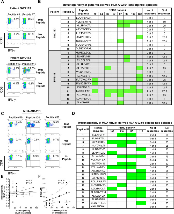
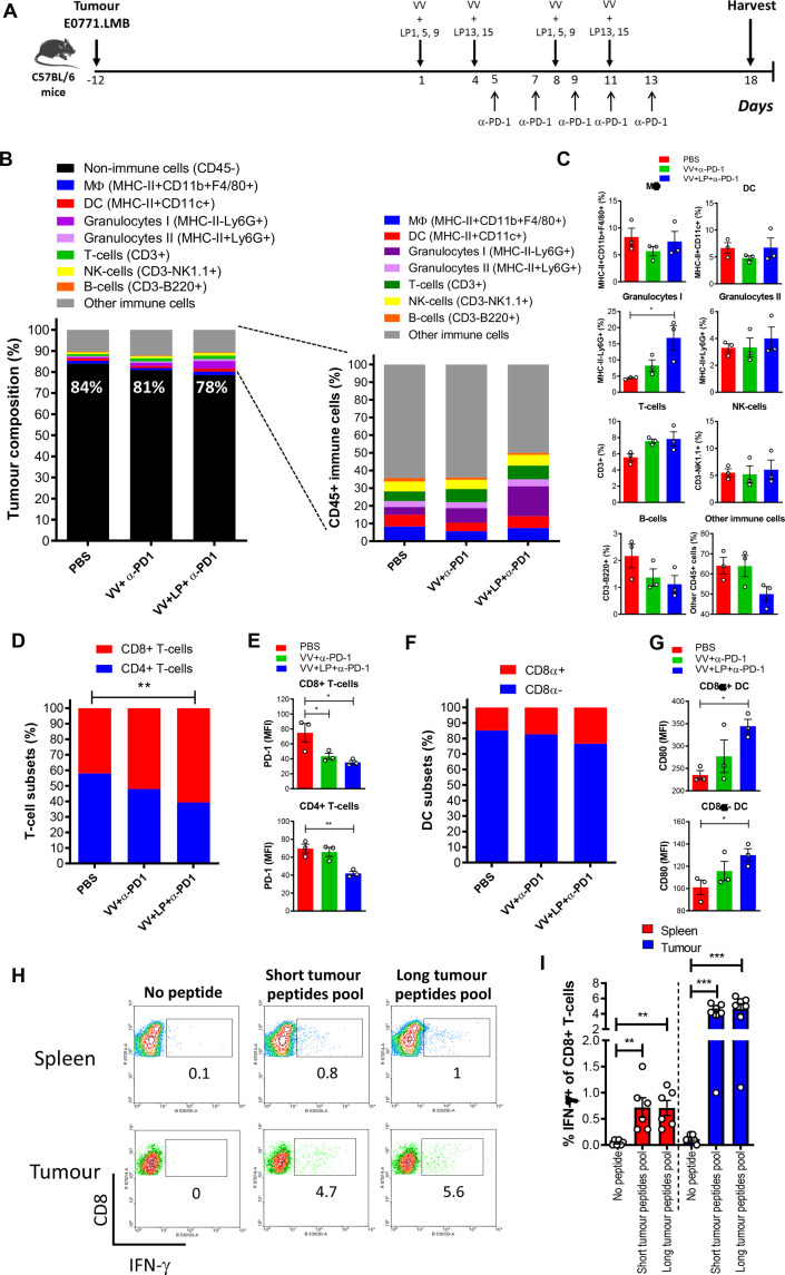
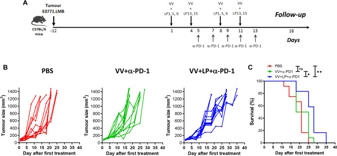
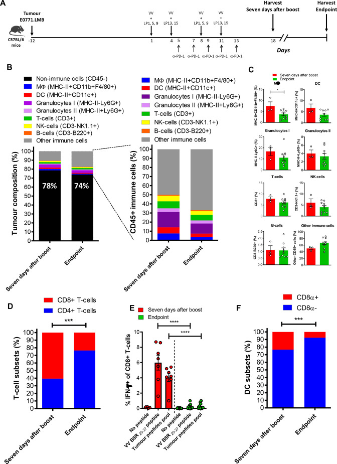
Methods: We used bioinformatic tools and cell-based assays to identify immunogenic neoantigens in TNBC patients' samples, human and murine cell lines. Immunogenicity of the neoantigens was tested in vitro (human) and ex vivo (murine) in T-cell assays. To assess the efficacy of our regimen, we used a preclinical model of TNBC where we treated tumor-bearing mice with neoantigens together with oncolytic VV and evaluated the effect on induction of neoantigen-specific CD8+T cells, tumor growth and survival.

Results: We successfully identified immunogenic neoantigens and generated neoantigen-specific CD8+T cells capable of recognizing a human TNBC cell line expressing the mutated gene. Using a preclinical model of TNBC, we showed that our tumor-specific oncolytic VV was able to change the tumor microenvironment, attracting and maintaining mature cross-presenting CD8α+dendritic cells and effector T-cells. Moreover, when delivered in a prime/boost regimen together with oncolytic VV, long peptides encompassing neoantigens were able to induce neoantigen-specific CD8+T cells, slow tumor growth and increase survival.

Conclusions: Our study provides a promising approach for the development of neoantigen-based immunotherapies for TNBC. By identifying immunogenic neoantigens and developing a delivery system through tumor-specific oncolytic VV, we have demonstrated that neoantigen-based vaccines could be effective in inducing neoantigen-specific CD8+T cells response with significant impact on tumor growth. Further studies are needed to determine the safety and efficacy of this approach in clinical trials.

Keywords: Breast Neoplasms; Immunogenicity, Vaccine; Immunotherapy; Oncolytic Virotherapy.


Conflict of interest statement

Competing interests: None declared.

Figures

Figure 1
Figure 1
TNBC-derived neoantigens are immunogenic. TNBC peptides were examined for their ability to promote immune responses in immunogenicity T-cell assays using healthy blood samples from nine donors positive for HLA-A*02:01. CD8+T cells isolated from healthy donor blood were stimulated three times with autologous DC pulsed with each of the HLA-A*02:01-binding peptides. Seven days after the last round of stimulation, CD8+T cells were challenged with T2 cells pulsed with the mutant, wild-type peptide (for MDA-MB-231 cells) or left without peptide. Intracellular IFN-γ was measured by FACS. The dot plots in (A, C) depict mutant peptides from TNBC patients and MDA-MB-231 cells, respectively, that elicited strong CD8+T cell responses from one representative donor and in (B, D) is shown the response of all donors analyzed; green represents the responses. (E) Correlation between the immunogenicity according to the IEDB score and T-cell assays; (F) Correlation between binding capacity of the peptides as per the EC50 values from cell-based binding assays and immunogenicity, expressed as the percentage of donors responding to each individual peptide. Responders in (B, D) were determined when percentage of IFN-γ-positive within the CD8+ population in the peptide-stimulated group was twice or higher compared with the non-stimulated T-cells. DCs, dendritic cells; IEDB, Immune Epitope DataBase; PBMC, peripheral blood mononuclear cell; TNBC, triple-negative breast cancer.
Figure 2
Figure 2
Validation of candidate MDA-MB-231-derived neoantigens. (A) For determination of cytotoxic activity of neoantigen-stimulated CD8+T cells, the bulk of CD8+T cells from donors 115 and 118 stimulated with the indicated neoantigen peptides were incubated overnight with carboxyfluorescein succinimidyl ester (CFSE)-labeled T2 cells pulsed with the neoantigen (red) or CFSE-labeled MDA-MB-231 cells (blue). Cell viability of target T-cells in these co-cultures was determined by staining with EthD-1 followed by flow cytometry analysis. Target T-cells gated on the CFSE-positive population that were positive for EthD-1 were considered dead cells. (B, C) CD8+T cells from a HLA-A*02:01-positive donor were stimulated as in (A) with peptide #22 and incubated overnight with peptide #22-negative/HLA-A*02:01-positive BT549 or peptide #22-positive/HLA-A*02:01-positive MDA-MB-231 cells with or without blocking anti-MHC-I antibodies. (B) Supernatants were collected and assayed for IFN-γ by ELISA and (B) cytotoxicity was determined as in (A). The data were analyzed by one-way ANOVA*p<0.05; **p<0.01; ***p<0.001. Graph shows mean±SEM. ANOVA, analysis of variance.
Figure 3
Figure 3
Induction of neoepitope specific CD8+T cells in E0771.LMB-bearing mice. Experimental setup of vaccination of E0771.LMB-bearing C57BL/6 mice with 31-mer long peptides admixed with VV is depicted in A. Seven days after the booster injection, spleens and tumors were harvested, processed and single cell suspensions were incubated ex vivo overnight with a pool of minimal short neoepitope peptides, VV B8R peptide or left with no peptide. Intracellular IFN-γ was measured by FACS and results are shown as the percentage of IFN-γ-positive cells gated on CD8+T cells. (B, D) Depict representative dot plots of the CD8+T cells response from spleen and tumor, respectively; and the graphs in C and E show the means with SEM of the percentage of IFN-γ-positive cells of CD8+T cells in spleen (C) and tumor (E) from all experiments combined. The data shown in (C, E) were analyzed by one-way ANOVA. *p<0.05; **p<0.01; ***p<0.001. ANOVA, analysis of variance; SEM, SE of the mean; VV, vaccinia virus.
Figure 4
Figure 4
Immune cell infiltration in the TME. (A) Treatment of E0771.LMB-bearing C57BL/6 mice with 31-mer long peptides admixed with VV. Seven days after the booster injection, tumors were harvested, processed and single cell suspensions were analyzed by FACS. (B) Percentage of immune and non-immune cells; (C) Percentage of immune cells subsets in the tumor; (D) Ratio between CD4+ and CD8+ T cells and (E) expression of PD-1 on CD4+ (E, upper graph) and CD8+ (E, bottom graph) T-cells; (F) Ratio between cross-presenting and conventional DC subsets and level of expression of the maturation marker CD80 on the cross-presenting CD8α+ (G, upper graph) and conventional CD8α- DC subset (G, bottom graph). Single cell suspensions of spleens and tumors were incubated overnight with a pool of short minimal neoepitopes or long peptides encompassing these neoepitopes and activation was measured by detecting IFN-γ in CD8+T cells by FACS. The dot plots in (H) depict CD8+T cell responses from one representative mouse and in (I) is shown the response of all mice analyzed. The data shown in (C, D, E, G, I) were analyzed by one-way ANOVA. *p<0.05; **p<0.01; ***p<0.001. ANOVA, analysis of variance; DC, dendritic cell; TME, tumor microenvironment; VV, analysis of variance.
Figure 5
Figure 5
In vivo VV+LP+α-PD-1 treatment delays tumor growth and increases survival. (A) Therapeutic regimen. (B) Evolution of tumor volume (MM3) after treatment as a function of time (days). (C) Kaplan-Meier survival analysis with log rank (Mantel-Cox) tests were used to assess survival. (n=12/group). *p<0.05; **p<0.01; n.s non-significant. VV, vaccinia virus.
Figure 6
Figure 6
Immune cell infiltration changes in the TME. (A) Treatment of E0771.LMB-bearing C57BL/6 mice with 31-mer long peptides admixed with VV. Seven days after the booster injection or at the endpoint, tumors were harvested, processed and single cell suspensions were analyzed by FACS. (B) Percentage of immune and non-immune cells; (C) Percentage of immune cells subsets in the tumor; (D) Ratio between CD4+ and CD8+ T cells; (E) Single cell suspensions of tumors were incubated overnight with VV B8R 20–27 peptide or a pool of short minimal neoepitopes and activation was measured by detecting IFN-γ in CD8+T cells by FACS; (F) Ratio between cross-presenting CD8α+ and conventional CD8α- DC subsets. The data shown in (C–F) were analyzed by one-way ANOVA. *p<0.05, ***p<0.001, ****p<0.0001. ANOVA, analysis of variance; DC, dendritic cell; VV, vaccinia virus.
Figure 7
Figure 7
Extended in vivo VV+LP+α-PD-1 treatment delays tumor growth and increases survival. (A) Therapeutic regimen. (B) Evolution of tumor volume (mm3) after treatment as a function of time (days). (C) Kaplan-Meier survival analysis with log rank (Mantel-Cox) tests were used to assess survival. (n=11/13/group). *p<0.05; **p<0.01; ***p<0.001; n.s, not significant. (D) VV armed with IL-21 (VVLΔTK-STC-ΔN1L-IL-21) is more efficacious than Poly I:C in increasing survival when administered in conjunction with neoantigen peptides and α-PD-1 antibodies. Kaplan-Meier survival analysis with log rank (Mantel-Cox) tests were used to assess survival. (n=8/10 mice per group). *p<0.05, **p<0.01, ****p<0.0001; n.s not significant. VV, vaccinia virus.

References

    1. Stagg J, Allard B. Immunotherapeutic approaches in triple-negative breast cancer: latest research and clinical prospects. Ther Adv Med Oncol 2013;5:169–81. 10.1177/1758834012475152 - DOI - PMC - PubMed
    1. Collignon J, Lousberg L, Schroeder H, et al. Triple-negative breast cancer: treatment challenges and solutions. Breast Cancer (Dove Med Press) 2016;8:93–107. 10.2147/BCTT.S69488 - DOI - PMC - PubMed
    1. Siroy A, Abdul-Karim FW, Miedler J, et al. MUC1 is expressed at high frequency in early-stage basal-like triple-negative breast cancer. Hum Pathol 2013;44:2159–66. 10.1016/j.humpath.2013.04.010 - DOI - PMC - PubMed
    1. Irvin WJ, Carey LA. What is triple-negative breast cancer Eur J Cancer 2008;44:2799–805. 10.1016/j.ejca.2008.09.034 - DOI - PubMed
    1. Hamdy S, Haddadi A, Hung RW, et al. Targeting dendritic cells with nano-particulate PLGA cancer vaccine formulations. Adv Drug Deliv Rev 2011;63:943–55. 10.1016/j.addr.2011.05.021 - DOI - PubMed

Publication types