Review
. 2023 Nov 27;60(12):1224-1234.
doi: 10.1136/jmg-2023-109141.

Deep phenotyping of the neuroimaging and skeletal features in KBG syndrome: a study of 53 patients and review of the literature

Francesca Peluso  1 Stefano G Caraffi  1 Gianluca Contrò  1 Lara Valeri  1   2 Manuela Napoli  3 Giorgia Carboni  4 Alka Seth  5 Roberta Zuntini  1 Emanuele Coccia  1 Guja Astrea  6 Anne-Marie Bisgaard  7 Ivan Ivanovski  8 Silvia Maitz  9 Elise Brischoux-Boucher  10 Melissa T Carter  11 Maria Lisa Dentici  12 Koenraad Devriendt  13 Melissa Bellini  14 Maria Cristina Digilio  15 Asif Doja  11 David A Dyment  11 Stense Farholt  16 Carlos R Ferreira  17 Lynne A Wolfe  17 William A Gahl  18 Maria Gnazzo  19 Himanshu Goel  10   11 Sabine Weller Grønborg  7   12 Trine Hammer  12   13 Lorenzo Iughetti  14   15 Tjitske Kleefstra  16 David A Koolen  16 Francesca Romana Lepri  19 Gabrielle Lemire  20 Pedro Louro  17 Gary McCullagh  21 Simona F Madeo  15 Annarita Milone  6 Roberta Milone  6 Jens Erik Klint Nielsen  22 Antonio Novelli  19 Charlotte W Ockeloen  16 Rosario Pascarella  3 Tommaso Pippucci  23 Ivana Ricca  6 Stephen P Robertson  24 Sarah Sawyer  20 Marie Falkenberg Smeland  25 Sander Stegmann  26 Constanze T Stumpel  26 Amy Goel  27 Juliet M Taylor  28 Domenico Barbuti  29 Annarosa Soresina  30 Maria Francesca Bedeschi  31 Roberta Battini  6   32 Anna Cavalli  33 Carlo Fusco  33 Maria Iascone  34 Lionel Van Maldergem  35 Sunita Venkateswaran  11 Orsetta Zuffardi  36 Samantha Vergano  37 Livia Garavelli  38 Allan Bayat  13   39
Affiliations
Review

Deep phenotyping of the neuroimaging and skeletal features in KBG syndrome: a study of 53 patients and review of the literature

Francesca Peluso et al. J Med Genet. .

Abstract

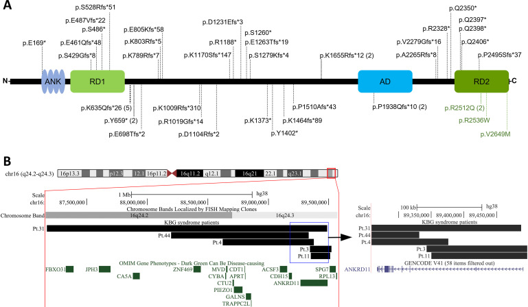
Background: KBG syndrome is caused by haploinsufficiency of ANKRD11 and is characterised by macrodontia of upper central incisors, distinctive facial features, short stature, skeletal anomalies, developmental delay, brain malformations and seizures. The central nervous system (CNS) and skeletal features remain poorly defined.

Methods: CNS and/or skeletal imaging were collected from molecularly confirmed individuals with KBG syndrome through an international network. We evaluated the original imaging and compared our results with data in the literature.

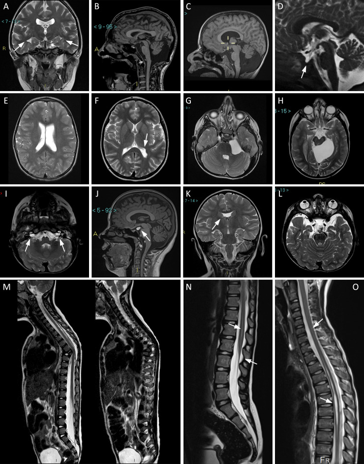
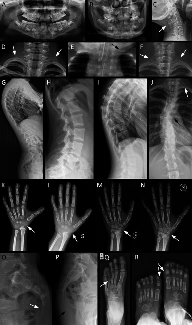
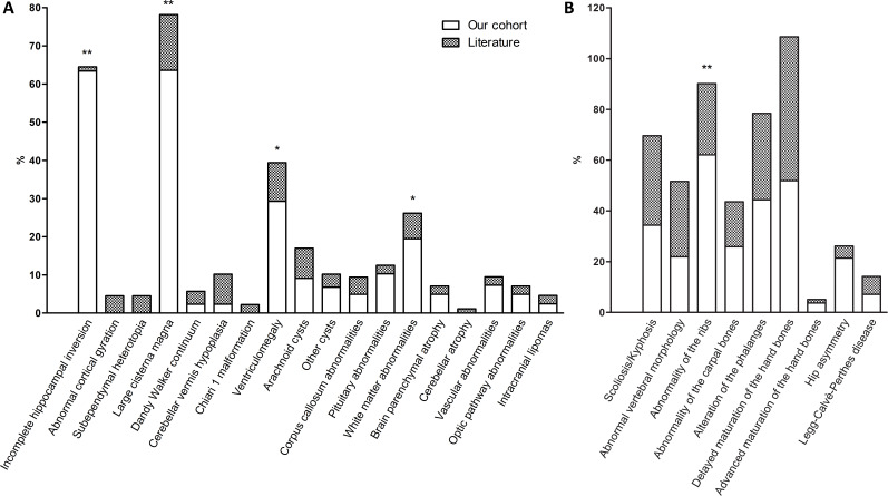
Results: We identified 53 individuals, 44 with CNS and 40 with skeletal imaging. Common CNS findings included incomplete hippocampal inversion and posterior fossa malformations; these were significantly more common than previously reported (63.4% and 65.9% vs 1.1% and 24.7%, respectively). Additional features included patulous internal auditory canal, never described before in KBG syndrome, and the recurrence of ventriculomegaly, encephalic cysts, empty sella and low-lying conus medullaris. We found no correlation between these structural anomalies and epilepsy or intellectual disability. Prevalent skeletal findings comprised abnormalities of the spine including scoliosis, coccygeal anomalies and cervical ribs. Hand X-rays revealed frequent abnormalities of carpal bone morphology and maturation, including a greater delay in ossification compared with metacarpal/phalanx bones.

Conclusion: This cohort enabled us to describe the prevalence of very heterogeneous neuroradiological and skeletal anomalies in KBG syndrome. Knowledge of the spectrum of such anomalies will aid diagnostic accuracy, improve patient care and provide a reference for future research on the effects of ANKRD11 variants in skeletal and brain development.

Keywords: Congenital, Hereditary, and Neonatal Diseases and Abnormalities; Genetic Research; Pathological Conditions, Signs and Symptoms; Patient Care; Radiology.


Conflict of interest statement

Competing interests: None declared.

Figures

Figure 1
Figure 1
Representative MRI images in our cohort. Arrows indicate the relevant details. (A) Pt. 16, coronal T2-weighted view: incomplete hippocampal inversion, bilateral. (B) Pt. 7, sagittal T1: large cisterna magna. (C) Pt. 4, sagittal T1: Dandy-Walker variant. (D) Pt. 25, sagittal T2: empty sella. (E) Pt. 23, axial T2 TSE: dilated and asymmetric lateral ventricles. (F) Pt. 10, axial T2 TSE: left trigonal enlargement. (G) Pt. 5, axial T2: arachnoid cyst in left cerebellopontine angle. (H) Pt. 31, axial T2: arachnoid cyst in left ambient cistern. (I) Pt. 12, axial T2: bilateral patulous internal auditory canal. (J) Pt. 16, sagittal T1: lipoma tuber cinereum. (K) Pt. 3, coronal T2: partial agenesis of septum pellucidum. (L) Pt. 12, axial T2: bilateral persistent hyperplastic primary vitreous. (M) Pt. 52, sagittal T2: focal dorsal kyphosis due to anomalous thoracic T6-T7 vertebral differentiation. (N) Pt. 51, sagittal T2 TSE: hydromyelia and low-lying Conus medullaris. (O) Pt. 38, sagittal T2 TSE: hydrosyringomyelia. Pt., participant.
Figure 2
Figure 2
Main radiological characteristics of selected patients from our cohort, representative of the skeletal features of KBG syndrome. Arrows indicate the most relevant details. (A) Pt. 10: orthopantomography showing macrodontia of the permanent upper incisors and dental crowding. (B) Pt. 32: macrodontia of the permanent upper incisors, dental crowding. (C) Pt. 1: cervical C2/C3 vertebral fusion. (D) Pt. 21: bilateral cervical ribs. (E) Pt. 12: supernumerary cervical rib on the left side. (F) Pt. 10: bilateral C7 hypertrophic transverse process. (G) Pt. 26: thoracic hyperkyphosis. (H–J) Spinal column anomalies of Pt. 39: tall lumbar vertebral bodies (H), kyphosis (I), left cervical rib and scoliosis due to thoracic hemivertebrae (J). (K–N) Evolution of the main anomalies of hand bones over time: delayed carpal ossification with absence of the proximal row at about 3 years (K) and 6 years of age (L); partial fusion of the lunate and triquetral carpal bones at 9 years (M); complete fusion of the lunate and triquetral bones at 10 years (N). (O) Pt. 32: agenesis of the coccyx: only the outline of the first coccygeal vertebra is present. (P) Pt. 26: supernumerary coccygeal vertebrae. (Q) Pt. 13: short and dysmorphic metatarsal and phalanges of fourth ray, bilaterally (right foot not shown). (R) Pt. 12: broad and short metatarsal and phalanges of first ray, bilaterally. Pt., participant.
Figure 3
Figure 3
Intragenic and structural variants in our cohort of patients with KBG. (A) ANKRD11 intragenic variants plotted on the protein diagram; numbers in parentheses indicate recurrence of a variant. (B) Large deletions in chromosome 16q24 visualised in UCSC Genome Browser (http://genome.ucsc.edu), assembly hg38. Zoom-in on the right shows the position of breakpoints relative to ANKRD11 exons. AD, activation domain; ANK, ankyrin repeats; RD, repressor domain.
Figure 4
Figure 4
Stacked histograms comparing the imaging findings in our cohort with those reported in the literature. (A) Brain abnormalities at diagnostic imaging. Cortical gyration anomalies consist solely of the four mild alterations reported in a volumetric study, and no macroscopic gyration defects were detected by standard MRI in any of the other publications. (B) Skeletal abnormalities at diagnostic imaging. Skeletal features mainly determined through physical examination rather than X-ray imaging, such as wide anterior fontanel and hip dysplasia, were not included because of the difficulty of establishing accurate ratios of evaluated patients. *Significant difference at p<0.05. **Significant difference at p<0.001.

References

    1. Sirmaci A, Spiliopoulos M, Brancati F, et al. . Mutations in Ankrd11 cause KBG syndrome, characterized by intellectual disability, skeletal malformations, and macrodontia. Am J Hum Genet 2011;89:289–94. 10.1016/j.ajhg.2011.06.007 - DOI - PMC - PubMed
    1. Skjei KL, Martin MM, Slavotinek AM. KBG syndrome: report of twins, neurological characteristics, and delineation of diagnostic criteria. Am J Med Genet A 2007;143A:292–300. 10.1002/ajmg.a.31597 - DOI - PubMed
    1. Gnazzo M, Lepri FR, Dentici ML, et al. . KBG syndrome: common and uncommon clinical features based on 31 new patients. Am J Med Genet A 2020;182:1073–83. 10.1002/ajmg.a.61524 - DOI - PubMed
    1. Herrmann J, Pallister PD, Tiddy W, et al. . The KBG syndrome-a syndrome of short stature, characteristic Facies, mental retardation, macrodontia and skeletal anomalies. Birth Defects Orig Artic Ser 1975;11:7–18. - PubMed
    1. Low K, Ashraf T, Canham N, et al. . Clinical and genetic aspects of KBG syndrome. Am J Med Genet A 2016;170:2835–46. 10.1002/ajmg.a.37842 - DOI - PMC - PubMed

Publication types

MeSH terms

Supplementary concepts