. 2023 Aug 16;14(1):4966.
doi: 10.1038/s41467-023-40531-3.

Integrin β3 directly inhibits the Gα13-p115RhoGEF interaction to regulate G protein signaling and platelet exocytosis

Affiliations

Integrin β3 directly inhibits the Gα13-p115RhoGEF interaction to regulate G protein signaling and platelet exocytosis

Yaping Zhang et al. Nat Commun. .

Abstract

The integrins and G protein-coupled receptors are both fundamental in cell biology. The cross talk between these two, however, is unclear. Here we show that β3 integrins negatively regulate G protein-coupled signaling by directly inhibiting the Gα13-p115RhoGEF interaction. Furthermore, whereas β3 deficiency or integrin antagonists inhibit integrin-dependent platelet aggregation and exocytosis (granule secretion), they enhance G protein-coupled RhoA activation and integrin-independent secretion. In contrast, a β3-derived Gα13-binding peptide or Gα13 knockout inhibits G protein-coupled RhoA activation and both integrin-independent and dependent platelet secretion without affecting primary platelet aggregation. In a mouse model of myocardial ischemia/reperfusion injury in vivo, the β3-derived Gα13-binding peptide inhibits platelet secretion of granule constituents, which exacerbates inflammation and ischemia/reperfusion injury. These data establish crucial integrin-G protein crosstalk, providing a rationale for therapeutic approaches that inhibit exocytosis in platelets and possibly other cells without adverse effects associated with loss of cell adhesion.


Conflict of interest statement

University of Illinois at Chicago holds patents related to this study. X.D. holds equity interests in DMT, Inc., which licenses UIC technology. The remaining authors declare no competing interests.

Figures
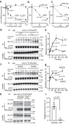
Fig. 1
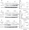
Fig. 1. Integrin β3 cytoplasmic domain inhibits Gα13-p115RhoGEF interaction in purified protein binding assays and in platelets.
a Binding of purified GST-tagged RGS domain of p115RhoGEF (GST-p115RGS) to purified Histidine-tagged Gα13 (His-Gα13) coated on microtiter plates is inhibited by increasing concentrations of purified GST-β3 cytoplasmic domain fusion protein (β3CD) compared with control GST, 3 independent experiments. b, c Inhibition of the binding of purified GST-p115RGS to microtiter well-coated His-Gα13 (b) and the binding of His-Gα13 to microtiter well-coated GST-p115RGS (c) by increasing concentration of mP6 peptide or a scrambled peptide with the same amino acid composition (mP6 Scr), 3 independent experiments. d, e Co-immunoprecipitation of p115RhoGEF and Gα13 in WT (C57BL/6J) and β3-/- mouse platelets stimulated with thrombin (0.035 U/ mL) for increasing lengths of time. d A representative Western blot; e, quantification of data from 4 independent experiments. f, g Co-immunoprecipitation of p115RhoGEF and Gα13 in resting (0) and 0.03 U/mL thrombin-stimulated WT mouse platelets pre-treated with RGDS or control RGES peptide (2 mM). f A representative western blot; g quantification of data from four independent experiments. h, i Co-immunoprecipitation of p115RhoGEF and Gα13 in wild-type mouse platelets pre-treated with control peptide (myr-FAAAKL) or mP6 peptide (200 μM, DMSO), stimulated with thrombin (0.03 U/ mL) for 10 seconds. h A representative Western blot; i Quantification of co-immunoprecipitation of Gα13. Data are from three independent experiments. Platelets used in (dg) were pre-treated with 500 μM aspirin to exclude the differential influence of secondary TXA2 production on Gα13 pathway of WT and β3-/- platelets. All data are shown as mean ± SEM. Statistical analysis was done using Student’s t-test, two tailed. Source data are provided as a Source Data file.
Fig. 2
Fig. 2. The role of β3 integrins in inhibiting thrombin-induced RhoA activation.
a, b Rhotekin-RBD bead pull down assay was used to analyze RhoA activation in WT or β3-/- mouse platelets stimulated with thrombin (0.03 U/mL) at various time points, 4 independent experiments. c, d RhoA pull down assay with RGDS or RGES-pre-treated (2 mM, 3 min, 22 °C) mouse platelets stimulated with thrombin (0.03 U/mL) for the indicated time points, four independent experiments. e, f RhoA pull down assay with 0.03 U/ mL thrombin-stimulated mouse platelets pre-treated with control peptide or mP6 peptide (100 μM, DMSO) for 3 min at 22 °C, three independent experiments. a, c, e Representative western blots; b, d, f quantification of western blot images as analyzed using ImageJ. All data are shown as mean ± SEM. Statistical significance was determined using Student’s t test, (f) paired Student’s t test, two tailed. Source data are provided as a Source Data file.
Fig. 3
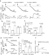
Fig. 3. β3 integrins negatively regulate thrombin-induced platelet granule secretion.
a Aggregation and ATP secretion traces of washed WT and β3-/- platelets stimulated with thrombin. b Quantification of ATP secretion for a, three independent experiments. c Aggregation and ATP secretion traces of washed human platelets pre-incubated with vehicle or 10 μg/mL Eptifibatide for 3 min at 22 °C and stimulated with thrombin. d Quantification of ATP secretion for c, three independent experiments. e Flow cytometry analysis of P-selectin expression on mouse platelets pre-treated with control vehicle or Eptifibatide (20 μg/mL) for 3 min at 22 °C and stimulated with 0.03 U/mL thrombin at 37 °C with stirring at 1000 rpm in an aggregometer. Three independent experiments. All data are shown as mean ± SEM. Statistical significance was determined using Student’s t test, two tailed. Source data are provided as a Source Data file.
Fig. 4
Fig. 4. β3 integrins negatively regulate integrin-independent platelet secretion but stimulate integrin-dependent secretion.
a Aggregation and ATP secretion traces of washed WT and β3-/- platelets induced by U46619 at 37 °C. Note the two waves of ATP secretion in WT platelets and a higher single (first) secretion wave in β3-/- platelets. b, c Quantification of the first (b) and second (c) waves of secretion as shown in a, three independent experiments. d Aggregation and ATP secretion traces of washed human platelets pre-incubated with vehicle or 10 μg/mL Eptifibatide for 3 min at 22 °C followed by stimulation with U46619 at 37 °C. e Quantification of secretion data from (d), 3 independent experiments. f U46619-induced ATP secretion traces of β3-/- platelets pre-incubated with control vehicle or 10 µM Y27632 for 3 min at 22 °C. All data are shown as mean ± SEM. Statistical significance was determined using Student’s t-test, two tailed. Source data are provided as a Source Data file.
Fig. 5
Fig. 5. Gα13 stimulates both integrin-independent and integrin-dependent granule secretion.
a Washed WT(Gα13fl/fl) and Gα13-/- mouse platelets were solubilized and immunoblotted with antibodies specifically recognizing mouse Gα13, and integrin β3. b Aggregation and ATP secretion traces of washed WT and Gα13-/- platelets stimulated with thrombin. c Quantification of secretion as shown in b, four independent experiments. d Aggregation and ATP secretion traces of WT and Gα13-/- platelets stimulated with 2 μmol/L U46619. e Quantification of ATP secretion as shown in d, six independent experiments. Aggregation assays were performed without exogenously added fibrinogen. All data are shown as mean ± SEM. Data were analyzed using Student’s t-test, (e) paired Student’s t-test, two tailed. Source data are provided as a Source Data file.
Fig. 6
Fig. 6. Gα13 antagonistic peptide mP6, inhibits both integrin-independent and integrin-dependent granule secretion.
a, c ATP secretion traces of washed mouse platelets pre-incubated with 20 μM control FAAAKL or mP6 high loading peptide nanoparticles (HLPNs) and then stimulated with 0.03 U/mL thrombin (a) or 3 μmol/L U46619 (c). b, d Quantification of ATP secretion as shown in a, n = 3 or (c), n = 4, independent experiments. e, f Effects of control FAAAKL or mP6 HLPNs (20 μM) on serotonin secretion (e), or on P-selectin expression (f), in mouse platelets stimulated with 0.03U/ mL thrombin in an aggregometer stirring at 37 °C and 1000 rpm. n = 3, independent experiments. The assays were performed using washed platelets without adding fibrinogen. g Plasma serotonin levels in sham, saline, and M3mP6 HLPN treatment groups 24 h after myocardial infarction with reperfusion. Sham group, n = 12; Saline group, n = 14; M3mP6 group, n = 13, independent animals. Data in b, df are shown as mean ± SEM and in g are shown as median ± SEM. Statistical significance was determined using Student’s t-test, two tailed. h A schematic of crosstalk between the (G protein-coupled receptor) GPCR-coupled Gα13 pathway and integrin αIIbβ3 in regulating RhoA activation and platelet granule secretion. Left panel: GPCR agonists such as thrombin induce Gα13-p115RhoGEF-dependent activation of RhoA, which mediates integrin-independent granule secretion. Center panel: following integrin ligation, Gα13 interacts with the β3 cytoplasmic domain ExE motif, which inhibits Gα13 binding to p115RhoGEF, resulting in inhibition of p115RhoGEF-mediated RhoA activation and integrin-independent granule secretion. Gα13 binding to β3, however, also mediates integrin outside-in signaling leading to Src activation and integrin-dependent granule secretion. Right Panel: The β3-derived mP6 peptide binds to Gα13 to inhibit Gα13 interacting with both p115-RhoGEF and β3. Thus, mP6 potently inhibits both integrin-independent and integrin-dependent secretion without affecting integrin ligation and primary platelet adhesion/aggregation necessary for normal hemostasis. Source data are provided as a Source Data file.

Similar articles

Cited by

References

    1. Wootten D, Christopoulos A, Marti-Solano M, Babu MM, Sexton PM. Mechanisms of signalling and biased agonism in G protein-coupled receptors. Nat. Rev. Mol. Cell Biol. 2018;19:638–653. - PubMed
    1. Hauser AS, Attwood MM, Rask-Andersen M, Schioth HB, Gloriam DE. Trends in GPCR drug discovery: new agents, targets and indications. Nat. Rev. Drug Discov. 2017;16:829–842. - PMC - PubMed
    1. Hauser AS, et al. Pharmacogenomics of GPCR drug targets. Cell. 2018;172:41–54.e19. - PMC - PubMed
    1. Hilger D, Masureel M, Kobilka BK. Structure and dynamics of GPCR signaling complexes. Nat. Struct. Mol. Biol. 2018;25:4–12. - PMC - PubMed
    1. Oldham WM, Hamm HE. Heterotrimeric G protein activation by G-protein-coupled receptors. Nat. Rev. Mol. Cell Biol. 2008;9:60–71. - PubMed

Publication types

Substances