

Comment
doi: 10.1038/s41467-023-40507-3.
Fluid signal suppression characteristics of 3D-FLAIR with a T2 selective inversion pulse in the skull base
Affiliations
- PMID: 37587125
- PMCID: PMC10432414
- DOI: 10.1038/s41467-023-40507-3
Item in Clipboard
Comment
Fluid signal suppression characteristics of 3D-FLAIR with a T2 selective inversion pulse in the skull base
Nat Commun.
.
No abstract available
Conflict of interest statement
The authors declare no competing interests.
Figures
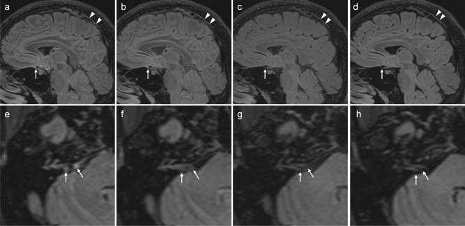
3D-FLAIR image obtained with T2-selective inversion pulse (a, e), that with T2-preparation pulses (b, f), that with conventional inversion pulse (c, g), and that with conventional inversion using longer repetition time of 8000 msec (d, h). MR imaging was performed at Siemens 3 Tesla using a 32-channel array head coil similar to that used in the article in ref. . MR imaging parameters are comparable to that in the article by Albayram et al.; TR/TE/TI: 5000/387/1700 for (a–c), and (e, f), 8000/387/2370 for (d, h), 0.9 mm resolution, using fat suppression. Sagittal images (a–d) and axial images (e–h) are presented. A similar contrast between gray/white matter as in the images in the article by Albayram et al. was achieved in (a, e), (b, f), and (d, h). Linear high signal along superior sagittal sinus, which is presumed to be lymphatic tissue, is visualized in all images (arrow heads in a–d). Linear high signal (arrow) presumed to be lymphatic tissue anterior to the pituitary gland is visualized most prominently in (a) as in the images in the article by Albayram et al., less prominently in (b) and far less prominently in (c, d). Nodular high signal areas (arrows) near the orifice of internal auditory canal which are presumed to be lymphatic tissue are visualized most prominently in (e), far less prominently in (f) and not in (g, h). Judging from these volunteer images, the images in the article by Albayram et al. is thought to be obtained with 3D-FLAIR using a T2-selective inversion pulse.
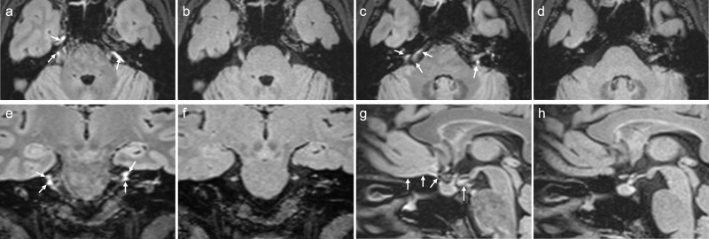
MR imaging was performed at Siemens 3 Tesla using a 32-channel array head coil similar to that used in the article by Albayram et al.. The scan parameters are also comparable to those used in the article by Albayram et al. In the 3D-FLAIR imaging with a T2-selective inversion pulse (a, c, e, g), similar contrast between gray/white matter is achieved as in the images shown in the article by Albayram et al. even with the same repetition time of 5000 msec. In 3D-FLAIR imaging with a conventional inversion pulse (b, d, f, h), less contrast between gray/white matter is provided than observed in the images of the article by Albayram et al. using the same repetition time of 5000 msec. High signal intensity, which corresponds to the areas presumed to be lymphatic tissues in Fig. 4 of the article by Albayram et al. (arrows in a, c, e, g) is only visible in the 3D-FLAIR images obtained with a T2-selective inversion pulse.
Comment in
-
Reply to: Fluid signal suppression characteristics of 3D-FLAIR with a T2 selective inversion pulse in the skull base.Nat Commun. 2023 Aug 16;14(1):4914. doi: 10.1038/s41467-023-40509-1. Nat Commun. 2023. PMID: 37587132 Free PMC article. No abstract available.
Comment on
-
Non-invasive MR imaging of human brain lymphatic networks with connections to cervical lymph nodes.Nat Commun. 2022 Jan 11;13(1):203. doi: 10.1038/s41467-021-27887-0. Nat Commun. 2022. PMID: 35017525 Free PMC article.
References
Publication types
MeSH terms
LinkOut - more resources
Full Text Sources
