. 2023 Aug 16;14(1):4375.
doi: 10.1038/s41467-023-39873-9.

Platelet-derived exerkine CXCL4/platelet factor 4 rejuvenates hippocampal neurogenesis and restores cognitive function in aged mice

Affiliations

Platelet-derived exerkine CXCL4/platelet factor 4 rejuvenates hippocampal neurogenesis and restores cognitive function in aged mice

Odette Leiter et al. Nat Commun. .

Abstract

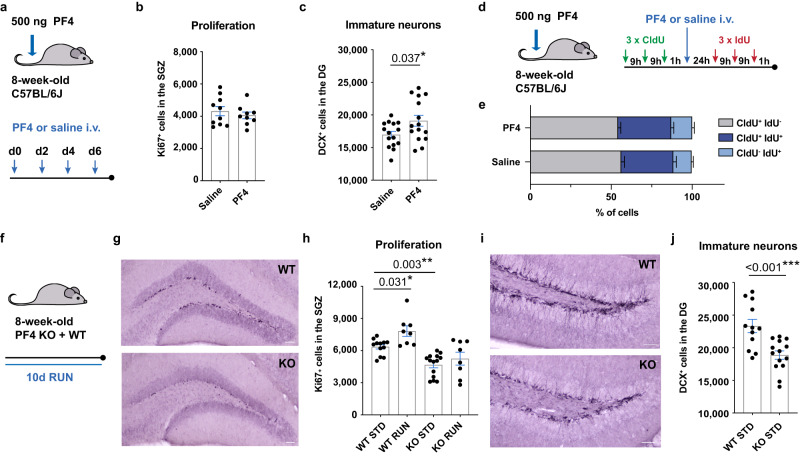
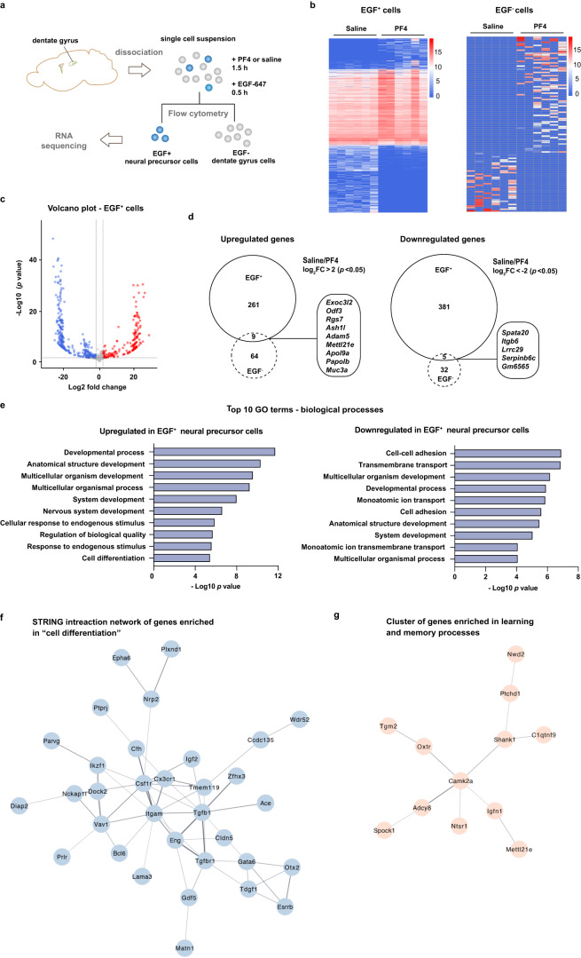
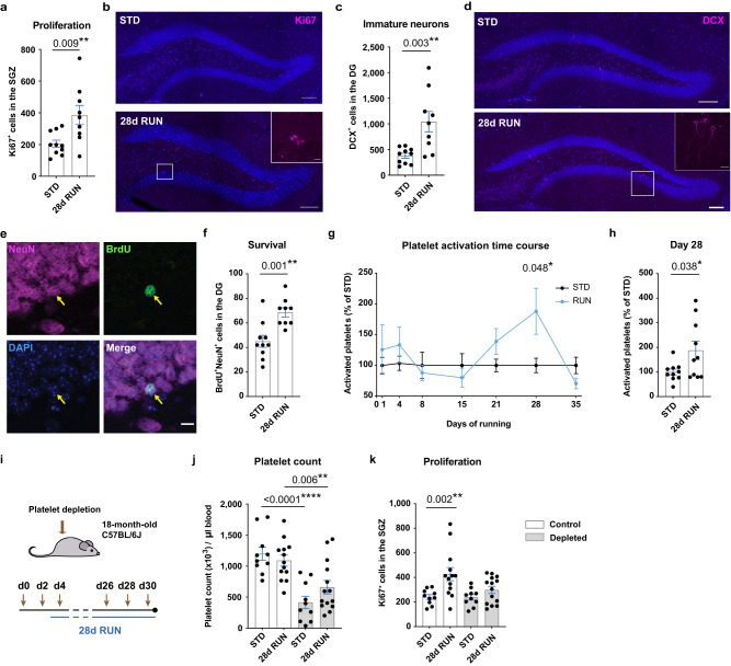
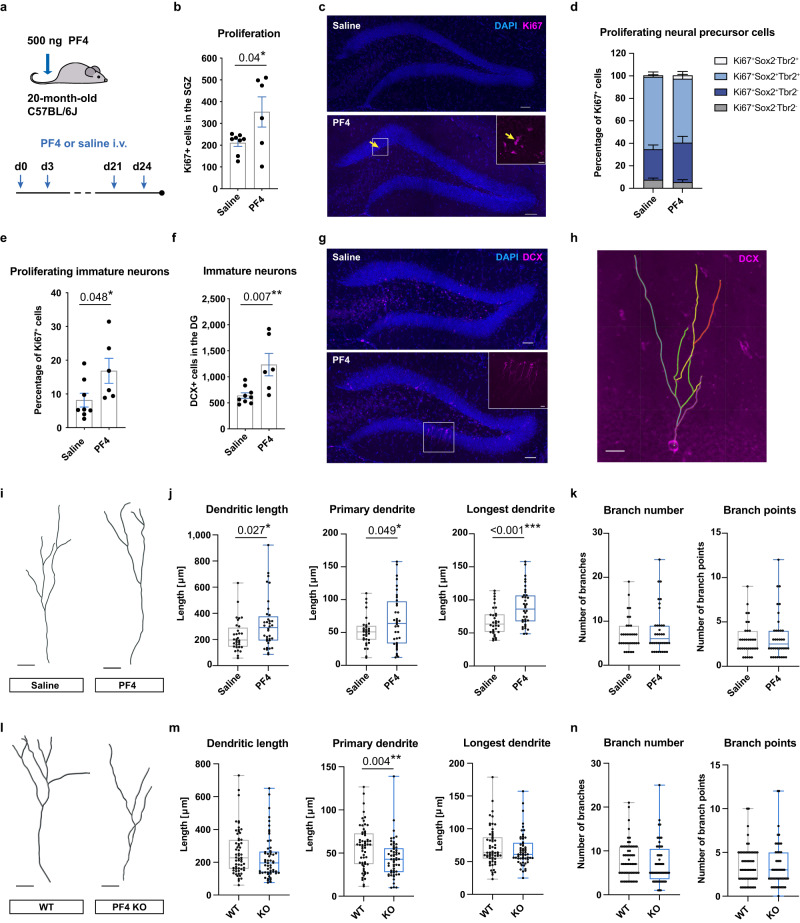
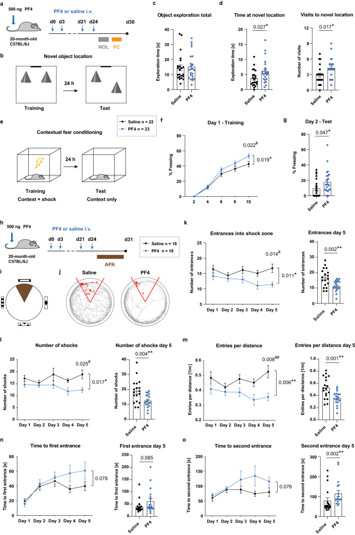
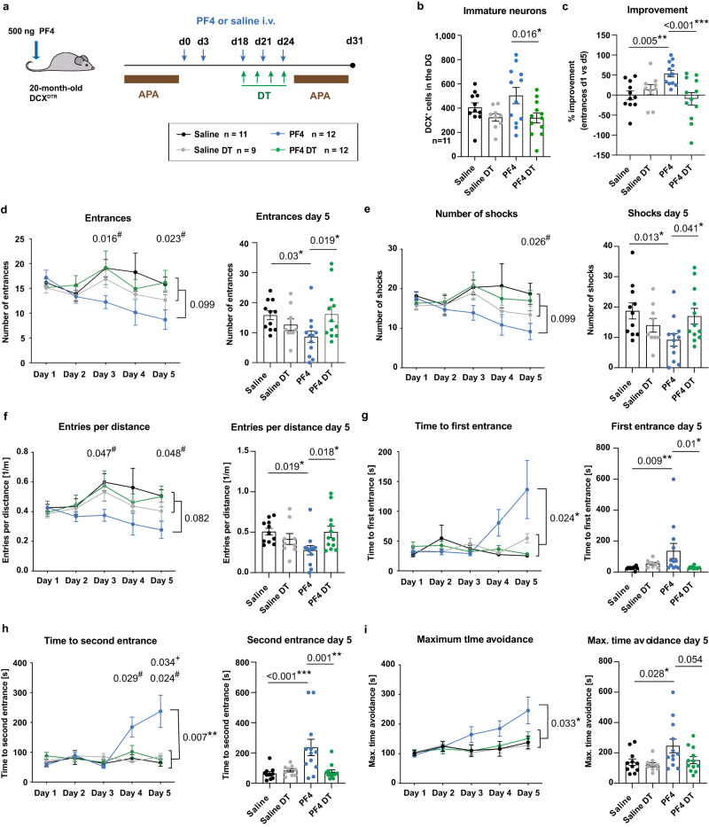
The beneficial effects of physical activity on brain ageing are well recognised, with exerkines, factors that are secreted into the circulation in response to exercise, emerging as likely mediators of this response. However, the source and identity of these exerkines remain unclear. Here we provide evidence that an anti-geronic exerkine is secreted by platelets. We show that platelets are activated by exercise and are required for the exercise-induced increase in hippocampal precursor cell proliferation in aged mice. We also demonstrate that increasing the systemic levels of the platelet-derived exerkine CXCL4/platelet factor 4 (PF4) ameliorates age-related regenerative and cognitive impairments in a hippocampal neurogenesis-dependent manner. Together these findings highlight the role of platelets in mediating the rejuvenating effects of exercise during physiological brain ageing.


Conflict of interest statement

The authors declare no competing interests.

Figures

Fig. 1
Fig. 1. Systemic PF4 enhances adult hippocampal neurogenesis in vivo.
a Experimental design of PF4 injection paradigm in young mice. b, c Intravenous (i.v.) PF4 injections for 1 week did not affect neural precursor cell proliferation in the subgranular zone (SGZ; n = 10 mice in saline group; n = 9 mice in PF4 group; counts of one hemisphere), but increased the number of doublecortin+ (DCX+) cells (n = 15 mice per group; counts of one hemisphere). d Experimental design of the double-labelling paradigm with CldU and IdU. e Acute administration of PF4 did not affect neural precursor cell proliferation, including the recruitment of cells from quiescence (n = 20 mice per group). f Running paradigm of PF4 knockout (KO) mice. g Representative images of Ki67+ cells in the SGZ of PF4 KO and wildtype (WT) mice. Scale bar: 50 μm. h PF4 KO mice show a significant reduction in the number of proliferating cells compared to wildtype mice. Physical exercise did not increase neural precursor proliferation in PF4 KO mice (WT STD n = 12; WT RUN n = 8; KO STD n = 14; KO RUN n = 8; counts of one hemisphere). i Representative images of DCX+ cells in the dentate gyrus (DG) of PF4 KO and wildtype mice. Scale bar: 50 μm. j PF4 KO mice have significantly lower levels of baseline neurogenesis compared to wildtype littermates (WT STD n = 12; KO STD n = 14; counts of one hemisphere). STD standard-housing, RUN 10-day running. Bars are mean ± SEM. Statistical analysis was performed using unpaired Student’s two-tailed t tests in (c) and (j), and one-way ANOVA with Sidak comparison in (h). *p < 0.05, **p < 0.01, ***p < 0.001. Source data are provided as Source Data file.
Fig. 2
Fig. 2. PF4 induces transcriptomic changes in adult neural precursor cells.
a Schematic illustration. Primary cells were isolated from the dentate gyrus of 8-week-old C57BL/6J mice and treated with either 100 ng/ml PF4 or saline, then stained with epidermal growth factor (EGF) conjugated with Alexa Fluor® 647 (EGF-647). EGF+ and EGF cells were collected using flow cytometry followed by RNA sequencing of six samples from each cell population. b Heatmaps of significantly changed genes induced by PF4 in EGF+ cells (left) and EGF cells (right). Colour code represents log2 normalised expression. c Volcano plot of differentially expressed genes in EGF+ cells treated with PF4 or saline, with significantly upregulated genes in red (log2 fold change >2 and adjusted p value < 0.05) and significantly downregulated genes in blue (log2 fold change < −2 and adjusted p value < 0.05). Wald test and Benjamini–Hochberg correction. d Euler diagrams comparing upregulated (left side) and downregulated (right side) genes between EGF+ cells and EGF cells (Wald test and Benjamini–Hochberg correction). e Gene ontology (GO) enrichment analysis of significantly upregulated (left panel) and significantly downregulated (right panel) genes in EGF+ cells treated with PF4 compared to saline-treated controls. Bar graphs show the top 10 significantly enriched GO terms for biological processes ranked by false discovery rate-adjusted p values (g:SCS multiple testing correction with significance threshold of 0.05). For a complete list of GO terms please see Supplementary Data 1. f A STRING interaction network of genes enriched in the GO category “cell differentiation” reveals a distinct gene cluster (genes with no connection are not depicted). g Markov clustering of the genes that were upregulated in EGF+ cells following PF4 treatment revealed a STRING network of 12 genes involved in learning and memory. FC fold change. Source data are provided as Source Data file.
Fig. 3
Fig. 3. Exercise alters the platelet proteomic signature in young and aged mice.
a Heatmap of platelet proteins significantly changed following 4 days of exercise in 8-week-old mice. b Volcano plots of quantified and differentially expressed proteins in the platelet lysate of 8-week-old (young) and 18-month-old (aged) mice following 4 days of running. Significantly upregulated proteins are labelled red (p < 0.05 and fold change >1.2) and significantly downregulated proteins are shown in blue (p < 0.05 and fold change < −1.2). Pairwise relative-abundance comparison using two-tailed t-tests. c Venn diagram showing the number of proteins significantly upregulated after 4 days of exercise in young and aged mice, including 5 proteins determined in both cohorts (p < 0.05 and fold change >1.2; pairwise relative-abundance comparison using two-tailed t-tests). d Gene ontology (GO) enrichment analysis of significantly increased proteins in the platelet lysate of young and aged running mice (for 4 days) compared to standard-housed controls. Bar graphs show the top 10 significantly enriched GO terms for biological processes ranked by Benjamini–Hochberg false discovery rate-corrected p values. For a complete list of GO terms please see Supplementary Data 6 and 7. e Venn diagram showing the number of enriched biological processes following a GO analysis with proteins significantly increased after 4 days of exercise in young and aged mice (p < 0.05 and fold change >1.2; pairwise relative-abundance comparison using two-tailed t-tests). The box highlights some of the 61 biological processes that were enriched in both cohorts. f Neurogenesis- and neuronal development-related GO terms resulting from the GO enrichment analysis of significantly increased proteins in the platelet lysate of young running mice compared to standard-housed controls. Bar graphs show significantly enriched neurogenesis-related GO terms for biological processes ranked by Benjamini–Hochberg false discovery rate-corrected p values. STD standard-housing, RUN running, FC fold change. Source data are provided as Source Data file.
Fig. 4
Fig. 4. Platelets are required for the exercise-induced increase in hippocampal neurogenesis in aged mice.
a Number of proliferating cells in the subgranular zone (SGZ) of 18-month-old C57BL/6J mice (STD n = 10 mice; 28 days RUN n = 9 mice). b Representative image of Ki67+ cells in the dentate gyrus (DG). Scale bars: 100 and 10 μm in box highlighting a cluster of proliferating cells. c Number of DCX+ cells in the DG of 18-month-old C57BL/6J mice (STD n = 10 mice; 28 days RUN n = 9 mice). d Representative image of DCX+ cells in the DG. Scale bars: 100 μm and 15 μm in box highlighting a cluster of DCX+ cells. e Representative image of BrdU/NeuN staining with yellow arrows indicating the same cell in each panel (BrdU—green; NeuN—magenta; DAPI—blue). Scale bar: 10 μm. f Number of BrdU+NeuN+ cells in aged C57BL/6J mice (STD n = 10 mice; 28 days RUN n = 9 mice). g Time course of platelet activation showing a defined peak at 28 days of exercise (n = 10 mice per group). h Individual values of platelet activation on day 28 (n = 10 mice per group). i Experimental design. j Platelet count in exercising and standard-housed mice receiving with control serum (white bars) or antiplatelet serum (grey bars; n = 10 mice in STD groups; n = 13 mice in 28 days RUN control group; n = 14 mice in 28 days RUN platelet-depleted group). k Number of proliferating cells in exercising and standard-housed mice receiving control serum (white bars) or antiplatelet serum (grey bars; n = 10 mice in STD groups; n = 13 mice in 28 days RUN control group; n = 14 mice in 28 days RUN platelet-depleted group). Bars are mean ± SEM. Statistical analysis was performed using unpaired Student’s two-tailed t tests in (a, c, f, h), two-way ANOVA with Sidak post hoc comparison in (g), and one-way ANOVA with Sidak post hoc comparison in (j) and (k). *p < 0.05, **p < 0.01, ****p < 0.0001. Source data are provided as Source Data file.
Fig. 5
Fig. 5. Systemic PF4 increases adult hippocampal neurogenesis in aged mice.
a Experimental design. b 20-month-old C57BL/6J mice receiving intravenous (i.v.) PF4 injections showed significant increases in Ki67+ cells in the subgranular zone (SGZ; saline n = 8 mice; PF4 n = 6 mice). c Representative image of Ki67+ cells. The yellow arrow shows a trio of proliferating cells. Scale bars: 50 μm and 10 μm in box highlighting three proliferating cells. d Phenotyping of the Ki67+ cells in saline- and PF4-treated mice (saline n = 8 mice; PF4 n = 6 mice). PF4 increased the percentage of proliferating DCX+ cells (e) and the total number of DCX+ cells (f) in the dentate gyrus (DG) compared to saline-treated controls (saline n = 8 mice (e) and n = 9 mice (f); PF4 n = 6 mice). g Representative images of DCX staining. Scale bar: 50 and 15 μm in box highlighting a cluster of DCX+ cells. h Representative image of dendritic branch tracing, showing a postmitotic DCX+ cell. Scale bar: 15 μm. i Representative reconstruction of DCX+ postmitotic cells. Scale bars: 20 μm. j Postmitotic DCX+ cells of mice receiving PF4 showed increased total dendritic length, longer primary dendrites, and length of the longest dendrite per traced cell (saline n = 32 cells; PF4 n = 36 cells). k PF4 did not affect dendritic branch numbers or the number of branch points (saline n = 32 cells; PF4 n = 36 cells). l Representative reconstruction of DCX+ postmitotic cells. Scale bars: 15 μm. m PF4 KO mice displayed significantly shorter primary dendrites in the postmitotic DCX+ cells compared to WT littermates (WT n = 59 cells; PF4 KO n = 56 cells). n Dendritic complexity was unaffected in mice lacking PF4 (WT n = 59 cells; PF4 KO n = 56 cells). Bars represent mean ± SEM. Box plots show median ± interquartile range, with whiskers defining minimum and maximum. Statistical analysis was performed using unpaired Student’s two-tailed t tests. *p < 0.05, **p < 0.01, ***p < 0.001. Source data are provided as Source Data file.
Fig. 6
Fig. 6. Systemic administration of PF4 rescues hippocampal learning and memory in aged mice.
a Experimental design of the novel object location (NOL) and fear conditioning (FC) tasks. b Schematic illustration of the NOL arena. c Mice from both treatment groups explored both objects during the first minute of the test session (saline n = 22 mice; PF4 n = 23 mice). d PF4-treated mice (blue) spent more time at the novel location and visited the moved object more frequently (saline n = 22 mice; PF4 n = 23 mice). e Schematic illustration of the FC paradigm. f PF4-treated mice showed a higher freezing response in the FC task (saline n = 22 mice; PF4 n = 23 mice). g PF4-treated mice showed higher freezing behaviour in the fear recall test (saline n = 22 mice; PF4 n = 23 mice). h Experimental design of the active place avoidance (APA) task. i Schematic illustration of the APA arena with angled shock zone (grey) and visual cues. j Representative paths of mice in the APA arena, with red circles indicating the shocks received when entering the shock zone (red lines). k PF4-treated mice (blue) showed a reduction of entrances into the shock zone compared to saline-treated mice (black). Performance of individual mice on day 5 is shown on the right (n = 18 mice per group). Further improvements were observed in the number of shocks (l), entries into the shock zone per distance travelled (m), and time to first (n) and second (o) entrance into the shock zone (n = 18 mice per group). Bars represent mean ± SEM. Statistical analysis was performed using unpaired Student’s two-tailed t tests in (d, g, km), and two-tailed Mann–Whitney U tests in (n, o) (right panels, *p < 0.05, **p < 0.01). Two-way repeated measures ANOVAs with Sidak post hoc comparison were performed in (f, ko) (left panels, effect of treatment *p < 0.05, **p < 0.01, Sidak comparison #p < 0.05, ##p < 0.01). i.v. intravenous. Source data are provided as Source Data file.
Fig. 7
Fig. 7. The beneficial effect of PF4 on cognition is neurogenesis-dependent.
a Experimental design of aged DCXDTR mice receiving intravenous (i.v.) PF4 injections every third day for 24 days and subsequent testing in the active place avoidance (APA) task, with an additional 4 injections of diphtheria toxin (DT) beginning on day 18. b The number of DCX+ cells following DT treatment and APA testing. c PF4-treated mice (blue) improved during the APA task, whereas this effect was absent in all other groups, including when PF4-treated mice received injections of DT, specifically ablating adult neurogenesis. d PF4-treated mice showed a reduction in the number of entries into the shock zone, whereas mice in the other three groups did not improve. Improvements following PF4 treatment were observed in all parameters of the test, including the number of shocks received (e), entries into the shock zone per distance travelled (f), and time to first (g) and second (h) entrance into the shock zone, as well as the maximum time spent avoiding the shock zone (i). Saline n = 11 mice; Saline DT n = 9 mice; PF4 n = 12 mice; PF4 DT n = 12 mice. Bars represent mean ± SEM. Statistical analysis was performed using one-way ANOVAs with Sidak post hoc comparison to compare groups on day 5 and two-way repeated measures ANOVAs with Dunnett’s post hoc comparison to compare groups throughout the 5-day test. ANOVA result *p < 0.05, **p < 0.01, ***p < 0.001, Dunnett’s comparison Saline vs. PF4 #p < 0.05, PF4 vs. PF4 DT +p < 0.05. Source data are provided as Source Data file.

Comment in

  • Platelets rejuvenate the aging brain.
    Todorovic V. Todorovic V. Nat Cardiovasc Res. 2023 Oct;2(10):859. doi: 10.1038/s44161-023-00355-2. Nat Cardiovasc Res. 2023. PMID: 39196256 No abstract available.

References

    1. De la Rosa A, et al. Physical exercise in the prevention and treatment of Alzheimer’s disease. J. Sport Health Sci. 2020;9:394–404. doi: 10.1016/j.jshs.2020.01.004. - DOI - PMC - PubMed
    1. Blondell SJ, Hammersley-Mather R, Veerman JL. Does physical activity prevent cognitive decline and dementia?: A systematic review and meta-analysis of longitudinal studies. BMC Public Health. 2014;14:510. doi: 10.1186/1471-2458-14-510. - DOI - PMC - PubMed
    1. Kennedy G, Hardman RJ, Macpherson H, Scholey AB, Pipingas A. How does exercise reduce the rate of age-associated cognitive decline? A review of potential mechanisms. J. Alzheimers Dis. 2017;55:1–18. doi: 10.3233/JAD-160665. - DOI - PubMed
    1. van Praag H, Kempermann G, Gage FH. Running increases cell proliferation and neurogenesis in the adult mouse dentate gyrus. Nat. Neurosci. 1999;2:266–270. doi: 10.1038/6368. - DOI - PubMed
    1. van Praag H, Christie BR, Sejnowski TJ, Gage FH. Running enhances neurogenesis, learning, and long-term potentiation in mice. Proc. Natl Acad. Sci. USA. 1999;96:13427–13431. doi: 10.1073/pnas.96.23.13427. - DOI - PMC - PubMed

Publication types