Clinical Trial
. 2023 Aug 16;14(1):4952.
doi: 10.1038/s41467-023-40324-8.

EGFR-targeted fluorescence molecular imaging for intraoperative margin assessment in oral cancer patients: a phase II trial

Affiliations
Clinical Trial

EGFR-targeted fluorescence molecular imaging for intraoperative margin assessment in oral cancer patients: a phase II trial

Jaron G de Wit et al. Nat Commun. .

Abstract

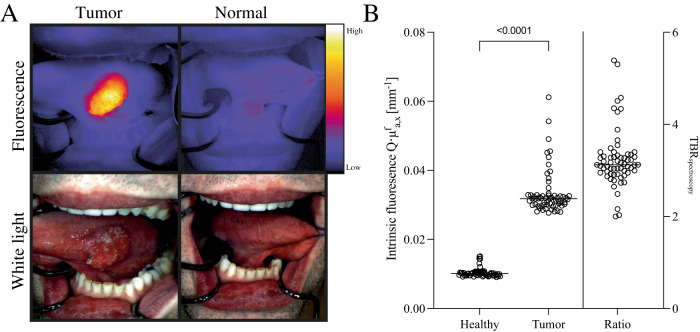
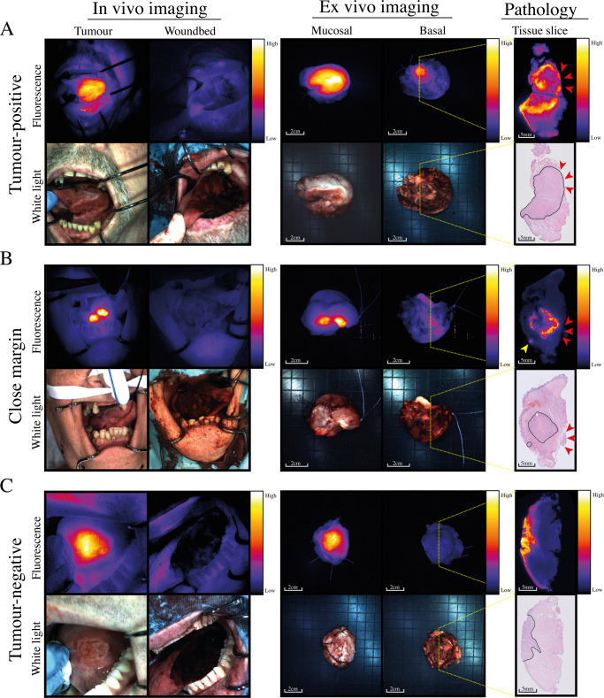
Inadequate surgical margins occur frequently in oral squamous cell carcinoma surgery. Fluorescence molecular imaging (FMI) has been explored for intraoperative margin assessment, but data are limited to phase-I studies. In this single-arm phase-II study (NCT03134846), our primary endpoints were to determine the sensitivity, specificity and positive predictive value of cetuximab-800CW for tumor-positive margins detection. Secondary endpoints were safety, close margin detection rate and intrinsic cetuximab-800CW fluorescence. In 65 patients with 66 tumors, cetuximab-800CW was well-tolerated. Fluorescent spots identified in the surgical margin with signal-to-background ratios (SBR) of ≥2 identify tumor-positive margins with 100% sensitivity, 85.9% specificity, 58.3% positive predictive value, and 100% negative predictive value. An SBR of ≥1.5 identifies close margins with 70.3% sensitivity, 76.1% specificity, 60.5% positive predictive value, and 83.1% negative predictive value. Performing frozen section analysis aimed at the fluorescent spots with an SBR of ≥1.5 enables safe, intraoperative adjustment of surgical margins.


Conflict of interest statement

G.M.v.D. is CEO of Tracer Europe B.V./AxelaRx. B.v.d.V. is a member of the Scientific Advisory Board of Visiopharm, for which compensation is received by the University Medical Centre Groningen. The remaining authors declare no competing interests.

Figures

Fig. 1
Fig. 1. Overview of study workflow.
A In vivo fluorescence imaging of the tumor. B Back table imaging of the excised specimen. Fluorescence imaging is performed from all surgical planes of the specimen. In the case of a complex specimen, multiple surgical planes can be identified and imaged, and in the case of a simple specimen, only one surgical plane per specimen is imaged. Fluorescent spots are observed in image 5 (top row) and image 1 (bottom row). C Bread loaf slicing of the specimen and fluorescence imaging of all bread loaf slices. D Correlation of the fluorescent spots relate to tumor-positive margins on histopathology.
Fig. 2
Fig. 2. In vivo imaging and spectroscopy results.
A In vivo fluorescence molecular imaging shows a sharp demarcation of a tumor on the lateral tongue. B In vivo multi-diameter single-fiber reflectance, single-fiber fluorescence contact measurements were performed in n = 63 tumors, showing significantly higher intrinsic fluorescence (Q.μfa,x) [mm−1]) in tumor (3.3 (2.7–6.1) × 10−2 mm−1) compared to normal tissue (1.0 (0.9–1.5) × 10−2), one-sided p = 0.0001 using Wilcoxon signed rank test. Source data are provided as a Source Data file.
Fig. 3
Fig. 3. Flowchart of patient inclusion and image acquisition.
OSCC oral squamous cell carcinoma, AE adverse event, SBR signal-to-background ratio. *One patient presented with two primary tumors.
Fig. 4
Fig. 4. Receiver operating characteristics of ex vivo margin assessment.
Receiver operating characteristics for the detection of tumor-positive margins (n = 14, panel A), close margins (n = 37, panel B). To further determine the accuracy of our approach, we subdivided close margins in 1–3 mm (n = 20 panel C) and 3–5 mm (n = 17, panel D). Source data are provided as a Source Data file. AUC area under the curve, CI confidence interval.
Fig. 5
Fig. 5. Representative examples of a tumor-positive margin, close margin and tumor-negative margin.
Representative examples of A a tumor-positive margin, B a close margin, and C a tumor-negative margin. In vivo fluorescence imaging shows sharply demarcated tumors compared to adjacent tissue (upper left images), and after excision, no fluorescence can be detected in the wound bed (upper right images). On the tissue slices, the tumor is delineated with a solid black line. Panel (A) shows a fluorescent spot with an SBR of 4.0 on the excised specimen, corresponding to a tumor-positive margin (red arrows). Panel (B) shows a fluorescence spot with an SBR of 2.3, revealing a close margin of 2.2 mm (red arrows). The yellow arrow indicates a fluorescent lesion in the mucosa, which corresponds to the tumor spreading mucosally. In panel (C), no fluorescent signal is seen in the margin, corresponding to a tumor-negative margin. Tumor tissue is demarcated with a solid black line on tissue slides.

References

    1. Sung H, et al. Global Cancer Statistics 2020: GLOBOCAN estimates of incidence and mortality worldwide for 36 cancers in 185 countries. CA Cancer J. Clin. 2021;71:209–249. doi: 10.3322/caac.21660. - DOI - PubMed
    1. Orosco RK, et al. Positive surgical margins in the 10 most common solid cancers. Sci. Rep. 2018;8:5686. doi: 10.1038/s41598-018-23403-5. - DOI - PMC - PubMed
    1. Woolgar JA, Triantafyllou A. A histopathological appraisal of surgical margins in oral and oropharyngeal cancer resection specimens. Oral. Oncol. 2005;41:1034–1043. doi: 10.1016/j.oraloncology.2005.06.008. - DOI - PubMed
    1. Smits RWH, et al. Resection margins in oral cancer surgery: room for improvement. Head Neck. 2016;38:E2197–E2203. doi: 10.1002/hed.24075. - DOI - PubMed
    1. Koo BS, Lim YC, Lee JS, Choi EC. Recurrence and salvage treatment of squamous cell carcinoma of the oral cavity. Oral. Oncol. 2006;42:789–794. doi: 10.1016/j.oraloncology.2005.11.016. - DOI - PubMed

Publication types

Associated data