. 2023 Aug 16;14(1):4967.
doi: 10.1038/s41467-023-40639-6.

IRX2 regulates angiotensin II-induced cardiac fibrosis by transcriptionally activating EGR1 in male mice

Affiliations

IRX2 regulates angiotensin II-induced cardiac fibrosis by transcriptionally activating EGR1 in male mice

Zhen-Guo Ma et al. Nat Commun. .

Abstract

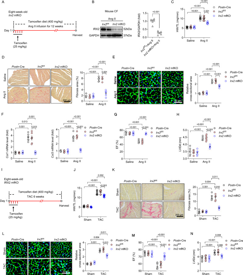
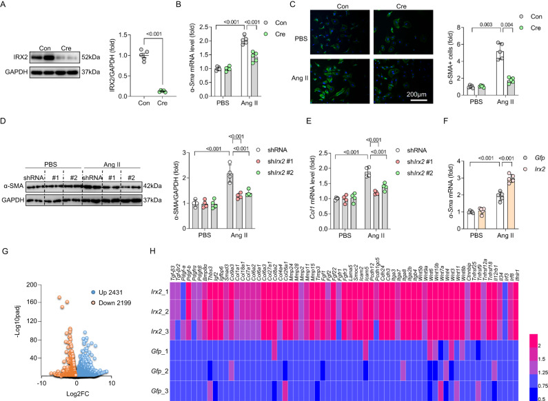
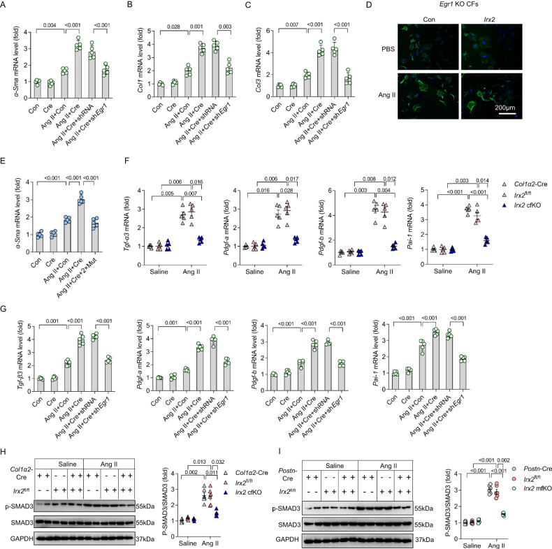
Cardiac fibrosis is a common feature of chronic heart failure. Iroquois homeobox (IRX) family of transcription factors plays important roles in heart development; however, the role of IRX2 in cardiac fibrosis has not been clarified. Here we report that IRX2 expression is significantly upregulated in the fibrotic hearts. Increased IRX2 expression is mainly derived from cardiac fibroblast (CF) during the angiotensin II (Ang II)-induced fibrotic response. Using two CF-specific Irx2-knockout mouse models, we show that deletion of Irx2 in CFs protect against pathological fibrotic remodelling and improve cardiac function in male mice. In contrast, Irx2 gain of function in CFs exaggerate fibrotic remodelling. Mechanistically, we find that IRX2 directly binds to the promoter of the early growth response factor 1 (EGR1) and subsequently initiates the transcription of several fibrosis-related genes. Our study provides evidence that IRX2 regulates the EGR1 pathway upon Ang II stimulation and drives cardiac fibrosis.


Conflict of interest statement

The authors declare that they have no competing interest.

Figures

Fig. 1
Fig. 1. IRX2 expression was increased in fibrotic hearts.
A Relative mRNA levels of 6 Irx family members in adult mouse cardiac fibroblasts (CFs) after angiotensin II (Ang II) treatment for 24 h (n = 5). B Relative mRNA levels of 6 IRX family members in human CFs after Ang II treatment for 24 h (n = 5). C Irx2 mRNA levels in the heart after Ang II infusion by an osmotic minipump (n = 6). D Representative western blots and statistical analysis of IRX2 expression in the heart after Ang II infusion for 12 weeks (n = 6). E Representative western blots and statistical analysis of IRX2 expression in heart samples obtained from patients with dilated cardiomyopathy (DCM) and control (Con) donors (Con, n = 6; DCM, n = 7). F Irx2 mRNA levels in endothelial cells (ECs), cardiomyocytes (CMs) and CFs isolated from hearts infused with Ang II for 12 weeks (n = 6). G Representative western blots and quantification of IRX2 protein expression in CFs isolated from hearts infused with Ang II for 12 weeks (n = 6). Data are shown as the mean ± SEM, and analysed using an unpaired two-tailed Student′s t test. For the analysis in (C), one-way ANOVA with Tamhane’s T2 test was conducted. Source data are provided as a Source Data file.
Fig. 2
Fig. 2. Conditional fibroblast-specific Irx2-deficient mice exhibited attenuated fibrotic remodelling in response to angiotensin II (Ang II) infusion.
A Conditional fibroblast-specific Irx2-deficient mice (Irx2 cfKO) were bred by crossing mice with a conditional knockout allele of Irx2 (Irx2fl/fl) with Col1α2-Cre mice. Irx2 cfKO mice and littermate controls were subjected to Ang II infusion for 12 weeks. B Representative western blots and statistical analysis of IRX2 protein expression in CFs isolated from Irx2 cfKO mice and littermate controls (n = 5). CE Irx2 cfKO mice exhibited an attenuated heart weight-to-tibia length (HW/TL) ratio (n = 10 mice, Col1α2-Cre+Saline; n = 12 mice, Irx2fl/fl+Saline; n = 12 mice, Irx2 cfKO+Saline; n = 12 mice, Col1α2-Cre+Ang II; n = 11 mice, Irx2fl/fl+Ang II; n = 12 mice, Irx2 cfKO+Ang II) (C), a reduced fibrosis area (n = 5) (D), and a decreased cell area of cardiomyocytes (n = 5) (E) after Ang II infusion. F Relative mRNA levels of Col1 and Col3 in hearts from Irx2 cfKO mice and littermate controls after Ang II infusion (n = 6). G Cardiac function (n = 6) was improved in Ang II-infused Irx2 cfKO mice. Data are shown as the mean ± SEM, and analysed using one-way ANOVA followed by Tukey post hoc test (E and G) or Tamhane’s T2 test (C, D and F). For the analysis in (B), an unpaired two-tailed Student′s t test was conducted. Source data are provided as a Source Data file.
Fig. 3
Fig. 3. Conditional myofibroblast-specific Irx2 depletion attenuated angiotensin II (Ang II) or pressure overload-induced cardiac fibrosis in mice.
A Conditional myofibroblast-specific Irx2-deficient mice (Irx2 mfKO) were bred by crossing mice with a conditional knockout allele of Irx2 (Irx2fl/fl) with Postn-Cre mice. Irx2 mfKO mice and littermate controls were subjected to Ang II infusion for 12 weeks. B Representative western blots and statistical analysis of IRX2 protein expression in CFs isolated from Irx2 mfKO mice and littermate controls after Ang II infusion for 12 weeks (n = 5). C Heart weight-to-tibia length (HW/TL) ratio (n = 10 mice, Postn-Cre+Saline; n = 11 mice, Irx2fl/fl+Saline; n = 11 mice, Irx2 mfKO+Saline; n = 10 mice, Postn-Cre+Ang II; n = 12 mice, Irx2fl/fl+Ang II; n = 12 mice, Irx2 mfKO+Ang II). D Cardiac fibrosis was determined by picrosirius red staining for Irx2 mfKO mice after Ang II infusion for 12 weeks (n = 5). E The cell area of cardiomyocytes was determined with wheat germ agglutinin (WGA) staining for Irx2 mfKO and littermate controls after Ang II infusion for 12 weeks (n = 5). F The relative mRNA levels of Col1 and Col3 were detected in hearts from Irx2 mfKO mice and control mice (n = 6). G, H The ejection fraction (EF) (G) and left ventricular end-diastolic dimension (LVIDd) (H) were detected in these groups (n = 6). I Irx2 mfKO mice and littermate controls were subjected to transverse aortic constriction (TAC) surgery for 6 weeks. J HW/TL ratio (n = 10 mice, Postn-Cre+Sham; n = 11 mice, Irx2fl/fl+Sham; n = 11 mice, Irx2 mfKO+Sham; n = 10 mice, Postn-Cre+TAC; n = 12 mice, Irx2fl/fl + TAC; n = 12 mice, Irx2 mfKO+TAC). K Histological staining showing cardiac fibrosis after TAC surgery in Irx2 mfKO mice (n = 5). L Cell area of cardiomyocytes determined with WGA staining after TAC surgery (n = 5). M, N EF (M) and LVIDd (N) detected in these groups (n = 8).The data were compared by one-way ANOVA. Data are shown as the mean ± SEM, and analysed using one-way ANOVA followed by Tukey post hoc test (DH and LN) or Tamhane’s T2 test (C, and JK). For the analysis in (B), an unpaired two-tailed Student′s t test was conducted. Source data are provided as a Source Data file.
Fig. 4
Fig. 4. Conditional myofibroblast-specific Irx2 overexpression promoted angiotensin II (Ang II)-induced fibrotic remodelling in mice.
A Conditional myofibroblast-specific Irx2-overexpressing mice (Irx2 mfTg) were bred by crossing conditional transgenic mice expressing the Irx2 gene (Irx2 Tgfl/fl) with Postn-Cre mice. Irx2 mfTg mice and their littermate controls were subjected to Ang II infusion for 4 weeks. B Representative western blots and statistical analysis of IRX2 protein expression in CFs isolated from Irx2 mfTg mice and littermate controls after Ang II infusion for 4 weeks (n = 5). C Heart weight to tibia length (HW/TL) ratio (n = 12 mice for all six groups). D Cardiac fibrosis was determined by picrosirius red staining in Irx2 mfTg mice after Ang II infusion (n = 5). E Cell area of cardiomyocytes was determined with wheat germ agglutinin (WGA) staining in Irx2 mfTg and littermate controls after Ang II infusion (n = 5). F Relative mRNA levels of Col1 and Col3 were detected in the hearts of Irx2 mfTg mice and their controls (n = 6). G, H Ejection fraction (EF) (G) and left ventricular end-diastolic dimension (LVIDd) (H) were detected in these groups (n = 6). The data were compared by one-way ANOVA. Data are shown as the mean ± SEM, and analysed using one-way ANOVA followed by Tukey post hoc test (CD and GH) or Tamhane’s T2 test (EF). For the analysis in (B), an unpaired two-tailed Student′s t test was conducted. Source data are provided as a Source Data file.
Fig. 5
Fig. 5. IRX2 is essential for fibroblast to myofibroblast transformation.
A Adult cardiac fibroblasts (CFs) were isolated from Irx2fl/fl mice and infected with an adenoviral vector carrying Cre to deplete Irx2. After 24 h of transfection, IRX2 protein expression was detected in CFs (n = 5). B Relative mRNA level of α-Sma in Irx2-depleted adult mouse CFs (n = 5). C Representative images and quantification of α-SMA (green) in Cre-infected Irx2fl/fl CFs in response to Ang II incubation for 24 h. Nuclei are stained with DAPI (blue). (n = 5, for each group, 50-60 fields were counted). D CFs were isolated from wild-type mice and then transfected with shRNAs targeting Irx2. After 24 h of transfection, the cells were subjected to Ang II treatment for 24 h to induce a fibrotic phenotype. After that, the cells were harvested for the detection of α-SMA with western blotting (n = 4). E Relative mRNA level of Col1 in IRX2-deficient CFs after Ang II incubation for 24 h (n = 4). F CFs were isolated from wild-type mice and then overexpressed Irx2 with an adenovirus vector, after which they were subjected to Ang II treatment for 24 h. The relative mRNA level of α-Sma was detected to reflect fibroblast to myofibroblast transformation (n = 5). G CFs were isolated from wild-type mice and then overexpressed Irx2 with an adenovirus vector, after which they were subjected to Ang II treatment for 24 h. Total RNA was extracted, and RNA sequencing was performed. Volcano plots indicating the differentially expressed genes (DEGs) in Irx2-overexpressing CFs relative to controls after Ang II administration for 24 h. H Heatmap of several differentially expressed genes in Irx2-overexpressing CFs relative to controls after Ang II infusion. Data are shown as the mean ± SEM, and analysed using one-way ANOVA followed by Tukey post hoc test (B and DF) or Tamhane’s T2 test (C). For the analysis in (A), an unpaired two-tailed Student′s t test was conducted. Source data are provided as a Source Data file.
Fig. 6
Fig. 6. EGR1 was identified as a direct downstream target gene of IRX2 in cardiac fibroblasts that regulates the angiotensin II (Ang II)-induced fibrotic response.
A CFs were infected with an adenovirus carrying IRX2 and then subjected to Ang II stimulation for 24 h. ChIP-seq was performed with two anti-IRX2 antibodies (IRX2-1B7 and IRX2-1C1). Similar spatial distributions of IRX2 peaks were identified by the two anti-IRX2 antibodies. B IRX2-regulated genes identified in the ChIP-seq data (bottom) were overlaid with upregulated differentially expressed genes revealed by RNA-seq (top). With this strategy, 70 genes were identified. C Two IRX2 binding sites were identified in the ChIP-seq peak distributed in the promoter site of Egr1. D The Egr1 promoter was stimulated by IRX2 overexpression. Egr1 promoter activity was measured by a luciferase assay upon Irx2 overexpression in CFs isolated from wild-type mice (n = 5 for each group). E The mutant (Mut) promoters (pro) deleting IRX2 binding sites cannot be stimulated by Irx2 overexpression (n = 5 for each group). F Independent ChIP-PCR was performed with Irx2-overexpressing CFs to verify IRX2 binding to the promoter of Egr1 (n = 5). GH Egr1 mRNA and EGR1 protein expression in CFs isolated from Irx2 cfKO mice and littermate controls with or without Ang II infusion for 12 weeks (n = 6). I Egr1 mRNA levels were detected in CFs with Irx2 deficiency caused by Cre expression (n = 5). JK Egr1 mRNA and EGR1 protein expression in CFs isolated from Irx2 mfTg mice and littermate controls with or without Ang II infusion for 4 weeks (n = 6). L Egr1 mRNA level in Irx2-overexpressing human CFs after Ang II treatment for 24 h (n = 6). Data are shown as the mean ± SEM, and analysed using one-way ANOVA followed by Tukey post hoc test (H, I and K) or Tamhane’s T2 test (DG, J and L). Source data are provided as a Source Data file.
Fig. 7
Fig. 7. EGR1 was responsible for the IRX2-promoted fibrotic response in angiotensin II (Ang II)-treated cardiac fibroblasts.
AC Cardiac fibroblasts (CFs) were isolated from Irx2 Tgfl/fl mice and infected with an adenovirus carrying Cre to overexpress Irx2. After that, Egr1 expression was knocked down, and the CFs were subjected to Ang II treatment for 24 h. After that, the cells were harvested to detect the mRNA levels of α-Sma (A), Col1 (B) and Col3 (C) (n = 5). D Representative images of α-SMA (green) in CFs isolated from Egr1-knockout mice after Ang II treatment for 24 h. Nuclei were stained with DAPI (blue) (n = 5, for each experiment, 50-60 fields were counted). E Relative mRNA levels of α-Sma in the indicated groups (n = 5). F Relative mRNA levels of fibrosis-related genes in CFs isolated from Irx2 cfKO mice and littermate controls with or without Ang II infusion for 12 weeks (n = 5). G CFs were isolated from Irx2 Tgfl/fl mice and infected with an adenovirus carrying Cre to overexpress Irx2. After that, Egr1 expression was knocked down, and the CFs were subjected to Ang II treatment for 24 h. After that, the cells were harvested to detect the mRNA levels of fibrosis-related genes (n = 5). H, I Western blot analysis was performed to analyse the phosphorylation of Smad3 in CFs isolated from Irx2 cfKO or Irx2 mfKO mice and corresponding littermate controls with or without Ang II infusion for 12 weeks (n = 6). Data are shown as the mean ± SEM, and analysed using one-way ANOVA followed by Tukey post hoc test (A, C, E and G) or Tamhane’s T2 test (B, F, H and I). Source data are provided as a Source Data file.
Fig. 8
Fig. 8. Genetic depletion of Egr1 attenuated fibrotic remodelling in mice with conditional myofibroblast-specific Irx2 overexpression after angiotensin II (Ang II) infusion.
Conditional myofibroblast-specific Irx2-overexpressing mice (Irx2 mfTg) were bred with Egr1 global knockout mice or wild type (WT). The resulting mouse line and littermate controls were subjected to Ang II infusion for 4 weeks. A Heart weight-to-tibia length (HW/TL) ratio (n = 10 mice, Irx2 Tgfl/fl + WT+Ang II; n = 10 mice, Irx2 Tgfl/fl+Egr1 KO+Ang II; n = 11 mice, Irx2 mfTg+WT+Ang II; n = 11 mice, Irx2 mfTg+Egr1 KO+Ang II). B Histological staining showed cardiomyocyte enlargement and cardiac fibrosis after Ang II infusion (n = 5). C Cardiac fibrosis was determined by picrosirius red staining after Ang II infusion (n = 5). D The cell area of cardiomyocytes was determined with wheat germ agglutinin (WGA) staining after Ang II infusion (n = 5). E, F The ejection fraction (EF) (E) and left ventricular end-diastolic dimension (LVIDd) (F) were detected in these groups (n = 6). Data are shown as the mean ± SEM, and analyzed using one-way ANOVA followed by Tukey post hoc test (A, C, and D). For the analysis in (E, F), repeated measures ANOVA was conducted. Source data are provided as a Source Data file.

References

    1. Chilton L, Giles WR, Smith GL. Evidence of intercellular coupling between co-cultured adult rabbit ventricular myocytes and myofibroblasts. J. Physiol.-London. 2007;583:225–236. - PMC - PubMed
    1. Spinale FG. Myocardial matrix remodeling and the matrix metalloproteinases: influence on cardiac form and function. Physiol. Rev. 2007;87:1285–1342. - PubMed
    1. Travers JG, Kamal FA, Robbins J, Yutzey KE, Blaxall BC. Cardiac Fibrosis: The Fibroblast Awakens. Circ. Res. 2016;118:1021–1040. - PMC - PubMed
    1. Gulati A, et al. Association of fibrosis with mortality and sudden cardiac death in patients with nonischemic dilated cardiomyopathy. JAMA-J. Am. Med. Asso. 2013;309:896–908. - PubMed
    1. Lal H, et al. Cardiac fibroblast glycogen synthase kinase-3beta regulates ventricular remodeling and dysfunction in ischemic heart. Circulation. 2014;130:419–430. - PMC - PubMed

Publication types