

Predictive value of cardiac magnetic resonance right ventricular longitudinal strain in patients with suspected myocarditis
- PMID: 37587516
- PMCID: PMC10433613
- DOI: 10.1186/s12968-023-00957-6
Predictive value of cardiac magnetic resonance right ventricular longitudinal strain in patients with suspected myocarditis
Abstract
Background: Recent evidence underlined the importance of right (RV) involvement in suspected myocarditis. We aim to analyze the possible incremental prognostic value from RV global longitudinal strain (GLS) by CMR.
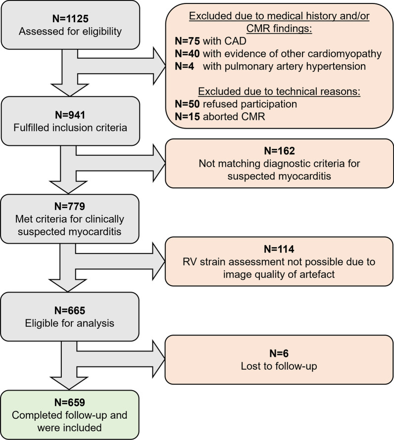
Methods: Patients referred for CMR, meeting clinical criteria for suspected myocarditis and no other cardiomyopathy were enrolled in a dual-center register cohort study. Ejection fraction (EF), GLS and tissue characteristics were assessed in both ventricles to assess their association to first major adverse cardiovascular events (MACE) including hospitalization for heart failure (HF), ventricular tachycardia (VT), recurrent myocarditis and death.
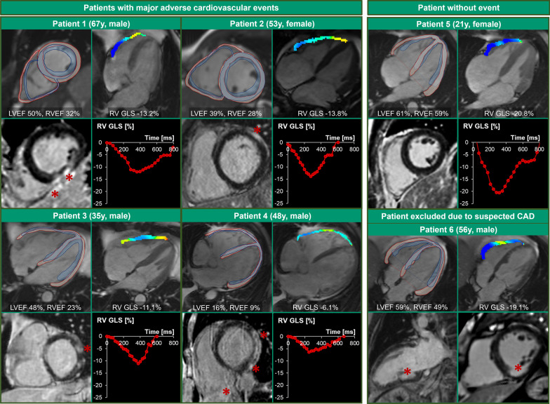
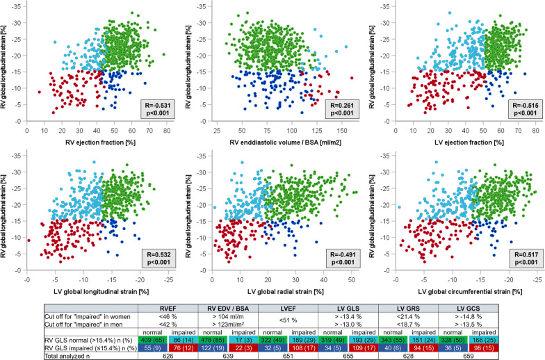
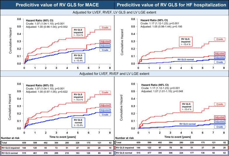
Results: Among 659 patients (62.8% male; 48.1 ± 16.1 years), RV GLS was impaired (> - 15.4%) in 144 (21.9%) individuals, of whom 76 (58%), 108 (77.1%), 27 (18.8%) and 40 (32.8%) had impaired right ventricular ejection fraction (RVEF), impaired left ventricular ejection fraction (LVEF), RV late gadolinium enhancement (LGE) or RV edema, respectively. After a median observation time of 3.7 years, 45 (6.8%) patients were hospitalized for HF, 42 (6.4%) patients died, 33 (5%) developed VT and 16 (2.4%) had recurrent myocarditis. Impaired RV GLS was associated with MACE (HR = 1.07, 95% CI 1.04-1.10; p < 0.001), HF hospitalization (HR = 1.17, 95% CI 1.12-1.23; p < 0.001), and death (HR = 1.07, 95% CI 1.02-1.12; p = 0.004), but not with VT and recurrent myocarditis in univariate analysis. RV GLS lost its association with outcomes, when adjusted for RVEF, LVEF, LV GLS and LV LGE extent.
Conclusion: RV strain is associated with MACE, HF hospitalization and death but has neither independent nor incremental prognostic value after adjustment for RV and LV function and tissue characteristics. Therefore, assessing RV GLS in the setting of myocarditis has only limited value.
Trial registration: ClinicalTrials.gov NCT04774549 NCT03470571.
Keywords: Cardiac magnetic resonance imaging; Feature tracking; Heart failure hospitalizations; Myocarditis; Right ventricle; Right ventricular global longitudinal strain.
© 2023. Society for Cardiovascular Magnetic Resonance.
Conflict of interest statement
Dr. Gräni received research funding from the Swiss National Science Foundation, Innosuisse, GAMBIT foundation and from the Center for Artificial Intelligence in Medicine Research Project Fund University Bern, outside of the submitted work. Dr. Bernhard and Dr. Benz report a career development grant from the Swiss National Science Foundation. Ms. Safarkhanlo received research funding from the Center for Artificial Intelligence in Medicine Research Project Fund University Bern, outside of the submitted work. Dr. Huber has received research grants from the Swiss National Science Foundation, the Swiss Academy of Medical Sciences, the Helmut-Hartweg Foundation and the Foundation to Fight against Cancer, all for work outside the submitted study. He has received speaker/consulting honoraria or travel support from Bayer, Bracco and Siemens, all for work outside the submitted study. All other authors report no conflicts.
Figures
References
-
- Caforio AL, Pankuweit S, Arbustini E, Basso C, Gimeno-Blanes J, Felix SB, Fu M, Heliö T, Heymans S, Jahns R, Klingel K, Linhart A, Maisch B, McKenna W, Mogensen J, Pinto YM, Ristic A, Schultheiss HP, Seggewiss H, Tavazzi L, Thiene G, Yilmaz A, Charron P, Elliott PM. Current state of knowledge on aetiology, diagnosis, management, and therapy of myocarditis: a position statement of the European Society of Cardiology Working Group on Myocardial and Pericardial Diseases. Eur Heart J. 2013;34(2636–48):2648a–2648d. doi: 10.1093/eurheartj/eht210. - DOI - PubMed
-
- Aquaro GD, Negri F, De Luca A, Todiere G, Bianco F, Barison A, Camastra G, Monti L, Dellegrottaglie S, Moro C, Lanzillo C, Scatteia A, Di Roma M, Pontone G, Perazzolo Marra M, Di Bella G, Donato R, Grigoratos C, Emdin M, Sinagra G. Role of right ventricular involvement in acute myocarditis, assessed by cardiac magnetic resonance. Int J Cardiol. 2018;271:359–365. doi: 10.1016/j.ijcard.2018.04.087. - DOI - PubMed
-
- Bernhard B, Schnyder A, Garachemani D, Fischer K, Tanner G, Safarkhanlo Y, Stark AW, Schütze J, Pavlicek-Bahlo M, Greulich S, Johner C, Wahl A, Benz DC, Kwong RY, Gräni C. Prognostic value of right ventricular function in patients with suspected myocarditis undergoing cardiac magnetic resonance. JACC Cardiovasc Imaging. 2023;16:28–41. doi: 10.1016/j.jcmg.2022.08.011. - DOI - PubMed
MeSH terms
Substances
Associated data
LinkOut - more resources
Full Text Sources
Medical
Research Materials
Miscellaneous
