Review
. 2023 Aug 17;10(1):36.
doi: 10.1186/s40779-023-00472-w.

Mesenchymal stem cell-derived extracellular vesicles in skin wound healing: roles, opportunities and challenges

Affiliations
Review

Mesenchymal stem cell-derived extracellular vesicles in skin wound healing: roles, opportunities and challenges

Jia-Yi Ding et al. Mil Med Res. .

Abstract

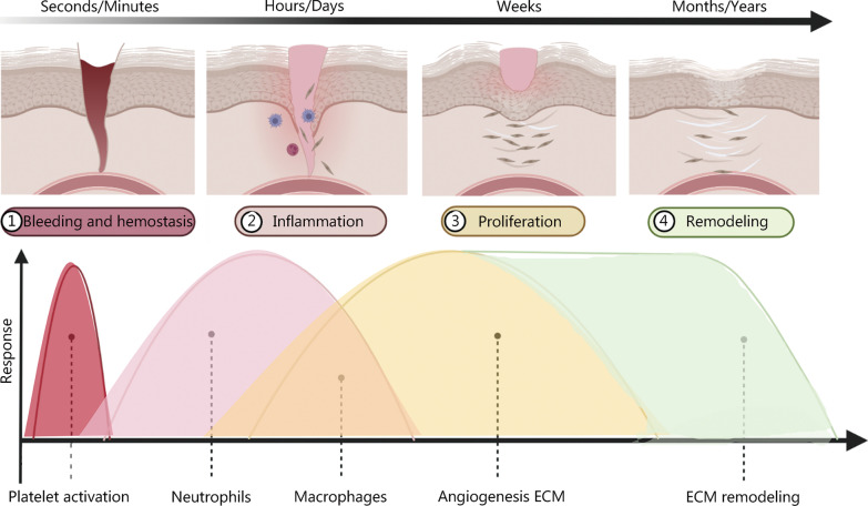
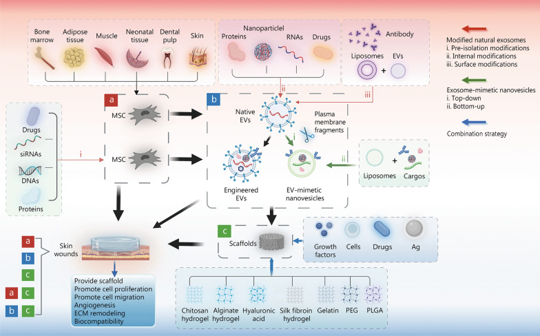
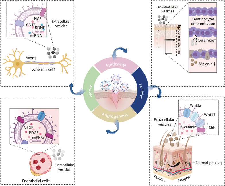
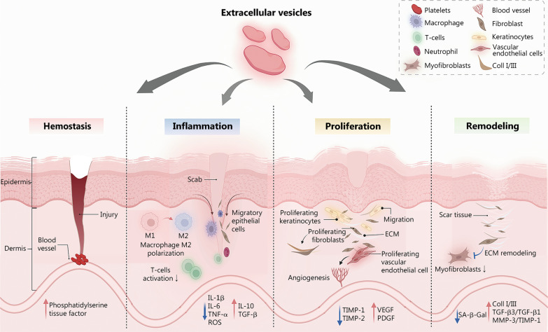
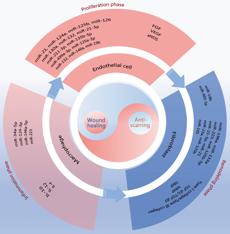
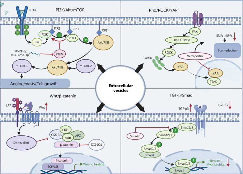
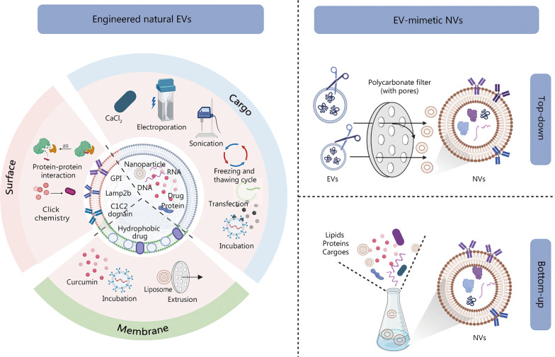
Skin wounds are characterized by injury to the skin due to trauma, tearing, cuts, or contusions. As such injuries are common to all human groups, they may at times represent a serious socioeconomic burden. Currently, increasing numbers of studies have focused on the role of mesenchymal stem cell (MSC)-derived extracellular vesicles (EVs) in skin wound repair. As a cell-free therapy, MSC-derived EVs have shown significant application potential in the field of wound repair as a more stable and safer option than conventional cell therapy. Treatment based on MSC-derived EVs can significantly promote the repair of damaged substructures, including the regeneration of vessels, nerves, and hair follicles. In addition, MSC-derived EVs can inhibit scar formation by affecting angiogenesis-related and antifibrotic pathways in promoting macrophage polarization, wound angiogenesis, cell proliferation, and cell migration, and by inhibiting excessive extracellular matrix production. Additionally, these structures can serve as a scaffold for components used in wound repair, and they can be developed into bioengineered EVs to support trauma repair. Through the formulation of standardized culture, isolation, purification, and drug delivery strategies, exploration of the detailed mechanism of EVs will allow them to be used as clinical treatments for wound repair. In conclusion, MSC-derived EVs-based therapies have important application prospects in wound repair. Here we provide a comprehensive overview of their current status, application potential, and associated drawbacks.

Keywords: Engineered nanoparticles; Extracellular vesicles (EVs); Mesenchymal stem cell (MSC); Wound repair.


Conflict of interest statement

The authors declare that they have no competing interests.

Figures

Fig. 1
Fig. 1
Schematic diagram of the skin repair process. The four phases of trauma repair, hemostasis, inflammation, proliferation, and remodeling phases, occur in order and can overlap. Platelets play a role in the hemostatic phase. Neutrophils play an anti-infection function primarily during the inflammatory phase. Macrophages are involved in both the inflammatory and proliferative phases and exert different roles depending on the phenotypic variation (M1 and M2). Vascularization as well as extracellular matrix (ECM) formation occurs during the proliferative phase, while ECM remodeling may take months or even years. Reproduced with permission from Ref. [7]. Copyright 2021, Elsevier B.V. Reproduced with permission from Ref. [8]. Copyright 2022 Cialdai, Risaliti, and Monici
Fig. 2
Fig. 2
Diagram illustrating the application of mesenchymal stem cell (MSC) in wound repair. a MSC therapy, including native MSC and pre-extracellular vesicles (EVs) isolation MSC modifications. b EV therapy, including native EVs, engineered EVs, and EV-mimetic nanovesicles. c Wound dressing therapy. For wound treatment, either a/b/c therapy alone or a combination of a/b with c can be used. The red arrows represent three modified natural EV methods, including pre-isolation modifications (i), internal modifications (ii), and surface modifications (iii). The green arrows represent EV-mimetic nanovesicle production methods such as top-down (i) and bottom-up (ii). The blue arrows show the combination strategies for wound dressing. It was created utilizing the templates on BioRender.com as a reference. Ag antibacterial material, ECM extracellular matrix, PEG polyethylene glycol, PLGA poly lactic-co-glycolic-acid
Fig. 3
Fig. 3
Process of extracellular vesicles (EVs) biogenesis and the molecular composition of EVs are shown as follows: (1) various components of the extracellular environment, including proteins, lipids, and small molecule metabolites, are endocytosed to form early endosomes; (2) the early endosomes are transformed into late endosomes that in turn form multivesicular bodies (MVBs); (3) MVBs are formed into EVs by fusion with microtubules and the cytoskeleton to the plasma membrane. Otherwise, a part of the MVB is transported and merged with the lysosomes to degrade the cargo. EVs enter the recipient cell through various pathways, including endocytosis, macrophage uptake, phagocytosis, and direct fusion with the plasma membrane. It was created utilizing the templates on BioRender.com as a reference. CD cluster of differentiation, HSP heat shock protein, Tsg tumor susceptibility gene, Alix apoptosis-linked gene 2-interacting protein X, RAB Ras-like proteins in brain, GTPases guanosine triphosphate hydrolases, ESCRT endosomal sorting complex required for transport, MHC major histocompatibility complex, mRNA messenger ribonucleic acid, miRNA micro ribonucleic acid, lncRNA long non-coding ribonucleic acid, mtRNA mitochondrial ribonucleic acid, tRNA transfer ribonucleic acid, dsRNA double-stranded ribonucleic acid, ssDNA single-stranded deoxyribonucleic acid, FAS tumor necrosis factor receptor superfamily member 6, DNA deoxyribonucleic acid, ER endoplasmic reticulum
Fig. 4
Fig. 4
Schematic diagram of the effect of extracellular vesicles (EVs) on wound repair. EVs promote axon and Schwann cell proliferation via BDNF, NGF, CNTF, and miRNAs. The VEGF, PDGF, and miRNAs within the EVs promote endothelial cell growth and angiogenesis. EVs also participate in keratinocyte differentiation and facilitate the de novo synthesis of ceramides. Mesenchymal stem cell (MSC)-derived EVs containing Wnt3a and Wnt11 facilitate the reconstruction and proliferation of hair follicles to promote the transition from the telogen to the anagen phase. It was created utilizing the templates on BioRender.com as a reference. BDNF brain-derived neurotrophic factor, NGF nerve growth factor, CNTF ciliary neurotrophic factor, VEGF vascular endothelial growth factor, PDGF platelet-derived growth factor, Wnt wingless/integrated, Shh sonic hedgehog
Fig. 5
Fig. 5
Major events in each phase of mesenchymal stem cell (MSC)-derived extracellular vesicles (EVs)-promoted skin repair. EVs work through the factors in the four phases to accelerate hemostasis, regulate inflammation via processes such as macrophage polarization, promote angiogenesis and cell proliferation, and exhibit anti-aging and anti-scarring abilities. It was created utilizing the templates on BioRender.com as a reference. M1 M1 macrophage, M2 M2 macrophage, IL interleukin, TNF-α tumour necrosis factor-α, TGF-β transforming growth factor-β, ROS reactive oxygen species, TIMP tissue inhibitors of metalloproteinase, VEGF vascular endothelial growth factor, PDGF platelet-derived growth factor, MMP matrix metalloproteinase, ECM extracellular matrix
Fig. 6
Fig. 6
Schematic diagram of the role of extracellular vesicles (EVs) in wound repair. EVs have pro-repair and anti-scarring roles through macrophages in the inflammatory phase, through endothelial cells in the proliferative phase, and through fibroblasts in the remodeling phase. EV-associated miRNAs and proteins regulate the activity of these three types of cells. miR micro ribonucleic acid, FGF fibroblast growth factor, VEGF vascular endothelial growth factor, eNOS endothelial nitric oxide synthase, IL interleukin, TGF-β transforming growth factor β, MMP matrix metalloproteinase, TIMP tissue inhibitors of metalloproteinase
Fig. 7
Fig. 7
Four major signaling pathways of mesenchymal stem cell (MSC)-derived extracellular vesicles (EVs) for skin repair. MSC-derived EVs affect the wound repair process through a series of signaling pathways, including the PI3K/Akt/mTOR pathway, the TGF-β/Smad signaling pathway, the Wnt/β-catenin pathway, and the Rho/ROCK/YAP axis signaling pathway. It was created utilizing the templates on BioRender.com as a reference. PI3K phosphoinositide 3-kinase, Akt protein kinase B, mTOR mammalian target of rapamycin, TGF-β transforming growth factor-β, Smad Drosophila mothers against decapentaplegic proteins, Wnt wingless/integrated, ROCK Rho-associated protein kinase, YAP Yes-associated protein, RTKs receptor tyrosine kinases, Ras Ras protein, PIP2 phosphatidylinositol 4,5-bisphosphate, PIP3 phosphatidylinositol 3,4,5-trisphosphate, PDK phosphoinositide-dependent kinases, PKB protein kinase B, PTEN phosphatase and tensin homolog deleted on chromosome 10, FAK focal adhesion kinase, Rho Ras homology, GTPase guanosine triphosphate hydrolases, TEAD YAP-transcriptional enhancer factor domain family member, ENFs Engrailed-1 lineage-negative fibroblasts, EPFs Engrailed-1 lineage-positive fibroblasts, LRP low-density lipoprotein receptor-related protein, GSK glycogen synthase kinase, APC adenomatous polyposis coli, ICG indocyanine green, TCF/LEF T-cell factor/lymphoid enhancer factor
Fig. 8
Fig. 8
Schematic diagram of engineered extracellular vesicles (EVs) modification. There are two main methods of EV modification: engineered natural EVs and EV-mimetic nanovesicles (NVs). The former comprises modifications of membranes, surfaces, and cargoes. The latter is divided into top-down and bottom-up methods. It was created utilizing the templates on BioRender.com as a reference. GPI glycosylphosphatidylinositol

Comment in

References

    1. Sun BK, Siprashvili Z, Khavari PA. Advances in skin grafting and treatment of cutaneous wounds. Science. 2014;346(6212):941–945. doi: 10.1126/science.1253836. - DOI - PubMed
    1. Wei S, Wang W, Li L, Meng HY, Feng CZ, Dong YY, et al. Recombinant human epidermal growth factor combined with vacuum sealing drainage for wound healing in Bama pigs. Mil Med Res. 2021;8(1):18. - PMC - PubMed
    1. Gantwerker EA, Hom DB. Skin: histology and physiology of wound healing. Facial Plast Surg Clin North Am. 2011;19(3):441–453. doi: 10.1016/j.fsc.2011.06.009. - DOI - PubMed
    1. Rodrigues M, Kosaric N, Bonham CA, Gurtner GC. Wound healing: a cellular perspective. Physiol Rev. 2019;99(1):665–706. doi: 10.1152/physrev.00067.2017. - DOI - PMC - PubMed
    1. Hassanshahi A, Moradzad M, Ghalamkari S, Fadaei M, Cowin AJ, Hassanshahi M. Macrophage-mediated inflammation in skin wound healing. Cells. 2022;11(19):2953. doi: 10.3390/cells11192953. - DOI - PMC - PubMed

Publication types