. 2023 Dec;38(1):2247579.
doi: 10.1080/14756366.2023.2247579.

Design, synthesis and evaluation of dihydro-1 H-indene derivatives as novel tubulin polymerisation inhibitors with anti-angiogenic and antitumor potency

Affiliations

Design, synthesis and evaluation of dihydro-1 H-indene derivatives as novel tubulin polymerisation inhibitors with anti-angiogenic and antitumor potency

Shengtao Xu et al. J Enzyme Inhib Med Chem. 2023 Dec.

Abstract

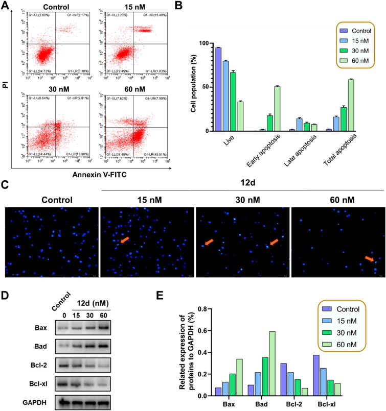
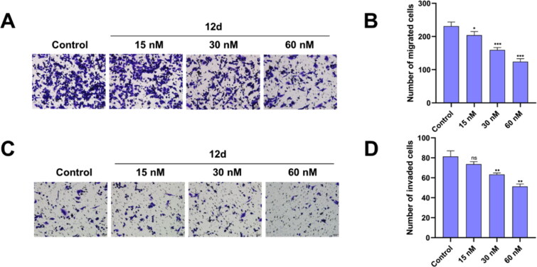
Angiogenesis plays an important role in tumour generation and progression, which is used to supply nutrients and metastasis. Herein, a series of novel dihydro-1H-indene derivatives were designed and evaluated as tubulin polymerisation inhibitors by binding to colchicine site, exhibiting anti-angiogenic activities against new vessel forming. Through structure-activity relationships study, compound 12d was found to be the most potent derivative possessing the antiproliferative activity against four cancer lines with IC50 values among 0.028-0.087 µM. Compound 12d bound to colchicine site on tubulin and inhibited tubulin polymerisation in vitro. In addition, compound 12d induced cell cycle arrest at G2/M phase, stimulated cell apoptosis, inhibited tumour metastasis and angiogenesis. Finally, the results of in vivo assay suggested that compound 12d could prevent tumour generation, inhibit tumour proliferation and angiogenesis without obvious toxicity. Collectively, all these findings suggested that compound 12d is a novel tubulin polymerisation inhibitor deserving further research.

Keywords: Anticancer; angiogenesis; colchicine binding site; indene; tubulin.


Conflict of interest statement

The authors declare that they have no known competing financial interests or personal relationships that could have appeared to influence the work reported in this paper.

Figures

Figure 1.
Figure 1.
Structures of typical CBSIs as tubulin polymerisation inhibitors.
Figure 2.
Figure 2.
Structure modification strategy of indene derivatives as CBSIs.
Scheme 1.
Scheme 1.
Synthesis of compounds 12a  12x. Reagents and conditions: (a) PPA, 90 °C, 2 h, 71.4%; (b) substituted benzaldehydes, KOH, MeOH, rt, overnight; (c) AlCl3, LiAlH4, THF, 0 °C, 2–12 h; (d) H2, Pd/C, MeOH, rt, overnight, 5.1–29.6% over three steps.
Scheme 2.
Scheme 2.
Synthesis of compounds 15a  15d. Reagents and conditions: (a) TfOH, 0 °C, 0.5 h, 56.2–91.8%; (b) substituted benzaldehyde, KOH, MeOH, rt, overnight, 78.4%; (c) AlCl3, LiAlH4, THF, 0 °C, 2–12 h; (d) H2, Pd/C, MeOH, rt, overnight, 23.6–71.6% over three steps.
Figure 3.
Figure 3.
Compound 12d disrupted tubulin distribution of K562 cells in vitro at different concentrations, and the scale bar = 10 µm.
Figure 4.
Figure 4.
Effect of 12d on the cell apoptosis. (A) K562 cells was stained with Annexin-V FITC/PI to analysis apoptosis. (B) Histograms showed the distribution of cell population. Error bars indicated SD of three independent experiments. (C) Apoptosis morphology and nucleus changes after treatment with compound 12d. Scale bar = 50 µm. (D) Expression of apoptosis associated proteins were evaluated by western blotting assay. (E) Histograms showed the western blotting assay.
Figure 5.
Figure 5.
Effect of 12d on the cell cycle. (A) K562 cells was stained with PI to analysis cell cycle. (B) Histograms showed that 12d induced cell cycle arrest at G2/M phase. Error bars indicated SD of three independent experiments. (C) Expression of G2/M phase associated proteins were evaluated by western blotting assay. (D) Histograms showed the western blotting assay.
Figure 6.
Figure 6.
Compound 12d influenced mitochondrial depolarisation and induced the generation of ROS. (A) MMP was measured using JC-1 staining assay after treatment with compound 12d on K562 cells. (B) Histograms showed the flow cytometry. (C) ROS was measured using DCF-DA assay after treatment with compound 12d on K562 cells. (D) Histograms showed the flow cytometry. Error bars indicated SD of three independent experiments. ***p < 0.001, ****p < 0.0001 vs control group.
Figure 7.
Figure 7.
12d inhibited the migration and invasion of MDA-MB-231 cells in (A) Transwell migration assay and (C) Transwell invasion assay. Scale bar = 50 µm. (B, D) Histograms showed the count of cells. Error bars indicated SD of three independent experiments. *P < 0.05, **P < 0.01, ***P < 0.001 vs control group.
Figure 8.
Figure 8.
Compound 12d inhibited angiogenesis of HUVECs in vitro. (A) Compound 12d prevented wound healing of HUVECs in a dose-dependence manner by comparing with wound closure percent after 24 h of culturation. Scale bar = 100 µm. (B) Histograms showed wound closure percent. Error bars indicated SD of three independent experiments. ***p < 0.001, ****p < 0.0001 vs control group. (C) Compound 12d destructed HUVECs forming normal capillary-like tubular networks. Scale bar = 100 µm.
Figure 9.
Figure 9.
Hypothesised binding interactions between compound 12d and tubulin (PDB id:5LYJ). (A) Supposed docking pose of 12d (green) in the colchicine binding site; (B) Overlap of CA-4 (orange) and compound 12d with tubulin. PyMOL was used to draw the figures.
Figure 10.
Figure 10.
Antitumor activity of compound 12d in H22 allograft mice model. (A) Tumours in each of group vehicle, compound 12d (10 or 20 mg/kg/day, i.v.), and CA-4 (20 mg/kg/day, i.v.) were shown. (B) Individual tumour weights were measured after treatment for three weeks. Error bars indicated SD of three independent experiments. ****p < 0.0001 vs control group, ###p < 0.001 vs CA-4 (20 mg/kg) group. (C) Tumour volumes and (D) mouse body weights were measured every other day for three weeks. Error bars indicated SD of three independent experiments.
Figure 11.
Figure 11.
Representative images of H&E-stained vital organ, including heart, liver, spleen, lung and kidney. Scale bar = 20 µm.
Figure 12.
Figure 12.
(A) Representative images of IHC-stained markers CD31 and Ki67 in tumour tissue. Scale bar = 20 µm. Histograms showed the relative percentages of (B) microvessel density assessed by prognostic angiogenic marker CD31 and (C) expression level of cell proliferation marker Ki67 in CA-4 (20 mg/kg) or 12d (20 mg/kg) treated group compared with the control group. Error bars indicated SD of three independent experiments. ***p < 0.001, ****p < 0.0001 vs control group.

References

    1. Jordan MA, Wilson L.. Microtubules as a target for anticancer drugs. Nat Rev Cancer. 2004;4(4):253–265. - PubMed
    1. Penna LS, Henriques JAP, Bonatto D.. Anti-mitotic agents: are they emerging molecules for cancer treatment? Pharmacol Ther. 2017;173:67–82. - PubMed
    1. Islam MN, Iskander MN.. Microtubulin binding sites as target for developing anticancer agents. Mini Rev Med Chem. 2004;4(10):1077–1104. - PubMed
    1. Dumontet C, Jordan MA.. Microtubule-binding agents: a dynamic field of cancer therapeutics. Nat Rev Drug Discov. 2010;9(10):790–803. - PMC - PubMed
    1. Mühlethaler T, Gioia D, Prota AE, Sharpe ME, Cavalli A, Steinmetz MO.. Comprehensive analysis of binding sites in tubulin. Angew Chem Int Ed Engl. 2021;60(24):13331–13342. - PMC - PubMed