Review
. 2023 Aug 6:43:102230.
doi: 10.1016/j.jcot.2023.102230. eCollection 2023 Aug.

Humerus shaft fractures, approaches and management

Affiliations
Review

Humerus shaft fractures, approaches and management

Wich Orapiriyakul et al. J Clin Orthop Trauma. .

Abstract

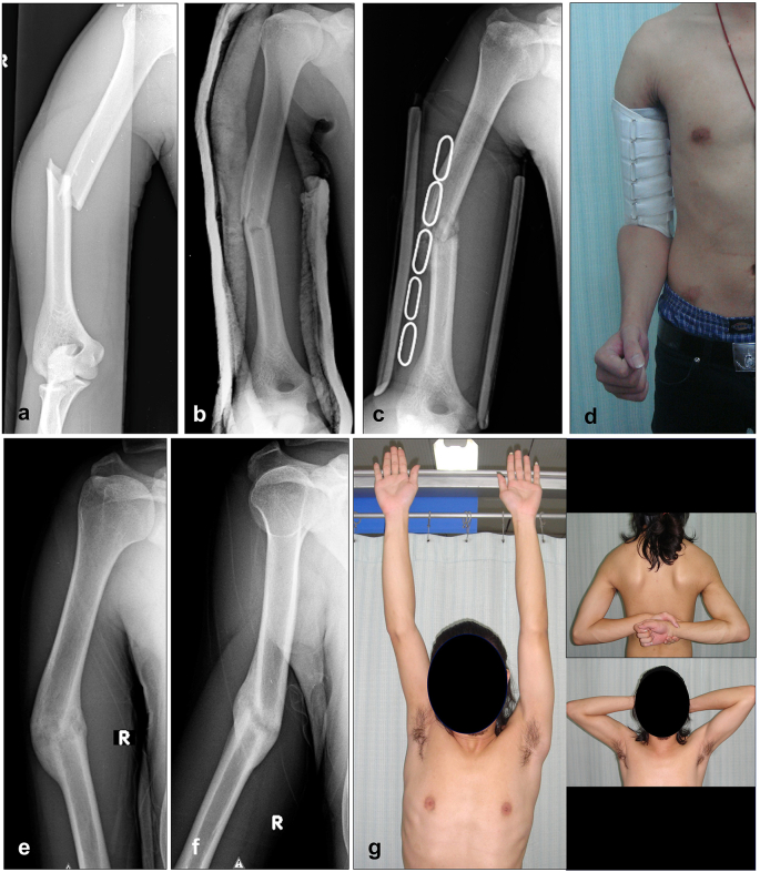
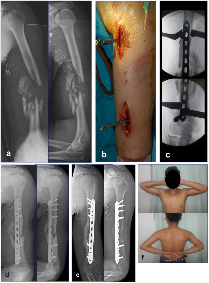
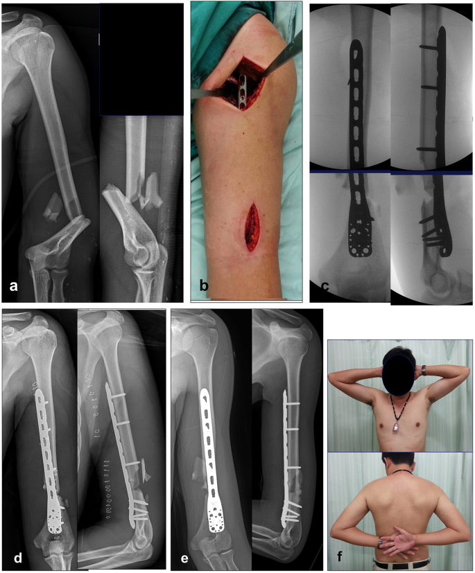
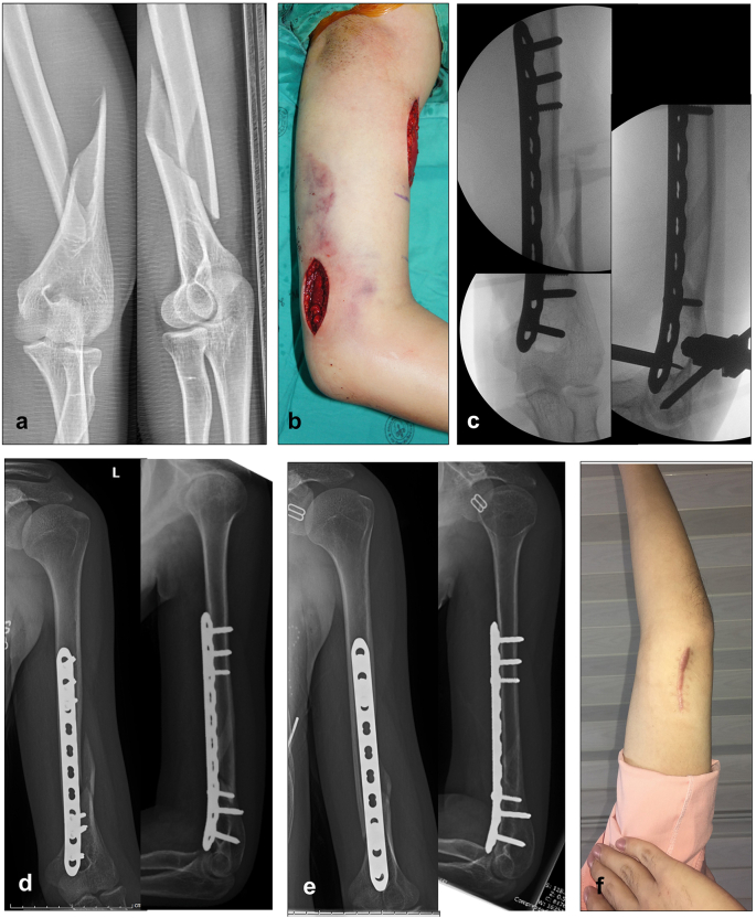
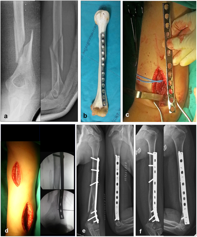
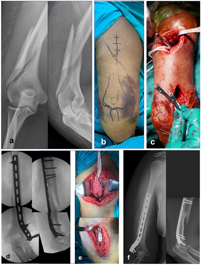
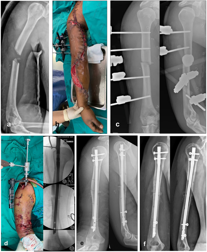
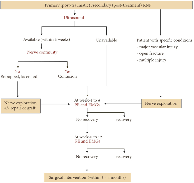
Humeral shaft fracture is a common injury which can be treated either conservatively with functional bracing or with surgical fixation. Current evidence shows an increase in the rate of nonunion after conservative treatment, suggesting that indications for conservative treatment may need to be re-examined. This article updates trends in treatment for humeral shaft fracture. Indications for surgery, both for plate osteosynthesis with open reduction and internal fixation (ORIF) as well as for minimally invasive plate osteosynthesis (MIPO) and intramedullary nailing (IMN) are described. Recognition of the advantages and disadvantages of each technique can better define the role of the plate or nail and can aid in the selection of an appropriate surgical approach. ORIF with compression plate continues to have a role in the treatment of simple or AO/OTA type A fractures. The primary goal of minimal invasive osteosynthesis, a surgical technique involving small incisions, closed reduction or mini-open reduction that minimizes soft tissue dissection and helps preserve the periosteal blood supply, is to achieve bone union and the best possible functional outcomes. MIPO of the humerus is now well accepted as being less invasive and providing relative stability to allow indirect (secondary) bone healing with callus formation. MIPO approaches can be performed circumferentially to the humerus, including the proximal, middle and distal shaft. The classic MIPO approach is anterior MIPO, followed by posterior, anterolateral and anteromedial MIPO. IMN is also an option for treating humerus fractures. In the past, IMN was not widely used due to the potential for complications such as shoulder impingement and elbow problems as well as the limited availability of implants and the steep learning curve of this surgical technique. Over the past decade, the launch of a new design of straight antegrade and retrograde IMN with established techniques has encouraged more surgeons to use IMN as an alternative option. Methods of dealing with concomitant and post-treatment radial nerve palsy have also been evolving, including the use of ultrasound for diagnosis of radial nerve conditions. Radial nerves with contusion, entrapment or laceration can be detected using ultrasound with reliability comparable to intraoperative findings. Trends in treatment of radial nerve palsy are described below. Future larger randomized controlled trials comparing conservative and operative management are necessary to further develop appropriate guidelines.

Keywords: Conservative; Fracture; Humeral shaft; Intramedullary nailing; MIPO; ORIF; Radial nerve injury; Treatment; Ultrasound.


Conflict of interest statement

The authors do not received funding from any source. We received no payments or other benefits from any commercial entity.

Figures

Fig. 1
Fig. 1
X-ray showing an oblique fracture of the midshaft humerus (a), X-ray after closed reduction with U slab (b), X-ray after change to functional brace (c), Humerus functional brace with adjustable strap (d), AP X-ray at 6 months showing complete healing with varus angulation (e), Lateral X-ray showing anterior angulation (f), Normal final functional outcome (g).
Fig. 2
Fig. 2
AP and lateral X-rays of the humerus showing comminuted fracture of the midshaft of the humerus from a gunshot injury (a), Proximal and distal incisions with drill sleeves controlling the alignment of the narrow LCP (b), The plate aligned in the center of the anterior surface of the proximal and distal humerus (c), Post op x-rays showing satisfactory alignment (d), X-rays at 6 months indicated good callus formation (e), Function outcome at 6 months (f).
Fig. 3
Fig. 3
AP and lateral X-rays of the humerus showing a comminuted fracture of the distal shaft of the humerus (a), Proximal and distal incisions of anterior MIPO using reverse PHILOS plate (b), Intraoperative images reveal satisfactory alignment of the plate in the center of the anterior surface of the proximal and distal humerus in AP and lateral (c), Post op x-rays show satisfactory alignment (d), X-rays at 6 months indicate posterior bridging callus (e), Functional outcome at 6 months (f).
Fig. 4
Fig. 4
AP and lateral X-rays of the humerus show a spiral fracture of the distal shaft of the humerus (a), Proximal and distal incisions of anteromedial MIPO (b), Intraoperative images reveal satisfactory alignment of the plate in the center of the anterior surface of the proximal and distal humerus in AP and lateral (c), Post op x-rays show satisfactory alignment (d), X-rays at 6 months show complete fracture healing (e), Distal incision scar hidden on the medial side of the elbow (f).
Fig. 5
Fig. 5
X-rays AP and lateral view of the humeral shaft showing spiral wedge fracture of the distal shaft of the humerus (a), Contoured anterolateral plate on the cadaveric bone (b), Distal incision between the brachialis and brachioradialis muscles to identify the radial nerve (c), Proximal and distal incisions of the anterolateral MIPO with the intraoperative alignment (d), Post op x-rays show satisfactory alignment (e), X-rays at 6 months indicated complete bone healing (f).
Fig. 6
Fig. 6
AP and lateral X-rays of the humeral shaft show a low spiral wedge fracture of the distal shaft of the humerus (a), Proximal and distal incisions of posterior MIPO are marked on the skin (b), Proximal and distal incisions with drill sleeves using the extra articular distal humerus locking plate (c), the intraoperative alignment in AP and lateral view (d), Radial nerve lies over the plate at the proximal incision (e), X-rays at 6 months indicated complete bone healing (f).
Fig. 7
Fig. 7
A 31-year-old man had a right humerus fracture with axillary artery and brachial plexus injury. X-rays showed a transverse fracture of the right humerus (a), Axillary artery was repaired and fasciotomy of the arm and forearm were followed by external fixator for temporary immobilization (b), X-rays after external fixator (c), Closed humeral nailing was done. The intraoperative X-rays show good reduction and fixation (d), Immediate post-op radiography showed a good alignment (e), At the 3-month follow-up, callus formation is seen at fracture site (f).
Fig. 8
Fig. 8
Flow diagram depicting of the management of radial nerve palsy associated with humerus fracture. The authors recommend utilizing ultrasound to evaluate nerve continuity in the initial phase. If evidence of nerve entrapment or nerve laceration is detected, immediate nerve exploration is advised. In certain conditions, such as major vascular injury, open fracture, or multiple injuries, early radial nerve exploration is required. When the ultrasound shows good continuity of the nerve, nerve contusion is suspected. Then, it is recommended to monitor clinical symptoms and perform electromyography (EMG) to evaluate the recovery status. The absence of signs and symptoms indicating recovery after 2–3 months suggests the requirement for surgical intervention, depending on the nature of the nerve pathology.

References

    1. Lode I., et al. Operative versus nonoperative treatment of humeral shaft fractures: a systematic review and meta-analysis. J Shoulder Elbow Surg. 2020;29(12):2495–2504. - PubMed
    1. Oliver W.M., et al. Surgical versus nonsurgical management of humeral shaft fractures: a systematic review and meta-analysis of randomized trials. J Am Acad Orthop Surg. 2023;31(2):e82–e93. - PubMed
    1. Sarmiento A., et al. Functional bracing of fractures of the shaft of the humerus. J Bone Joint Surg Am. 1977;59(5):596–601. - PubMed
    1. Rämö L., et al. Effect of surgery vs functional bracing on functional outcome among patients with closed displaced humeral shaft fractures: the FISH randomized clinical trial. JAMA. 2020;323(18):1792–1801. - PMC - PubMed
    1. Serrano R., et al. Modern results of functional bracing of humeral shaft fractures: a multicenter retrospective analysis. J Orthop Trauma. 2020;34(4):206–209. - PubMed

LinkOut - more resources