. 2022 Apr 26;2(3):360-368.
doi: 10.1016/j.xrrt.2022.04.004. eCollection 2022 Aug.

Elbow lateral ulnar collateral ligament reconstruction by transposition of the local extensor fascia septum: surgical technique and preliminary results

Affiliations

Elbow lateral ulnar collateral ligament reconstruction by transposition of the local extensor fascia septum: surgical technique and preliminary results

Andrea Celli et al. JSES Rev Rep Tech. .

Abstract

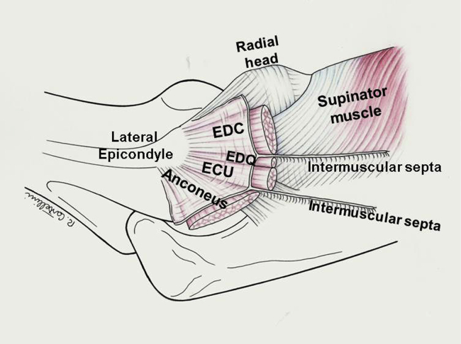
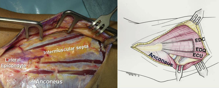
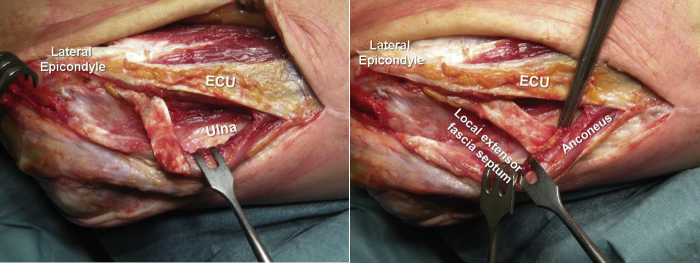
The lateral ulnar collateral ligament (LUCL) is a primary lateral stabilizer of the elbow that originates from the isometric center of the capitulum and inserts into the supinator crest of the ulna. LUCL injury may be due to trauma, chronic strain, or iatrogenic lesion. In patients with symptomatic LUCL insufficiency and recurrent posterolateral rotatory instability, surgical reconstruction can restore elbow stability. In primary acute treatment, the injured LUCL is reattached to the lateral epicondyle with transosseous sutures and anchors placed at the isometric origin of the ligament. If the ligament quality is poor, patients with chronic elbow instability may require reconstruction with a tendon autograft or allograft. Alternatively, the LUCL can be reconstructed by transposition of the local extensor fascia septum, a local flap that exploits the common extensor fascia connected to a thin strip of extensor digitorum quinti or the extensor digitorum communis intermuscular septum. We describe a new LUCL reconstruction technique based on the transposition of the local extensor fascia septum and report the preliminary result in a series of patients aged 50 years or less.

Keywords: Chronic strain; Iatrogenic lesion; Lateral ulnar collateral ligament; Soft tissue lesions; Transposition of the local extensor fascia septum; Trauma.


Figures

Figure 1
Figure 1
The superficial antebrachial fascia is a dense membranous layer covering the extensor muscles and tendons. It originates from the supracondylar ridge of the lateral epicondyle and is attached posteriorly to the olecranon and the dorsal border of the ulna. Numerous intermuscular septa that enclose and separate the muscles and provide insertions for muscle fibers originate from its depth. The EDQ is a slender muscle that together with the ECU lies lateral to the EDC. A well-developed, thicker deep septum divides the two muscles and inserts proximally into the lateral epicondyle, where its fibers merge with the underlying LCL. EDQ, extensor digitorum quinti; ECU, extensor carpi ulnaris; EDC, extensor digitorum communis; LCL, lateral collateral ligament.
Figure 2
Figure 2
Development of the Kocher interval between the anconeus and the ECU allows visualization of the remainder of the ruptured LCL. ECU, extensor carpi ulnaris; LCL, lateral collateral ligament.
Figure 3
Figure 3
The septum between the EDQ and the EDC is identified by palpation anterior to the ECU insertion, whereas the EDQ is identified by passive movement of the small finger with the wrist blocked. EDQ, extensor digitorum quinti; ECU, extensor carpi ulnaris; EDC, extensor digitorum communis.
Figure 4
Figure 4
A triangular fragment of common extensor fascia (ca. 2 × 6 cm wide and 8 cm long) is marked and collected. EDQ, extensor digitorum quinti; ECU, extensor carpi ulnaris; EDC, extensor digitorum communis.
Figure 5
Figure 5
(A, B, and C) The superficial fascial flap is mobilized off the underlying muscles, preserving its continuity with the deep intermuscular septum. The muscle fiber insertions on the septa are raised and divided longitudinally, leaving 2-3 mm in continuity with the fascial flap. EDQ, extensor digitorum quinti; ECU, extensor carpi ulnaris; EDC, extensor digitorum communis.
Figure 6
Figure 6
The fascial flap with the split septum is mobilized, preserving its epicondylar insertion. EDQ, extensor digitorum quinti; ECU, extensor carpi ulnaris; EDC, extensor digitorum communis.
Figure 7
Figure 7
The two long sides of the flap are folded around the split of the septum, and an imbricated Krackow locking suture is placed along the anterior and posterior aspects of the new ligament.
Figure 8
Figure 8
The EDC is divided; the EDQ and the ECU tendons are split as a unit from the underlying supinator muscle, capsule, and annular ligament using blunt scissors. The oblique fibers of the supinator muscle allow identifying the deep plane of dissection. EDQ, extensor digitorum quinti; ECU, extensor carpi ulnaris; EDC, extensor digitorum communis.
Figure 9
Figure 9
The graft is rotated posterior to the ulnar insertion under the ECU-EDQ tendon unit and above the supinator muscle. EDQ, extensor digitorum quinti; ECU, extensor carpi ulnaris.
Figure 10
Figure 10
To ensure its alignment with the native ligament at the isometric point, the new ligament is fixed with a suture anchor at the center of the lateral aspect of the capitulum.
Figure 11
Figure 11
The supinator crest is excavated with a high-speed burr to receive the rotational flap (about 1-1.5 cm × 0.5 cm), and three holes are drilled into the ulnar border of the excavated site. EDQ, extensor digitorum quinti; ECU, extensor carpi ulnaris; EDC, extensor digitorum communis.
Figure 12
Figure 12
The graft sutures are passed through the ulnar holes, the new ligament is introduced into the groove, and the sutures are tied with correct tension with the elbow in 40° of flexion and the forearm fully pronated. The remaining native ligament and the annular ligament are repaired in continuity with the LUCL. LUCL, lateral ulnar collateral ligament.
Figure 13
Figure 13
The extensor fascia is closed and the Kocher interval is reapproximated with absorbable sutures. ECU, extensor carpi ulnaris.
Figure 14
Figure 14
X-ray scans (case #2).

References

    1. Arvind C.H., Hargreaves D.G. Table top relocation test: New clinical test for postero-lateral rotatory instability of the elbow. J Shoulder Elbow Surg. 2006;15:500–501. doi: 10.1016/j.jse.2005.11.014. - DOI - PubMed
    1. Badhrinarayanan S., Desai A., Watson J.J., White C.H.R., Phadnis J. Indications, Outcomes, and Complications of Lateral Ulnar Collateral Ligament Reconstruction of the Elbow for Chronic Posterolateral Rotatory Instability: A Systematic Review. Am J Sports Med. 2021;49:830–837. doi: 10.1177/0363546520927412. - DOI - PubMed
    1. Baumfeld J.A., Van Riet R.P., Zobitz M.E., Eygendaal D., Kai-Nan A.N., Steinmann S.P. Triceps tendon properties and its potential as an autograft. J Shoulder Elbow Surg. 2010;19:607–609. doi: 10.1016/j.jse.2009.12.001. - DOI - PubMed
    1. Cohen M.S., Hastings H.H. Rotatory instability of the elbow, the anatomy and role of the lateral stabilizers. J Bone Joint Surg Am. 1997;79:225–233. - PubMed
    1. Finkbone P.R., O’Driscoll S.W. Box loop ligament reconstruction of the elbow for medial and lateral instability. J Shoulder Elbow Surg. 2015;24:647–654. doi: 10.1016/j.jse.2014.12.008. - DOI - PubMed

LinkOut - more resources