. 2023 Jul;18(4):100831.
doi: 10.1016/j.ajps.2023.100831. Epub 2023 Jul 26.

Dual pH and microbial-sensitive galactosylated polymeric nanocargoes for multi-level targeting to combat ulcerative colitis

Affiliations

Dual pH and microbial-sensitive galactosylated polymeric nanocargoes for multi-level targeting to combat ulcerative colitis

Mahira Zeeshan et al. Asian J Pharm Sci. 2023 Jul.

Abstract

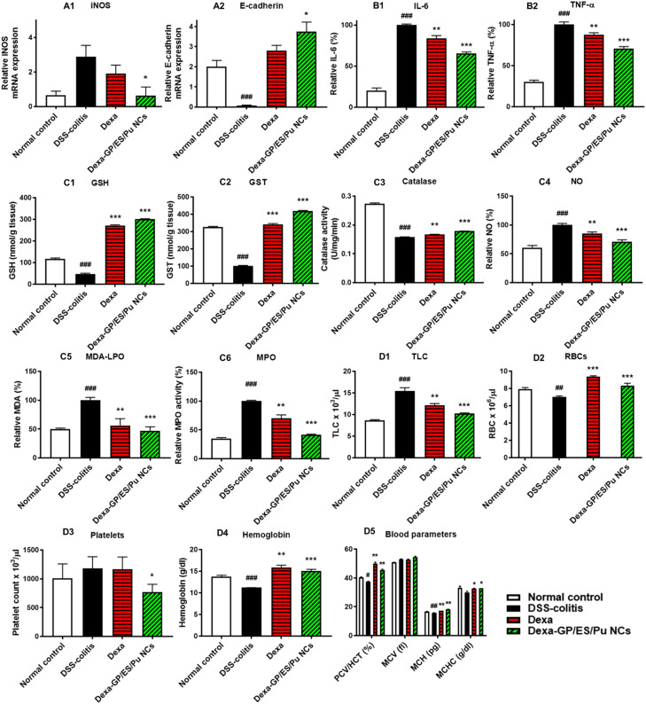
Ulcerative colitis (UC) is a type of inflammatory bowel disease characterized by inflammation, ulcers and irritation of the mucosal lining. Oral drug delivery in UC encounters challenges because of multifaceted barriers. Dexamethasone-loaded galactosylated-PLGA/Eudragit S100/pullulan nanocargoes (Dexa-GP/ES/Pu NCs) have been developed with a dual stimuli-sensitive coating responsive to both colonic pH and microbiota, and an underneath galactosylated-PLGA core (GP). The galactose ligand of the GP preferentially binds to the macrophage galactose type-lectin-C (MGL-2) surface receptor. Therefore, both stimuli and ligand-mediated targeting facilitate nanocargoes to deliver Dexa specifically to the colon with enhanced macrophage uptake. Modified emulsion method coupled with a solvent evaporation coating technique was employed to prepare Dexa-GP/ES/Pu NCs. The nanocargoes were tested using in vitro, ex vivo techniques and dextran sodium sulfate (DSS) induced UC model. Prepared nanocargoes had desired physicochemical properties, drug release, cell uptake and cellular viability. Investigations using a DSS-colitis model showed high localization and mitigation of colitis with downregulation of NF-ĸB and COX-2, and restoration of clinical, histopathological, biochemical indices, antioxidant balance, microbial alterations, FTIR spectra, and epithelial junctions' integrity. Thus, Dexa-GP/ES/Pu NCs found to be biocompatible nanocargoes capable of delivering drugs to the inflamed colon with unique targeting properties for prolonged duration.

Keywords: Galactosylated nanocargoes; Macrophage galactose type-lectin C; Microbial sensitive; Pullulan; Ulcerative colitis; pH-sensitive drug delivery.


Conflict of interest statement

The authors declared no conflict of interest.

Figures

Image, graphical abstract
Graphical abstract
Fig 1
Fig. 1
Physicochemical characterization of Dexa-GP/ES/Pu NCs. (A) Particle size analysis, (B) SEM analysis, (C) ATR-FTIR, (D) XPRD, and (E) TGA analysis of drug, excipients and nanocargoes.
Fig 2
Fig. 2
Dexa release from GP/Pu NPs, GP/ES NPs and GP/ES/Pu NCs (n = 3) at acidic pH (pH 1.2–4.5) and pH 7.4, without or with cecal contents (A, B) (Note: Dexa-GP/ES/Pu NCs vs GP/Pu NPs: *P<0.05, **P<0.01, ***P<0.001; and Dexa-GP/ES/Pu NCs vs GP/ES NPs: σP<0.05, σσP<0.01, σσσP<0.001); Rheological investigations of mucin and nanocargoes interaction (C); Spectrophotometric analysis of percent mucin binding to nanocargoes (D); Size analysis of Mucin (E); Size analysis after mucin-nanocargoes interaction (F).
Fig 3
Fig. 3
Hemolytic activity of nanoformulations after 24 h (A) and 48 h (B)(n = 3, Statistical significance between nano-formulations and Triton-X if *P<0.05, **P<0.01, ***P<0.001); Macrophage cell viability assay after 24 h (C) and 48 h (D); Colon cell viability assay after 24 h (E) and 48 h (F), (For C-F: n = 3; Statistical significance between nano-formulations and Normal cells control if *P<0.05, **P<0.01, ***P<0.001 and between Triton-X and Normal control if #P<0.05, ##P<0.01, ###P<0.001); Macrophage and colon cells uptake of dye-labelled nanoformulations (n = 3) (G), Fluorescence intensity of the dye-labelled nanoformulation up taken by the macrophage and colon cells, scale= 25 µm (H), (n = 3; Statistical significance between nano-formulations and plain dye control if *P<0.05, **P<0.01, ***P<0.001).
Fig 4
Fig. 4
(A) Cross section of goat intestine illuminating under dye released from Rho-GP/ES/Pu NCs retention (i-iv) and free Rho dye control (v-viii) over 8 h, scale = 100 µm; (B) Relative percentage retention in the large intestine; (C) Relative percentage permeation across intestine into the buffer (pH 7.4); (D) Retention to permeation ratio of the Rho-GP/ES/Pu NCs and free dye for 8 h. (B-D: n = 3; Statistical significance between Rho-GP/ES/Pu NCs and free Rho if *P<0.05, **P<0.01, ***P<0.001); (E) Percentage drug localization after 6-h administration of Dexa-GP/ES/Pu NCs to the inflamed (DSS-induced colitis) and normal mice (n = 5; Statistical significance between DSS and Normal group if *P<0.05, **P<0.01, ***P<0.001); (F) Percentage drug retention or biodistribution to the vital organs after 6-h administration of Dexa-GP/ES/Pu NCs, n = 5; Statistical significance between Dexa-GP/ES/Pu NCs and Dexa*P<0.05, **P<0.01, ***P<0.001); (G) Vascular integrity test: Evans blue permeation across vessels into the inflamed colon (n = 3; Statistical significance between treated mice Vs DSS-colitis group if *P<0.05, **P<0.01, ***P<0.001 and Normal vs DSS: #P<0.05, ##P<0.01, ###P<0.001); (H) Flowcytometry to analyze fluorescein loaded GP/ES/Pu NCs uptake by the colon-derived macrophages, untreated control (a), GP treated (b), GP/ES/Pu NCs treated (c) (n = 3).
Fig 5
Fig. 5
Therapeutic evaluation of Dexa-GP/ES/Pu NCs; Scheme of experimental design (A), body weight assessment (B), DAI scores (C), vital organs weight assessment (F), distress scores (D) and mortality (E), colon weight to length ratio (G), length of intestines (H), pictorial representation of colon and spleen (I). For statistical significance between treatment groups vs DSS colitis: *P<0.05, **P<0.01, ***P<0.001; the significance levels were same for Normal VS DSS-colitis except asterisks were changed to sign# (n = 5).
Fig 6
Fig. 6
(A) Histology of colon, small intestine, and spleen from the representatives of normal control, DSS-colitis, Dexa and Dexa-GP/ES/Pu NCs treated mice groups (scale = 100 µm); (B) Colon cumulative histopathological score, n = 3; (C) Expression of NF-ĸB and COX-2 at the colon tissues in the normal control, DSS-colitis, Dexa and Dexa-GP/ES/Pu NCs treated groups by immunohistochemistry, n = 3; (D) Relative immunoreactivity (%) of NF-ĸB; (E) Relative immunoreactivity of COX-2; (F) ATR-FTIR spectra of colon tissues excised from normal control, DSS-colitis, Dexa and Dexa-GP/ES/Pu NCs treated groups, n = 3; (G) Descriptive analysis of ATR-FTIR peaks with percent transmittance at a particular wavenumber (cm−1) and functional groups; (H) Microbial colony forming units (logCFU/ml) from the fecal suspension obtained from different groups under investigation, n = 3/group; (I) Blood glucose levels of the mice of different groups under study, n = 3. Statistical significance between treatment groups and DSS colitis is defined as *P<0.05, **P<0.01, ***P<0.001; the significance levels between Normal control and DSS-colitis are #P<0.05, ##P<0.01, ###P<0.001#.
Fig 7
Fig. 7
Relative mRNA expression of iNOS (a1) and E-cadherin (a2) (n = 3) from the colon tissues of different groups under study; Relative percentage of pro-inflammatory cytokines IL-6 (b1) and TNF- α (b2) in the colon tissues of comparative groups (n = 3); Concentration of antioxidants GSH (c1), GST (c2), catalase (c3), and oxidants NO (c4), MDA (c5) and MPO (c6) in the excised colon tissues from normal control, DSS-colitis, Dexa and Dexa-GP/ES/Pu NCs treated mice (n = 3); Levels of hematological parameters TLC (d1), RBC (d2), Platelets (d3), Hemoglobin (d4), PCV/HCT, MCV, MCH and MCHC (d5) in normal control, DSS-colitis, Dexa and Dexa-GP/ES/Pu NCs treated mice (n = 3). Statistical significance between treatment groups and DSS colitis is defined as *P<0.05, **P<0.01, ***P<0.001; the significance levels between Normal control and DSS-colitis are #P<0.05, ##P<0.01, ###P<0.001.
Fig 8
Fig. 8
Assessment of in-vivo biocompatibility and toxicology during and after 7-d administration of Dexa-GP/ES/Pu to the healthy mice (n = 3): (A) Body weight (%), (B) Average organ weight (g), (C) Intestine lengths (cm), (D) Colon weight to length ratio, (E) Fecal microbial growth (log CFU/ml), (F) Hematological analysis, (G) Platelet count, (H) Histopathological investigations of vital organs (kidney, heart, liver, lung, stomach, small intestine, colon and spleen), (I) Colon cells apoptosis study with Annexin-V/Propidium iodide in the untreated normal healthy control and Dexa-GP/ES/Pu NCs treated healthy mice.

References

    1. Tatiya-aphiradee N., Chatuphonprasert W., Jarukamjorn K. Immune response and inflammatory pathway of ulcerative colitis. J Basic Clin Physiol Pharmacol. 2019;30(1):1–10. - PubMed
    1. Mukhtar M., Ali H., Ahmed N., Munir R., Talib S., Khan A.S., et al. Drug delivery to macrophages: a review of nano-therapeutics targeted approach for inflammatory disorders and cancer. Expert Opin Drug Deliv. 2020;17(9):1239–1257. - PubMed
    1. Zeeshan M., Ali H., Khan S., Khan S.A., Weigmann B. Advances in orally-delivered pH-sensitive nanocarrier systems; an optimistic approach for the treatment of inflammatory bowel disease. Int J Pharm. 2019;558:201–214. - PubMed
    1. Chen F., Liu Q., Xiong Y., Xu L. Current strategies and potential prospects of nanomedicine-mediated therapy in inflammatory bowel disease. Int J Nanomedicine. 2021;16:4225. - PMC - PubMed
    1. Xu Y., Zhu B.W., Li X., Li Y.F., Ye X.M., Hu J.N. Glycogen-based pH and redox sensitive nanoparticles with ginsenoside Rh2 for effective treatment of ulcerative colitis. Biomaterials. 2021 - PubMed