. 2023 Aug 1;16(8):dmm049762.
doi: 10.1242/dmm.049762. Epub 2023 Aug 17.

An interaction between OTULIN and SCRIB uncovers roles for linear ubiquitination in planar cell polarity

Affiliations

An interaction between OTULIN and SCRIB uncovers roles for linear ubiquitination in planar cell polarity

Stephanie M Almeida et al. Dis Model Mech. .

Abstract

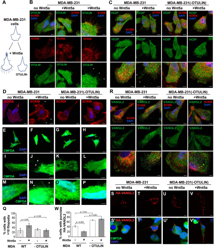
Planar cell polarity (PCP) plays critical roles in developmental and homeostatic processes. Membrane presentation of PCP complexes containing Van Gogh-like (VANGL) transmembrane proteins is central to PCP and can be directed by the scaffold protein scribble (SCRIB). The role atypical linear ubiquitin (Met1-Ub) chains might play in PCP is unknown. Here, HEK293 cell-based interactomic analyses of the Met1-Ub deubiquitinase OTULIN revealed that OTULIN can interact with SCRIB. Moreover, Met1-Ub chains associated with VANGL2 and PRICKLE1, but not SCRIB, can direct VANGL2 surface presentation. Mouse embryos lacking Otulin showed variable neural tube malformations, including rare open neural tubes, a deficit associated with PCP disruption in mice. In Madin-Darby canine kidney cells, in which the enrichment of VANGL2-GFP proteins at cell-cell contacts represents activated PCP complexes, endogenous OTULIN was recruited to these sites. In the human MDA-MB-231 breast cancer cell model, OTULIN loss caused deficits in Wnt5a-induced filopodia extension and trafficking of transfected HA-VANGL2. Taken together, these findings support a role for linear (de)ubiquitination in PCP signaling. The association of Met1-Ub chains with PCP complex components offers new opportunities for integrating PCP signaling with OTULIN-dependent immune and inflammatory pathways.

Keywords: Linear ubiquitin; Linear ubiquitin assembly complex; Mouse genetics; OTULIN; Planar cell polarity; Scribble.


Conflict of interest statement

Competing interests The authors declare no competing or financial interests.

Figures

Fig. 1.
Fig. 1.
OTULIN uses its PBM to interact with SCRIB. (A) Schematic diagram showing FLAG-tagged OTULIN constructs used in AP-MS experiments: full-length OTULIN (WT), catalytically inactive OTULIN (W96R), OTULIN lacking its PBM (ΔPBM) or N-terminal 54 amino acids (Δ54), and OTULIN truncated after amino acid 105 (C105X). PIM, PUB domain-interacting motif; PBM, PDZ domain-binding motif. (B) Spectral counts recovered in AP-MS experiments. OTULINΔPBM did not recover SNX27, SCRIB or SLC9A3R2. The N-terminal region of OTULIN is required for its interactions with HOIP (Rivkin et al., 2013). Notably, reduced SCRIB and no HOIP peptides were recovered by full-length OTULIN treated with the proteasomal inhibitor MG132 (WT+MG) or by OTULINΔ54. (C) Immunoprecipitation (IP) of FLAG-SCRIB with an anti-FLAG antibody recovered equivalent levels of WT HA-OTULIN, HA-OTULINW96R (‘W’) and HA-OTULINC129S (‘C’), as detected by immunoblotting with an anti-HA antibody. Conversely, immunoprecipitation of HA-OTULIN using an anti-HA antibody recovered FLAG-SCRIB when the OTULIN PBM was present, but this was not seen for HA-OTULINΔPBM constructs (‘P’). Deletion of the N-terminal HOIP-interacting region (HA-OTULINΔ54 constructs) did not affect interactions with SCRIB. HA-OTULINC105X (‘105’) did not interact with FLAG-SCRIB. Input amounts for each construct and tubulin levels are shown. Immunoblots are representative of three independent experiments.
Fig. 2.
Fig. 2.
Localization of OTULIN to juxtamembrane positions depends on its PBM and SCRIB. (A-E) Representative images show immunofluorescence staining of endogenous SCRIB (red) in HEK293 cells co-stained for (A) endogenous (‘endo’) OTULIN (green); (B) transfected HA-OTULIN constructs (magenta) – HA-OTULIN (WT), HA-OTULINC129S (C129S), HA-OTULINΔPBM (no PBM) and HA-OTULINY56A (Y56A); (C) endogenous HOIP (green); (D) transfected myc-HOIP (green); and (E) co-transfected myc-HOIP (green) and HA-OTULIN constructs (WT, C129S, ΔPBM and Y56A) (magenta). (F-H) Representative images show immunofluorescence staining of Neuro2A cells (F) transfected with HA-OTULIN constructs (WT, C129S, ΔPBM and Y56A) (magenta), (G) co-transfected with FLAG-SCRIB (red) and HA-OTULIN constructs (magenta), and (H) transfected with FLAG-SCRIB (red). (I) A schematic diagram summarizes subcellular localization of HA-OTULIN constructs. Images are representative of three independent experiments. Scale bars: 10 µm.
Fig. 3.
Fig. 3.
Endogenous OTULIN is recruited to GFP-VANGL1- and GFP-VANGL2-containing PCP complexes in MDCK cells. (A-A″,D,D′) In untransduced MDCK cells, endogenous OTULIN expression is punctate and distributed uniformly throughout the cell. (B-C″,E-F′) MDCK cells transduced with (B-B″,E,E′) GFP-VANGL1 or (C-C″,F,F′) GFP-VANGL2 display characteristic VANGL1/2 localization to the cell membrane at sites of cell-cell contact when cells are grown to 80% confluency. Endogenous OTULIN is enriched at these sites where GFP-VANGL1 or GFP-VANGL2 is co-localized but not at cell-cell contacts of untransduced cells. D-F show higher magnification views of outlined regions shown in A″-C″, respectively. Images are representative of three independent experiments. Scale bars: 50 µm (A-C″); 25 µm (D-F); 10 µm (D′-F′). (G) Quantification of the percentage of total cells with endogenous OTULIN at free edges or localized to edges in contact with neighboring cells is shown for untransduced MDCK cells (MDCK), and GFP-VANGL1 (VANGL1:MDCK) and GFP-VANGL2 (VANGL2:MDCK) transduced MDCK cells. Error bars show the s.e.m. Statistical significance values between the localization of OTULIN in untransduced cells compared to GFP-VANGL1- or GFP-VANGL2-expressing cells were determined using two-way ANOVA and the Bonferroni post test. ns, not significant; **P<0.001; ***P<0.0001.
Fig. 4.
Fig. 4.
VANGL2, but not SCRIB, is modified with Met1-Ub chains, and linear ubiquitination correlates with VANGL2 surface presentation. (A-C) A Met1-Ub antibody detected proteins modified with Met1-Ub chains in immunoprecipitates of (A) FLAG-SCRIB and (B) FLAG-NEMO recovered with an anti-FLAG antibody, and (C) GFP-VANGL2 recovered with an anti-GFP antibody from HEK293T cells transfected with HA-HOIL and myc-HOIP. Adding HA-OTULIN (‘wt’), but not HA-OTULINC129S (‘C’), eliminated Met1-Ub immunoreactivity. Input amounts for each construct and tubulin levels are shown. (D-F) Purification of Met1-Ub-conjugated (D) FLAG-SCRIB, (E) FLAG-NEMO and (F) GFP-VANGL2 using GST protein, GST-coupled to the Met1-Ub-binding UBAN domain of NEMO (GST-UBAN) or to a mutated non-Met1-Ub-binding UBAN domain (GST-RRE). Pull-down of GST-UBAN recovered (E) FLAG-NEMO and (F) GFP-VANGL2, but not (D) FLAG-SCRIB from HA-HOIL/myc-HOIP-expressing HEK293 cells. The addition of HA-OTULIN (‘wt’), but not catalytically inactive HA-OTULINC129S (‘C’), abrogated recovery of FLAG-NEMO and GFP-VANGL2. (G) A cell surface biotinylation assay recovered biotinylated GFP-VANGL2 from HEK293T cells transfected with HA-HOIL and myc-HOIP, as shown in a representative immunoblot probed with anti-GFP antibody; ‘Bio-PD’ indicates biotin pulldown. The presence of FLAG-OTULIN (‘wt’), but not FLAG-OTULINC129S (‘C’), eliminated surface biotinylation of GFP-VANGL2. Immunoblots are representative of three independent experiments.
Fig. 5.
Fig. 5.
Mice homozygous for the OtulinΔex3 null allele show variable neural tube deficits and VANGL2 localization. (A) Schematic diagram of the OtulinΔex3 allele. (B) An immunoblot using a polyclonal anti-OTULIN antibody detected the 34 kDa OTULIN protein in Otulin+/+ E10.5 embryos, but not in their OtulinΔex3/Δex3 littermates. (C-F) At E10.5, OtulinΔex3/Δex3 embryos (D-F) showed collapsed hindbrain roofplates and variable head morphologies relative to a representative Otulin+/+ littermate (C). (G) The table summarizes the number of OtulinΔex3 allele-carrying embryos exhibiting neural tube malformations or laterality turning deficits at E10.5. Resorbing embryos were excluded from scoring. (H-J) At E13.5, relative to OtulinΔex3/+ littermates (H), OtulinΔex3/Δex3 embryos (I,J) appeared smaller, exhibited curly tails, widespread hemorrhages (I) and sometimes an open neural tube (J). (K) SCRIB protein levels in E10.5 OtulinΔex3/+ and OtulinΔex3/Δex3 embryonic lysates, as detected in an immunoblots probed with anti-SCRIB and anti-tubulin antibodies. (L,M) Immunofluorescence using an anti-SCRIB antibody detected SCRIB localized to cell boundaries in neural tubes of E10.5 OtulinΔex3/Δex3 embryos with open (L) and closed (M) neural tubes. (N-S′) VANGL2 localization in hindbrains of Otulin+/+ (N-Q′) and OtulinΔex3/Δex3 (R-S′) embryos at E10.5. (N,O) Secondary antibody only control. (P-S′) Anti-VANGL2 staining in OtulinΔex3/Δex3 embryos (R-S′) with closed neural tubes appeared similar to that in WT littermates (P-Q′), except for more punctate appearance in some mutant embryos or occasional subtle differences in localized concentration of VANGL2 in cells facing the hindbrain ventricle (asterisks in Q′). P′-S’ show higher magnification views of P-S. Images are representative of three or more sections from six embryos per genotype. Scale bars: 2 mm (C-F,H-J); 10 µm (L-S,P′,R′); 5 µm (Q′,S′).
Fig. 6.
Fig. 6.
OTULIN loss disrupts Wnt5a responses in MDA-MB-231 cells. (A) Schematic diagrams depict Wnt5a-induced filopodia extension in MDA-MB-231 cells with and without OTULIN. (B,C) Immunofluorescence analyses reveal (B) OTULIN (green) and (C) HOIP (green) presence throughout MDA-MB-231 cells and along with SCRIB (red) in processes upon Wnt5a treatment. Righthand panels in B and bottom panels in C show higher-magnification views. (D) Immunofluorescence analyses with a rabbit anti-SCRIB antibody show SCRIB (red) localization in cells with or without OTULIN. (E-P) Impact of OTULIN loss on Wnt5a-dependent filopodia formation in MDA-MB-231 cells, as detected with green Cell Tracker Green CMFDA. In contrast to control medium-treated MDA-MB-231 cells (E,I,M), Wnt5a-stimulated MDA-MB-231 cells (F,J,N) extended many fine filopodia. Neither unstimulated (G,K,O) nor Wnt5a-stimulated (H,L,P) MDA-MB-231 (−OTULIN) cells showed increased filopodia extension. Asterisks (B-D,J,N) mark examples of filopodia. (Q) Quantification of the percentage of cells with more than ten filopodia. Data were analyzed using Student’s one-tailed paired t-test, type 2 (two-samples; assuming equal variance). Error bars show standard deviation of error. (R) VANGL2 (green) was broadly expressed in MDA-MB-231 cells regardless of OTULIN presence and was present in Wnt5a-induced processes in MDA-MB-231 cells with OTULIN. SCRIB immunofluorescence is shown in red. (S,S′) Upon treatment with control medium, transfected HA-VANGL2 (red) was present in a few puncta in the cytoplasm. (T,T′) Upon treatment with Wnt5a, HA-VANGL2 was distributed in the cytoplasm and towards the membrane. (U-V′) MDA-MB-231 cells lacking OTULIN showed punctate cytoplasmic distribution in unstimulated (U,U′) and Wnt5a-stimulated (V,V′) cells. Cells were incubated with CellTracker Green CMFDA (green) and immunostained for HA-VANGL2 (red), and the nucleus was stained with DAPI. (W) Quantification of percentage of cells with punctate VANGL2 localization for unstimulated and Wnt5-stimulated MDA-MB-231 and MDA-MB-231 (−OTULIN) cells. Images are representative of three or more independent experiments. Scale bars: 10 µm (B, left and middle columns; C, top and middle rows; D,S-V); 5 µm (B, right column; C, bottom row; I-P,R); 20 µm (E-H).

Similar articles

Cited by

  • Synergistic modelling of human disease.
    Hmeljak J, Hooper KM. Hmeljak J, et al. Dis Model Mech. 2024 Feb 1;17(2):dmm050755. doi: 10.1242/dmm.050755. Epub 2024 Feb 28. Dis Model Mech. 2024. PMID: 38415825 Free PMC article.

References

    1. Bailly, E., Walton, A. and Borg, J. P. (2018). The planar cell polarity Vangl2 protein: From genetics to cellular and molecular functions. Semin. Cell Dev. Biol. 81, 62-70. 10.1016/j.semcdb.2017.10.030 - DOI - PubMed
    1. Bassuk, A. G., Wallace, R. H., Buhr, A., Buller, A. R., Afawi, Z., Shimojo, M., Miyata, S., Chen, S., Gonzalez-Alegre, P., Griesbach, H. L.et al. (2008). A homozygous mutation in human PRICKLE1 causes an autosomal-recessive progressive myoclonus epilepsy-ataxia syndrome. Am. J. Hum. Genet. 83, 572-581. 10.1016/j.ajhg.2008.10.003 - DOI - PMC - PubMed
    1. Bastock, R., Strutt, H. and Strutt, D. (2003). Strabismus is asymmetrically localised and binds to Prickle and Dishevelled during Drosophila planar polarity patterning. Development 130, 3007-3014. 10.1242/dev.00526 - DOI - PubMed
    1. Behrends, C. and Harper, J. W. (2011). Constructing and decoding unconventional ubiquitin chains. Nat. Struct. Mol. Biol. 18, 520-528. 10.1038/nsmb.2066 - DOI - PubMed
    1. Belotti, E., Polanowska, J., Daulat, A. M., Audebert, S., Thome, V., Lissitzky, J. C., Lembo, F., Blibek, K., Omi, S., Lenfant, N.et al. (2013). The human PDZome: a gateway to PSD95-Disc large-zonula occludens (PDZ)-mediated functions. Mol. Cell. Proteomics 12, 2587-2603. 10.1074/mcp.O112.021022 - DOI - PMC - PubMed

Publication types