. 2023 Oct;43(10):2042-2057.
doi: 10.1161/ATVBAHA.123.319099. Epub 2023 Aug 17.

Cell Surface Platelet Tissue Factor Expression: Regulation by P2Y12 and Link to Residual Platelet Reactivity

Affiliations

Cell Surface Platelet Tissue Factor Expression: Regulation by P2Y12 and Link to Residual Platelet Reactivity

Marta Brambilla et al. Arterioscler Thromb Vasc Biol. 2023 Oct.

Abstract

Background: ADP-induced platelet activation leads to cell surface expression of several proteins, including TF (tissue factor). The role of ADP receptors in platelet TF modulation is still unknown. We aimed to assess the (1) involvement of P2Y1 and P2Y12 receptors in ADP-induced TF exposure; (2) modulation of TFpos-platelets in anti-P2Y12-treated patients with coronary artery disease. Based on the obtained results, we revisited the intracellular localization of TF in platelets.

Methods: The effects of P2Y1 or P2Y12 antagonists on ADP-induced TF expression and activity were analyzed in vitro by flow cytometry and thrombin generation assay in blood from healthy subjects, P2Y12-/-, and patients with gray platelet syndrome. Ex vivo, P2Y12 inhibition of TF expression by clopidogrel/prasugrel/ticagrelor, assessed by VASP (vasodilator-stimulated phosphoprotein) platelet reactivity index, was investigated in coronary artery disease (n=238). Inhibition of open canalicular system externalization and electron microscopy (TEM) were used for TF localization.

Results: In blood from healthy subjects, stimulated in vitro by ADP, the percentage of TFpos-platelets (17.3±5.5%) was significantly reduced in a concentration-dependent manner by P2Y12 inhibition only (-81.7±9.5% with 100 nM AR-C69931MX). In coronary artery disease, inhibition of P2Y12 is paralleled by reduction of ADP-induced platelet TF expression (VASP platelet reactivity index: 17.9±11%, 20.9±11.3%, 40.3±13%; TFpos-platelets: 10.5±4.8%, 9.8±5.9%, 13.6±6.3%, in prasugrel/ticagrelor/clopidogrel-treated patients, respectively). Despite this, 15% of clopidogrel good responders had a level of TFpos-platelets similar to the poor-responder group. Indeed, a stronger P2Y12 inhibition (130-fold) is required to inhibit TF than VASP. Thus, a VASP platelet reactivity index <20% (as in prasugrel/ticagrelor-treated patients) identifies patients with TFpos-platelets <20% (92% sensitivity). Finally, colchicine impaired in vitro ADP-induced TF expression but not α-granule release, suggesting that TF is open canalicular system stored as confirmed by TEM and platelet analysis of patients with gray platelet syndrome.

Conclusions: Data show that TF expression is regulated by P2Y12 and not P2Y1; P2Y12 antagonists downregulate the percentage of TFpos-platelets. In clopidogrel good-responder patients, assessment of TFpos-platelets highlights those with residual platelet reactivity. TF is stored in open canalicular system, and its membrane exposure upon activation is prevented by colchicine.

Keywords: P2Y12 antagonists; P2Y12 receptor; blood platelets; coronary artery disease; thromboplastin.


Conflict of interest statement

Disclosures M. Camera, M. Brambilla, and P. Canzano are inventors of a patent application covering the work described in the study. The other authors report no conflicts.

Figures

Figure 1.
Figure 1.
Effect of P2Y receptor inhibition on platelet-associated TF (tissue factor) expression and activity in healthy subjects (HS) and in a P2Y12-deficient patient. Platelet-associated TF expression was analyzed by whole blood flow cytometry while TF-dependent thrombin generation was measured in isolated platelets by calibrated automated thrombogram (CAT) assay, in HS (A–G) and in a P2Y12-deficient patient (H–N). Effect of the P2Y1 antagonist MRS-2500 and of P2Y12 antagonist AR-C69931MX (blue bars), incubated 30 minutes before ADP stimulation, on platelet-associated TF expression (A and C, percentage of positive platelets; B and D, mean fluorescence intensity [MFI]) and on the lag time of thrombin generation (E) in HS (n=6). The specificity of TF contribution to thrombin generation was evaluated by preincubating resting and ADP-stimulated platelets with a neutralizing anti-TF antibody. AR-C69931MX and MRS-2500 effects on TF expression were evaluated also upon TRAP-6 (thrombin receptor-activating peptide) and U46619 stimulation (F and G; n=3). Platelet purinergic receptor expression was analyzed by flow cytometry in a P2Y12-deficient patient (red dot) in comparison with a reference group of HS (H: percentage of positive platelets; I, MFI). Platelet-associated TF expression upon stimulation with ADP, TRAP-6, and U46619 in the presence or absence of the P2Y1 receptor antagonist MRS-2500 in P2Y12-deficient patient (J and K). TF intracellular expression (L, percentage of positive platelets; M, MFI; red dot, P2Y12-deficient patient). Thrombin generation of untreated platelets from the P2Y12-deficient patient (red lines) was assessed in comparison to that of n=3 HS (N). Curves generated by preincubating platelets with a neutralizing αTF antibody to evaluate TF-dependent thrombin formation are also reported. Data are expressed as mean±SD from the indicated numbers of independent experiments and were analyzed by Student paired t test or Mann-Whitney U test as appropriate. IC indicates intracellular; and PLT, platelets.
Figure 2.
Figure 2.
Platelet activation marker expression in clopidogrel-treated patients with coronary artery disease (CAD). Clopidogrel (75 mg/die)-treated patients were divided according to their platelet reactivity index (PRI) measured by VASP (vasodilator-stimulated phosphoprotein) phosphorylation assay by flow cytometry: patients with PRI <60% were defined as responders (n=144; red bars), and those with PRI >60% were defined as poor responders (n=49; blue bars). Flow cytometry analysis of platelet (PLT)-associated TF (tissue factor) expression (A, percentage of positive platelets; B, mean fluorescence intensity [MFI]), activated GPIIbIIIa (glycoprotein IIbIIIa; aGPIIbIIIa; C), and P-selectin (D) assessment upon ADP stimulation (10 µM, 15 min) was reported for the 2 groups of patients and compared with a group of CAD patients (n=39) free of anti P2Y12 treatment (no P2Y12 antagonist; gray bar). Green dots highlight patients who, despite a good clopidogrel response, show a percentage of TFpos-platelets higher than the median value measured in the poor responder group. Dot line in A and B indicates the mean value measured in healthy subject as reference. Results are shown as mean±SD of the percentage and MFI of TFpos-platelets and as percentage of aGPIIbIIIa-positive and P-selectin–positive platelets. Data were analyzed by 1-way ANOVA followed by Dunnet post hoc analysis. Concentration-response curves of platelet TF (E), platelet-aGPIIbIIIa (F), platelet-Psel (G) and VASP (H) induced by ADP (black squares) and upon AR-C69931MX treatment (open triangles). Agonist EC50 was calculated using a 4-parameter logistic model while antagonist pA2s were calculated as described in Materials and Methods. Results are presented as percentage of protein expression over baseline (s/b; n=4–11). All curves shown were computer generated.
Figure 3.
Figure 3.
TF (tissue factor ) expression in prasugrel-, ticagrelor-, and clopidogrel-treated patients with coronary artery disease (CAD). Flow cytometry analysis of VASP (vasodilator-stimulated phosphoprotein) platelet reactivity index (PRI; A) and platelet-associated TF (B), Psel (P-selectin; C), and activated GPIIbIIIa (glycoprotein IIbIIIa; aGPIIbIIIa; D) expression upon ADP stimulation (10 µM, 15 min) was analyzed in prasugrel- (n=34) and ticagrelor-treated (n=31) patients and compared with that measured in clopidogrel-treated patients. Results are shown as mean±SD of the percentage of antigen-positive platelets (PLT) and were analyzed by 1-way ANOVA followed by Dunnet post hoc analysis.
Figure 4.
Figure 4.
Platelet activation marker expression in patients with gray platelet syndrome (GPS). Platelet-Psel (P-selectin; A and B) and TF (tissue factor; E and F) expression under resting conditions and upon ADP (10 µM, 15 min), TRAP-6 (thrombin receptor-activating peptide; 10 µM, 15 min), and U46619 (1 µM, 15 min) stimulation was assessed by flow cytometry and reported as surface (A and E) and intracellular (IC; B and F) percentage of positive cells in 2 patients with GPS and in 2 healthy subjects studied in parallel. Percentage of monocyte-platelet aggregates (mon-plt aggre; C) and of granulocyte-platelet aggregates (gran-plt; D) was also reported. The effect of the Gαi inhibitor Bordetella pertussis toxin (PTx, 5 µg/mL; G and I) and the PI3 (phosphoinositide 3) kinase inhibitor LY294002 (10 µM; H and J) was analyzed upon ADP (10 µM) stimulation as percentage of TF-positive and Psel-positive platelets. Data are expressed as mean±SD.
Figure 5.
Figure 5.
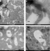
Localization of TF (tissue factor) in human platelets. Ultrathin Tokuyasu cryosections were single immunolabeled for TF (10 nm gold). Only a fraction of the platelet population expresses TF (A); when expressed, TF localizes in the open canalicular system (OCS; B–D).
Figure 6.
Figure 6.
Involvement of actin polymerization in platelet activation marker expression. Whole blood was incubated with cytochalasin D (CytD; 10 µmol/L, 15 min). Surface expression of platelet-associated TF (tissue factor; A and B), P-selectin (E and F), CD36 (G and H), CD40L (I and J) and activated GPIIbIIIa (glycoprotein IIbIIIa; aGPIIbIIIa; K and L) was measured by flow cytometry upon stimulation with ADP (10 µM, 15 min). Intracellular (IC) expression of platelet-associated TF was also assessed (C and D). Data are expressed as mean±SD (n=3–8) of percentage of positive platelets (PLT; A, C, E, G, I, and K) or mean fluorescence intensity (MFI; B, D, F, H, J, and L). Results were analyzed by Student paired t test or Mann-Whitney U test as appropriate.
Figure 7.
Figure 7.
Involvement of microtubule disruption in platelet activation marker expression. Whole blood was incubated with colchicine (20 nM to 100 µM, 15 min) and platelet-associated TF (tissue factor; A and B), Psel (P-selectin; C and D), and activated GPIIbIIIa (glycoprotein IIbIIIa; aGPIIbIIIa; E and F) expression was measured by flow cytometry upon stimulation with ADP (10 µM, 15 min). Data are expressed as mean±SD (n=4–7) of percentage of positive cells (A, C, and E) or mean fluorescence intensity (MFI; B, D, and F) and were analyzed by Student paired t test or Mann-Whitney U test as appropriate.

References

    1. Drake TA, Morrissey JH, Edgington TS. Selective cellular expression of tissue factor in human tissues. Implications for disorders of hemostasis and thrombosis. Am J Pathol. 1989;134:1087–1097. - PMC - PubMed
    1. Camera M, Frigerio M, Toschi V, Brambilla M, Rossi F, Cottell DC, Maderna P, Parolari A, Bonzi R, De Vincenti O, et al. . Platelet activation induces cell-surface immunoreactive tissue factor expression, which is modulated differently by antiplatelet drugs. Arterioscler Thromb Vasc Biol. 2003;23:1690–1696. doi: 10.1161/01.ATV.0000085629.23209.AA - PubMed
    1. Mann KG, Brummel K, Butenas S. What is all that thrombin for? J Thromb Haemost. 2003;1:1504–1514. doi: 10.1046/j.1538-7836.2003.00298.x - PubMed
    1. Brummel KE, Paradis SG, Butenas S, Mann KG. Thrombin functions during tissue factor-induced blood coagulation. Blood. 2002;100:148–152. doi: 10.1182/blood.v100.1.148 - PubMed
    1. Falanga A, Marchetti M, Vignoli A, Balducci D, Russo L, Guerini V, Barbui T. V617f jak-2 mutation in patients with essential thrombocythemia: relation to platelet, granulocyte, and plasma hemostatic and inflammatory molecules. Exp Hematol. 2007;35:702–711. doi: 10.1016/j.exphem.2007.01.053 - PubMed

Publication types

MeSH terms