Review
. 2025 Jan;93(1):11-25.
doi: 10.1002/prot.26571. Epub 2023 Aug 17.

Empowering canonical biochemicals with cross-linked novelty: Recursions in applications of protein cross-links

Affiliations
Review

Empowering canonical biochemicals with cross-linked novelty: Recursions in applications of protein cross-links

Jinam Ravindra Bora et al. Proteins. 2025 Jan.

Abstract

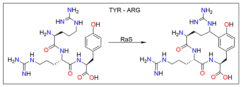
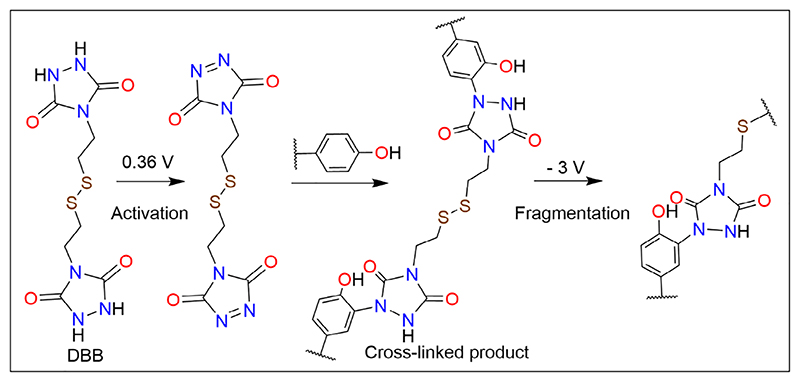
Diversity in the biochemical workhorses of the cell-that is, proteins-is achieved by the innumerable permutations offered primarily by the 20 canonical L-amino acids prevalent in all biological systems. Yet, proteins are known to additionally undergo unusual modifications for specialized functions. Of the various post-translational modifications known to occur in proteins, the recently identified non-disulfide cross-links are unique, residue-specific covalent modifications that confer additional structural stability and unique functional characteristics to these biomolecules. We review an exclusive class of amino acid cross-links encompassing aromatic and sulfur-containing side chains, which not only confer superior biochemical characteristics to the protein but also possess additional spectroscopic features that can be exploited as novel chromophores. Studies of their in vivo reaction mechanism have facilitated their specialized in vitro applications in hydrogels and protein anchoring in monolayer chips. Furthering the discovery of unique canonical cross-links through new chemical, structural, and bioinformatics tools will catalyze the development of protein-specific hyperstable nanostructures, superfoods, and biotherapeutics.

Keywords: hyperstable structures; novel protein function; side chain modification; unusual cross‐link.


Conflict of interest statement

Conflict Of Interest Statement

The authors declare no conflicts of interest.

Figures

Figure 1
Figure 1
Radical-radical reactions forming di-Tyr, di-Trp, and Trp−Tyr cross-links. Tyrosine and tryptophan, in the presence of free radical generating agents, forsm resonating structures (center), and subsequently undergo cross-linking by the formation of C−O, C−C, C−N, or O−N bonds. Di-Tyr and di-Trp cross-links are shown on the left, and Trp-Tyr on the right. Di-Tyr cross-links have also been observed in the presence of peroxidase and laccase enzymes. Figure was redrawn from fig. 7 of Figueroa et al. (2020), with permission from the Royal Society of Chemistry under the CC BY-NC 3.0 license, and was generated using ChemDraw (® PerkinElmer Informatics Inc.).
Figure 2
Figure 2
Plaque formation in Alzheimer's disease. Tyrosine present in the Aβ fibers forms di-Tyr cross-links in the presence of Cu2+ ions and ROS stress. These cross-links are central to stabilizing the Aβ oligomers. The reaction chemistry for the di-Tyr formation is similar to that shown in Figure 1. Image inspired from Al-Hilaly et al. (2013), and created with BioRender.com.
Figure 3
Figure 3
7-F indole-mediated aryl cross-linking. Crystal structures of cyclophilin A before (left) and after (right) the cross-linking of W121 with F. The process is chemically driven by first converting the indole side chain of W121 to its 7-fluoro analogue (left; fluorine in red), followed by irradiation of the protein with 282 nm light. 7-Fluoro-tryptophan is usually incorporated metabolically by growing the cells in media carrying indole analogues. Image inspired and reprinted (adapted) with permission from Lu et al., (2022). Copyright (2022) American Chemical Society. Structure coordinates PDB ID: 3K0N rendered using PyMOL (The PyMOL Molecular Graphics System, Version 1.2r3pre, Schrödinger, LLC).
Figure 4
Figure 4
Autoregulation of MMPs by W-G cross-link. HOCl inactivates human MMP-7 by first oxidizing Trp to 3-chloroindolenine. This transient intermediate generates a cyclic six-membered indole-amide species by reacting with the backbone of the succeeding Gly. On the left is the proposed mechanism of the cross-linking, and on the right is the model Trp−Gly-containing peptide and the cyclized product. Note how the formation of the cyclized product dramatically lowers the conformational flexibility of the peptide backbone, which consequently affects substrate binding at the catalytic pocket of MMP-7. MMP-7 activity is regulated by the oxidants produced by the surrounding phagocytes, which causes sequential activation and inactivation of MMP-7 during inflammation. Figure on the left is redrawn with permission from Fu et al. (2004). Figure was generated using ChemDraw (® PerkinElmer Informatics Inc.) and PyMOL (The PyMOL Molecular Graphics System, Version 1.2r3pre, Schrödinger, LLC).
Figure 5
Figure 5
Peptide-modifying K-W cyclase. A RaS enzyme with a characteristic SPASM domain post-translationally cross-links the side chains of Lys and Trp in the KGDGW motif of the 21 residue peptide substrate, with the first eight residues serving as leader sequence and enzymebinding site. The pentapeptide segment highlighting the cross-link is represented on the top, and spatial location of the Lys−Trp in the 21-residue peptide is illustrated at the bottom. The peptide structure was generated in silico using I-TASSER. The top panel was generated using ChemDraw (® PerkinElmer Informatics Inc.) and the bottom panel using PyMOL (The PyMOL Molecular Graphics System, Version 1.2r3pre, Schrödinger, LLC).
Figure 6
Figure 6
Tyr−Arg cross-linking by the RaS enzyme RrrB. The RrrB enzyme, belonging to the RRR subfamily of RaS, recognizes the RRY motif in a co-translationally expressed peptide, and catalyzes its macro-cyclization via an Arg−Tyr cross-link. Unlike the K-W cyclase, RrrB can work independent of a leader sequence in the substrate. Figure is reprinted (adapted) with permission from Caruso et al. (2019). Copyright (2019) American Chemical Society. Figure was generated using ChemDraw (® PerkinElmer Informatics Inc.).
Figure 7
Figure 7
Novel electrochemically cleavable tyrosine cross-linker. DBB catalyzes the cross-linking of tyrosines without requirement of UV or enzyme catalysis. The N−N bond of DBB is first oxidized to N=N bond at 0.36 V. This activated form of DBB can then cross-link with tyrosine residues in a peptide or protein. The cross-linking is reversible, and the cross-linked product can be fragmented by reducing the disulfide bond in DBB at -3 V. Figure is reprinted (adapted) with permission from Cui et al. (2021). Copyright (2021) American Chemical Society. Figure was generated using ChemDraw (® PerkinElmer Informatics Inc.).
Figure 8
Figure 8
A C−C cross-link between two unfunctionalized side chains. A valine−phenylalanine cross-link is produced in an oxygen-dependent reaction in symerythrin protein in C. paradoxa. The crosslink formation requires the diiron metallocenter and O2 as the reductant, with the di-iron abstracting a hydrogen from Cγ1 of V127, forming a primary alkyl radical. This radical then reacts with F forming a C−C cross-link, without additional involvement of any functional groups. Structure coordinates PDB ID: 3QHB rendered using PyMOL (The PyMOL Molecular Graphics System, Version 1.2r3pre, Schrödinger, LLC).
Figure 9
Figure 9
NOS bridge as an allosteric redox switch. The transaldolase enzyme of the gonorrhea-causing pathogen, N. gonorrhoeae, is regulated by an allosteric redox switch. Activation of C in the presence of reactive oxygen species oxidizes the cysteine thiolate, which then forms a covalent conjugate with K residue by the attack of the Lys Nε on the thio-(hydro)peroxy intermediate. The Lys−NOS−Cys bridge (shown here) can be reversed under reducing conditions, relaxing the covalent restraint and reconfiguring the surrounding residues, thereby activating the enzyme. Image inspired from Wensien et al. (2021). Structure coordinates PDB ID: 6ZX4 rendered using PyMOL (The PyMOL Molecular Graphics System, Version 1.2r3pre, Schrödinger, LLC).
Figure 10
Figure 10
BF4112 forms a Cys−Tyr cross-link. Proteomics studies confirm the formation of a 3-(S-cysteinyl)-tyrosine cross-link between Y and C when the protein BF4112 is first prepared with one Cu2+ ion per monomer, reacted with sodium dithionite under anaerobic conditions, followed by O2 exposure. The additional formation of Q-Y H-bond increases tyrosine oxidation, but also restricts its rotation. The cupin fold topology is vital in ensuring that all components to generate the cross-link are located within a 3.0-4.0 Å vicinity of the metal-binding center. Image inspired from Martinie et al. (2012). Structure coordinates PDB ID: 3CEW (10.2210/pdb3CEW/pdb) rendered using PyMOL (The PyMOL Molecular Graphics System, Version 1.2r3pre, Schrödinger, LLC).

Similar articles

References

    1. Lee D, Redfern O, Orengo C. Predicting protein function from sequence and structure. Nat Rev Mol Cell Biol. 2007;8(12):995–1005. - PubMed
    1. Dill KA, MacCallum JL. The protein-folding problem, 50 years on. Science. 2012;338(6110):1042–1046. - PubMed
    1. Kahn A, Boivin P, Rubinson H, Cottreau D, Marie J, Dreyfus JC. Modifications of purified glucose-6-phosphate dehydrogenase and other enzymes by a factor of low molecular weight abundant in some leukemic cells. Proc Natl Acad Sci U S A. 1976;73(1):77–81. doi: 10.1073/pnas.73.1.77. - DOI - PMC - PubMed
    1. Kivirikko KI, Risteli L. Biosynthesis of collagen and its alterations in pathological states. Med Biol. 1976;54(3):159–186. - PubMed
    1. Risteli J, Kivirikko KI. Intracellular enzymes of collagen biosynthesis in rat liver as a function of age and in hepatic injury induced by dimethylnitrosamine. Changes in prolyl hydroxylase, lysyl hydroxylase, collagen galactosyltransferase and collagen glucosyltransferase activities. Biochem J. 1976;158(2):361–367. doi: 10.1042/bj1580361. - DOI - PMC - PubMed

LinkOut - more resources