Review
. 2023 Aug 17;13(1):35.
doi: 10.1186/s12348-023-00342-5.

Challenges in posterior uveitis-tips and tricks for the retina specialist

Affiliations
Review

Challenges in posterior uveitis-tips and tricks for the retina specialist

Manuel Paez-Escamilla et al. J Ophthalmic Inflamm Infect. .

Abstract

Purpose: Posterior uveitis is a common chorioretinal pathology affecting all ages worldwide and is a frequent reason for referral to the retina clinic. The spectrum of etiologies for uveitis is very broad and includes infectious and auto-immune diseases. Inflammation can be confined to the eye or may be a part of systemic disease. A useful outline is therefore proposed to aid in the correct diagnosis of these challenging entities. The situation is further complicated by the fact that many neoplastic conditions resemble features of posterior uveitis; they are known as "masqueraders of uveitis". Here, we summarize different posterior uveitides that present with rare findings, along with masqueraders that can be difficult to distinguish. These conditions pose a diagnostic dilemma resulting in delay in treatment because of diagnostic uncertainty.

Methods: An extensive literature search was performed on the MEDLINE/PUBMED, EBSCO and Cochrane CENTRAL databases from January 1985 to January 2022 for original studies and reviews of predetermined diagnoses that include posterior uveitic entities, panuveitis and masquerade syndromes.

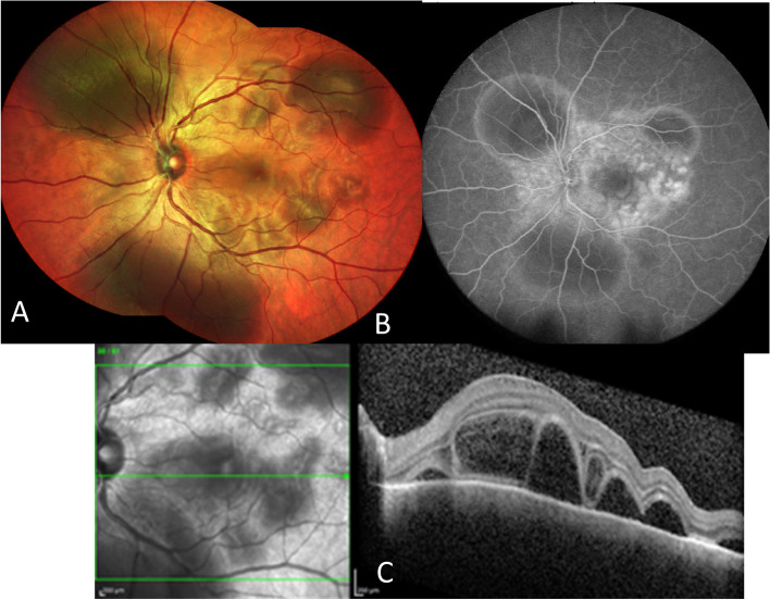
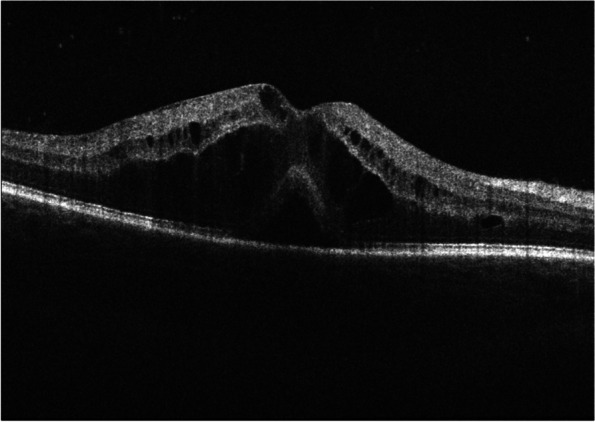
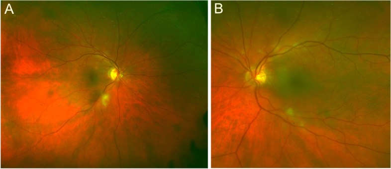
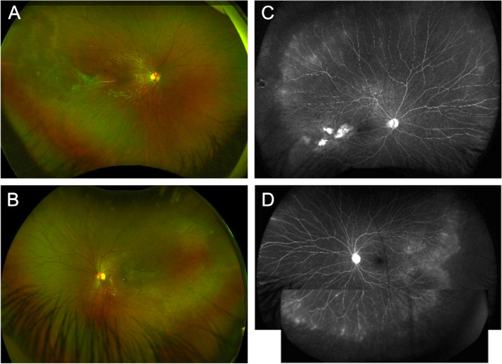
Results: We described conditions that can present as mimickers of posterior uveitis (i.e., immune check-points inhibitors and Vogt-Koyanagi-Harada-like uveitis; leukemia and lymphoma associated posterior uveitis), inflammatory conditions that present as mimickers of retinal diseases (i.e., Purtscher-like retinopathy as a presentation of systemic lupus erythematosus; central serous chorioretinopathy masquerading inflammatory exudative retinal detachment), and uveitic conditions with rare and diagnostically challenging etiologies (i.e., paradoxical inflammatory effects of anti-TNF-α; post vaccination uveitis; ocular inflammation after intravitreal injection of antiangiogenic drugs).

Conclusion: This review of unique posterior uveitis cases highlights the overlapping features of posterior uveitis (paradoxical inflammatory effects of anti -TNF α and uveitis; Purtscher-like retinopathy as a presentation of systemic lupus erythematosus, …) and the nature of retinal conditions (ischemic ocular syndrome, or central retinal vein occlusion, amyloidosis, inherited conditions like retinitis pigmentosa, autosomal dominant neovascular inflammatory vitreoretinopathy (ADNIV), etc.…) that may mimic them is represented. Careful review of past uveitis history, current medications and recent vaccinations, detailed examination of signs of past or present inflammation, eventually genetic testing and/ or multimodal retinal imaging (like fluorescein angiography, EDI-OCT, OCT-angiography for lupus Purtscher-like retinopathy evaluation, or ICG for central serous retinopathy, or retinal amyloid angiopathy) may aid in correct diagnosis.

Keywords: Birdshot choroidopathy; Check-point inhibitors retinopathy; MEK inhibitors retinopathy; Masquerades; Multifocal choroiditis uveitis; Ocular inflammation; Posterior uveitis; Punctate inner choroidopathy; Serpiginous choroiditis; Vogt- Koyanagi-Harada; White dot syndromes.


Conflict of interest statement

The authors declare no competing interests.

Figures

Fig. 1
Fig. 1
Multimodal imaging in a 51-year-old female with acute pseudo-Vogt-Koyanagi-Harada (VKH) following one month treatment with a combination of ipilimumab/nivolumab (CTLA-4 and PD-1 checkpoint blockade inhibitors) for a malignant melanoma of the skin. A The fundus photograph of the left eyes reveals multiple deep yellow choroidal lesions with areas of the subretinal detachments in the peripapillary region. B Note multiple hyperfluorescent retinal pigment epithelium leaks on fluorescein angiography with hyperfluorescent dye pooling beneath subretinal fluid. C Optical coherence tomography (OCT) in pseudo-VKH showed a multilobular serous macular detachments, with subretinal hyper-reflective material within the subretinal fluid that likely represents fibrin and a part of the outer segment layer
Fig. 2
Fig. 2
OCT showing a cystoid macular edema and a serous neuroretinal detachment of the fovea in a 39-year-old male with widespread metastatic melanoma, stage IV disease, treated by BRAF/MEK inhibitor therapy (ecorafenib/binimetinib) since the last 3 months
Fig. 3
Fig. 3
Fundus color pictures of a 75-year-old female with Giant cell arteritis (GCA) proven by a bilateral temporal artery biopsy. Note bilateral cotton-wool spots without optic disc edema seen in the right (A) and left (B) eyes
Fig. 4
Fig. 4
Multimodal image of a patient with familial exudative vitreoretinopathy. Wide field Optos fundus photos of the right (A) and left (B) eye show perivascular sheathing, worse on the right eye. Fluorescein angiogram shows focal venous leakage with staining of lesions temporal to the macula (C). Both the right and the left eye show diffuse peripheral leakage with capillary dropout in the periphery (D)
Fig. 5
Fig. 5
Fundus color picture of a 52-year-old female with systemic lupus erythematosus and lupus nephrosis. She was diagnosed as a case of lupus retinopathy owing (A) to patches of polygonal retinal whitening nasal to the nerve and on the fovea and (B) occlusion vasculitis of the retinal capillaries and arterioles on fluorescein angiography
Fig. 6
Fig. 6
Fundus color picture of a 46-year-old female with transthyretin amyloidosis. She initially was referred for blurry vision as a case of intermediate uveitis (vitritis) and retinal vasculitis in the right eye and a pars plana vitrectomy was performed in the right eye. She later presented with a left eye involvement. The pictures demonstrate retinal vascular sheathing as shown in arrows in the right eye (A) and intermediate uveitis with vitritis in the left eye (B). The genetic diagnosis of transthyretin amyloidosis was made. Fluorescein angiography showed focal vascular staining in intermediate phase (arrow) (C). The Spectral Domain OCT (SD-OCT) showed a deposit perpendicular to the retinal surface towards the vitreous from the retinal vessel (arrow) (D), and hyperfluorescent foci and staining along choroidal and retinal vessels in the supero-, infero- and temporal periphery on ICG in late phase (E)

Similar articles

Cited by

References

    1. Gritz DC, Wong IG. Incidence and prevalence of uveitis in Northern California; the Northern California Epidemiology of Uveitis Study. Ophthalmology. 2004;111:491–500. doi: 10.1016/j.ophtha.2003.06.014. - DOI - PubMed
    1. Gaucher D, Bodaghi B, Charlotte F, et al. MALT-type B-cell lymphoma masquerading as scleritis or posterior uveitis. J Fr Ophtalmol. 2005;28:31–38. doi: 10.1016/s0181-5512(05)81022-8. - DOI - PubMed
    1. Dedieu D, Bouayed E, Brezin AP. Hodgkin Lymphoma Associated Retinopathy: Report of a Case. Ocul Immunol Inflamm. 2022;30(1974–1976):20210513. doi: 10.1080/09273948.2021.1912360. - DOI - PubMed
    1. Fredrick DR, Char DH, Ljung BM, et al. Solitary intraocular lymphoma as an initial presentation of widespread disease. Arch Ophthalmol. 1989;107:395–397. doi: 10.1001/archopht.1989.01070010405034. - DOI - PubMed
    1. Ramsay A, Lightman S. Hypopyon uveitis. Surv Ophthalmol. 2001;46:1–18. doi: 10.1016/s0039-6257(01)00231-4. - DOI - PubMed

LinkOut - more resources