. 2023 Sep 22;381(6664):eadi3448.
doi: 10.1126/science.adi3448. Epub 2023 Sep 22.

In silico protein interaction screening uncovers DONSON's role in replication initiation

Affiliations

In silico protein interaction screening uncovers DONSON's role in replication initiation

Yang Lim et al. Science. .

Abstract

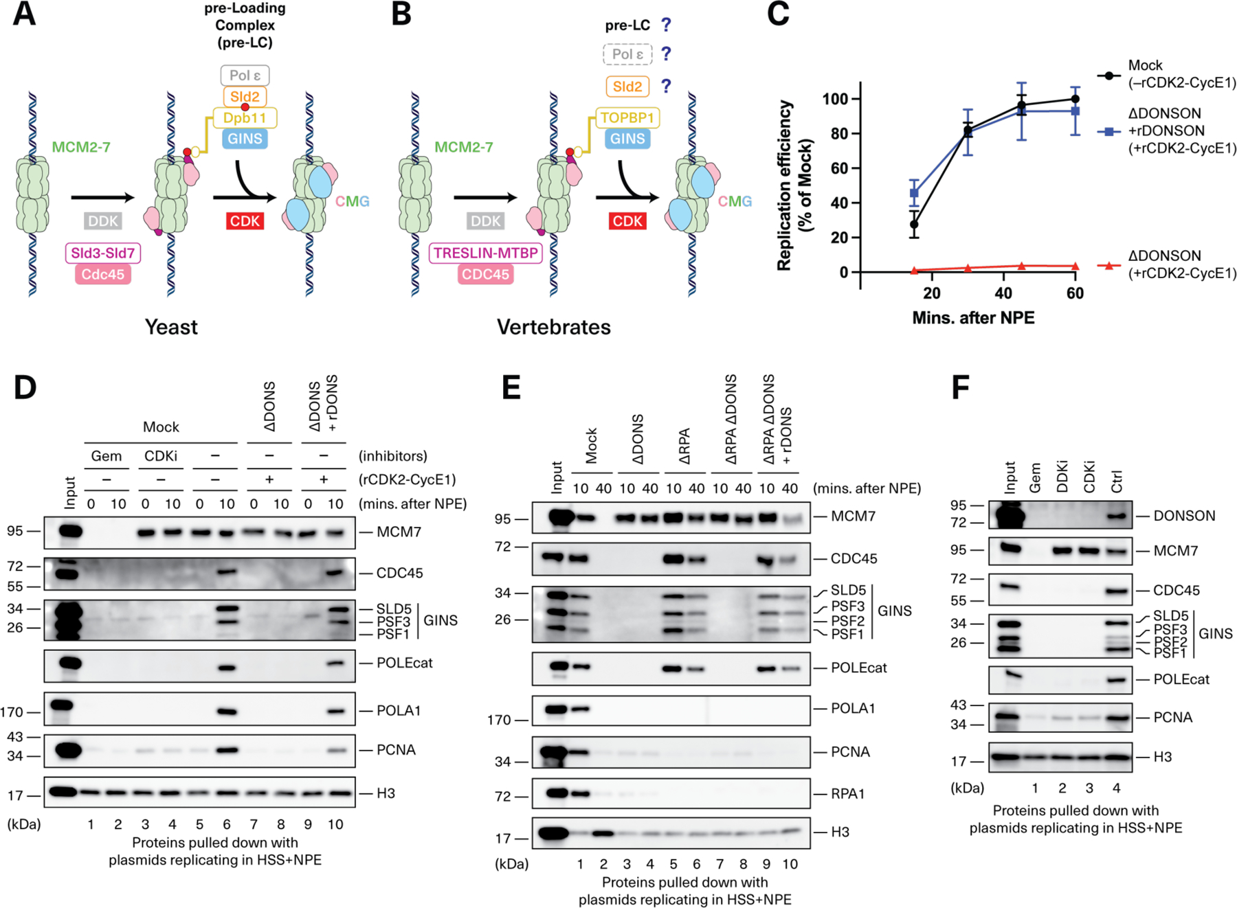
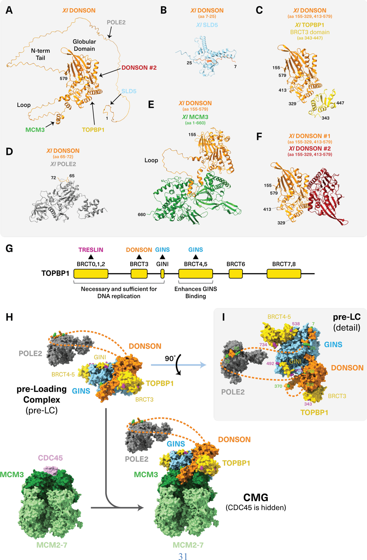
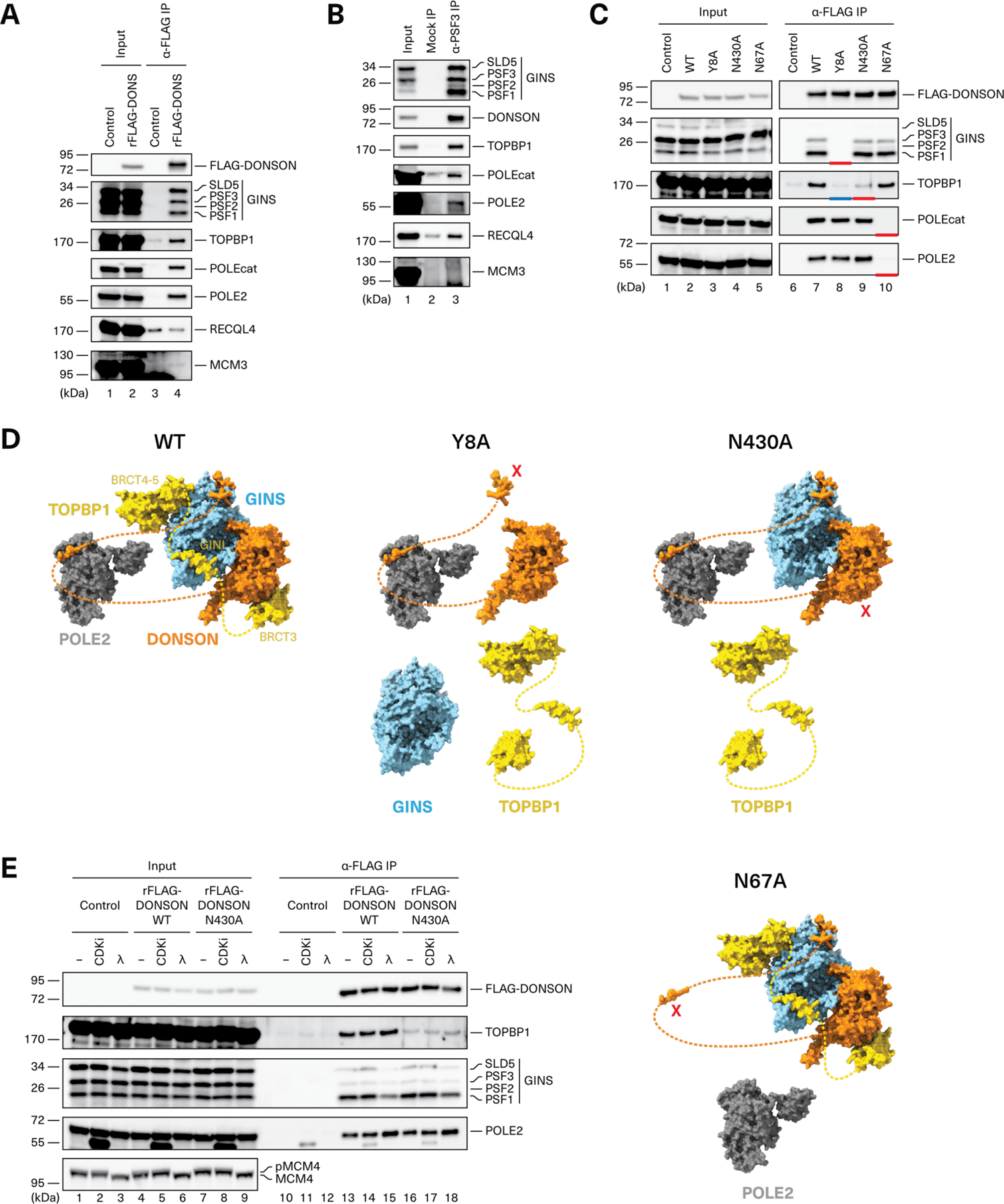
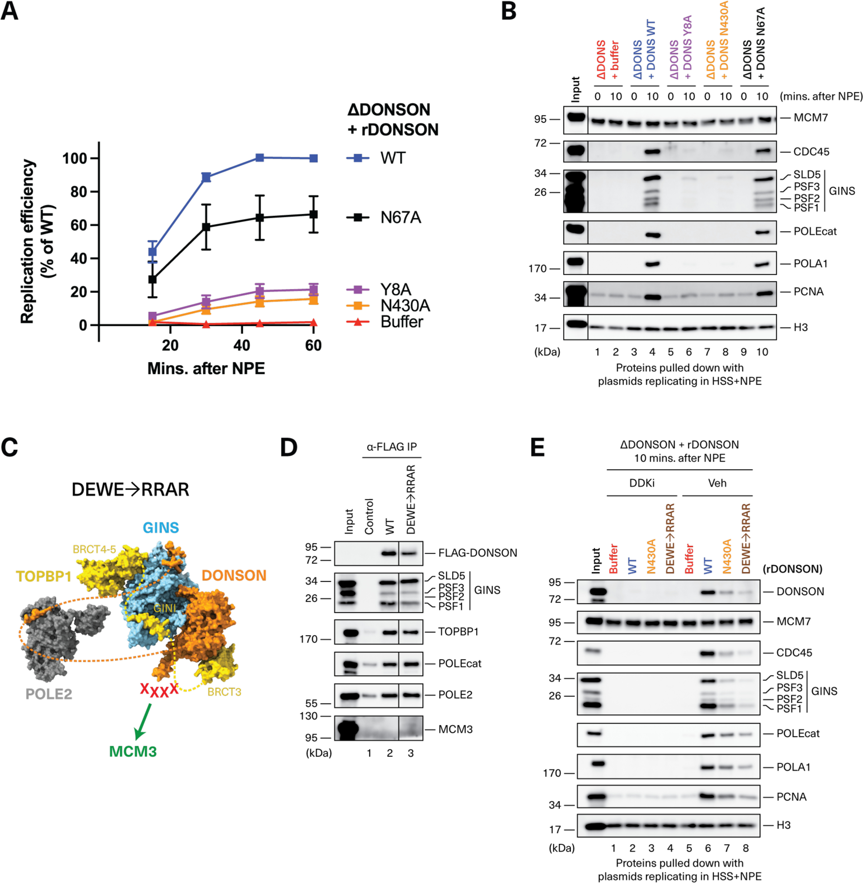
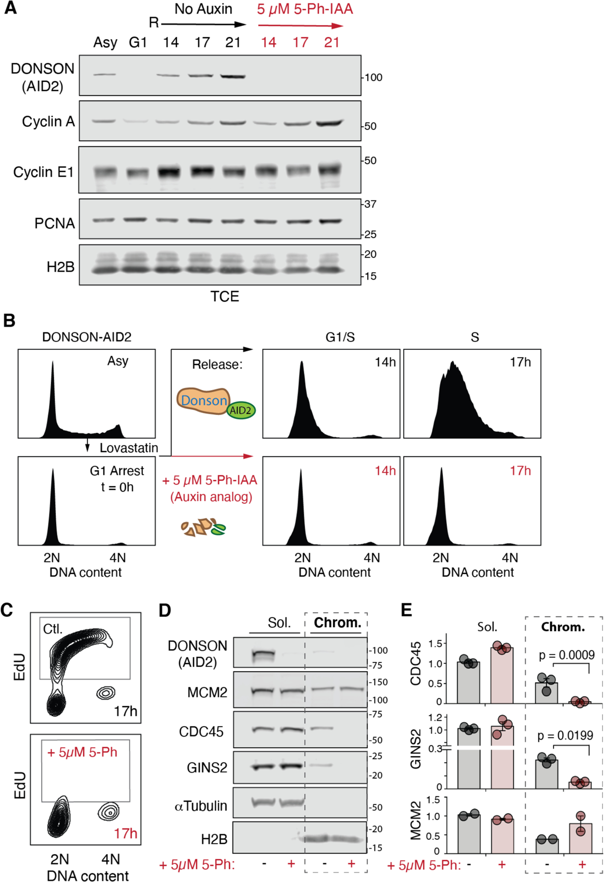
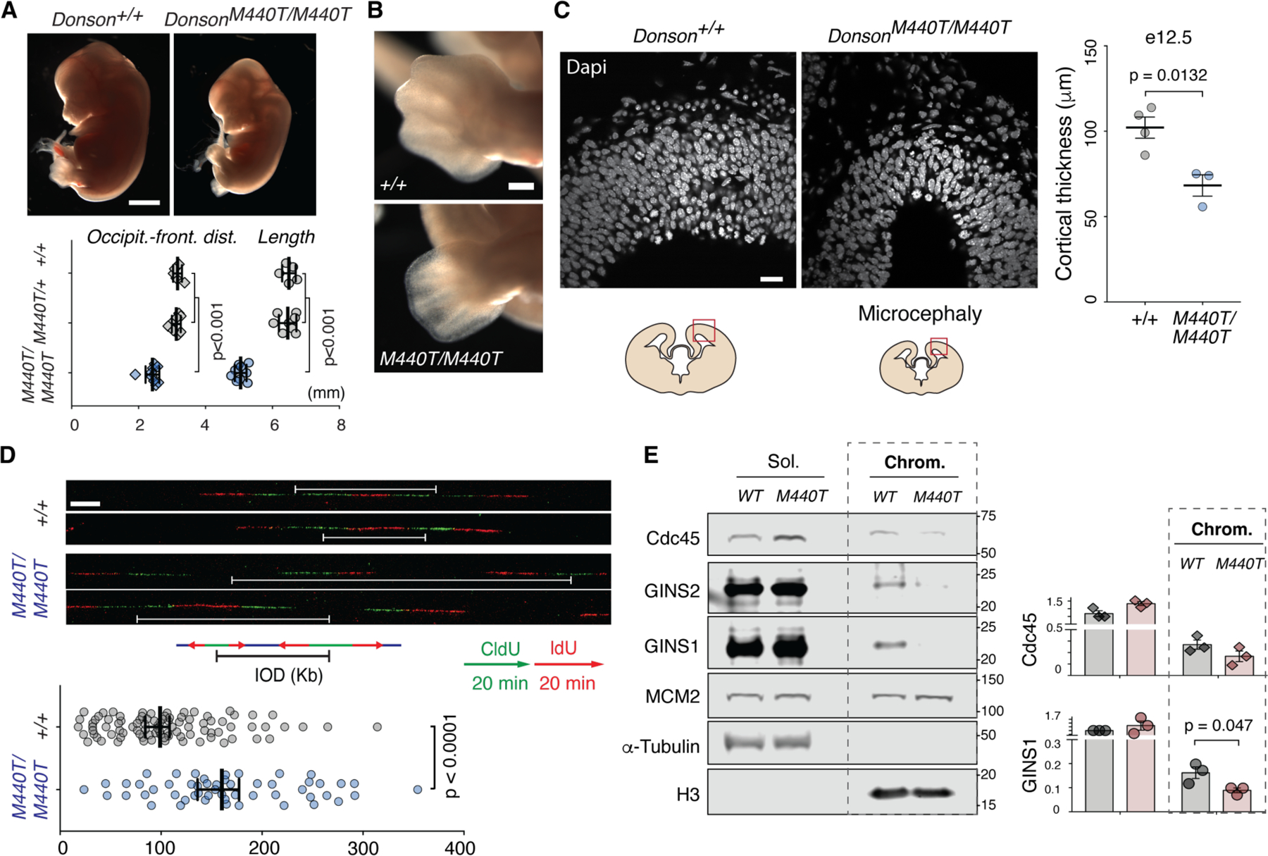
CDC45-MCM2-7-GINS (CMG) helicase assembly is the central event in eukaryotic replication initiation. In yeast, a multi-subunit "pre-loading complex" (pre-LC) accompanies GINS to chromatin-bound MCM2-7, leading to CMG formation. Here, we report that DONSON, a metazoan protein mutated in microcephalic primordial dwarfism, is required for CMG assembly in vertebrates. Using AlphaFold to screen for protein-protein interactions followed by experimental validation, we show that DONSON scaffolds a vertebrate pre-LC containing GINS, TOPBP1, and DNA pol ε. Our evidence suggests that DONSON docks the pre-LC onto MCM2-7, delivering GINS to its binding site in CMG. A patient-derived DONSON mutation compromises CMG assembly and recapitulates microcephalic dwarfism in mice. These results unify our understanding of eukaryotic replication initiation, implicate defective CMG assembly in microcephalic dwarfism, and illustrate how in silico protein-protein interaction screening accelerates mechanistic discovery.


Figures

Fig. 1.
Fig. 1.. DONSON is required for CMG assembly and DNA replication.
(A-B) Models of CMG assembly in budding yeast and vertebrates. (C) Relative DNA replication efficiency in the indicated egg extracts. Because depletion of DONSON co-depletes roughly half of the 0.5 to 1 µM endogenous CDK2-Cyclin E, DONSON-depleted (ΔDONSON) extracts but not mock-depleted extracts were supplemented with 0.3 µM recombinant human CDK2-Cyclin E1. Recombinant DONSON (rDONSON, fig. S1B) was added where indicated. Datapoints, n=3 experiments. Mean±SD. A representative western blot of total protein levels in these reactions is shown in fig. S1E. (D) Plasmid DNA was incubated in the indicated egg extracts. At the specified times following NPE addition, chromatin was recovered and blotted for the indicated proteins. DONS, DONSON; Gem, geminin; CDKi, p27Kip. A representative western blot of total protein levels in these reactions is shown in fig. S1E. (E) Plasmid DNA was incubated in extracts depleted of DONSON and/or RPA. At the specified times following NPE addition, chromatin was recovered and blotted for the indicated proteins. Western blot of total protein levels in these reactions is shown in fig. S1H. (F) In the presence of the indicated inhibitors of replication initiation, plasmid DNA was recovered 10 minutes after NPE addition and blotted for the indicated proteins. DDKi, PHA-767491.
Fig. 2.
Fig. 2.. Hypothetical model of DONSON function in CMG assembly.
(A) AlphaFold-Multimer (AF-M) prediction of DONSON’s structure. Sites predicted to bind interacting proteins are indicated with arrows. All proteins shown are from Xenopus, but the predicted human complexes appear almost identical. (B to F) AF-M predictions of relevant DONSON domains complexed with SLD5 (B), TOPBP1 (C), POLE2 (D), MCM3 (E), and a second copy of DONSON (F). The amino acids (aa) of each protein shown are indicated in brackets. (G) Functional domains of TOPBP1. (H) AF-M predictions suggest that a pre-LC consisting of DONSON, GINS, Pol ε, and TOPBP1 docks onto the MCM2-7 complex through the predicted DONSON-MCM3 interaction. (Top) The predicted Xenopus pre-LC is shown with only the POLE2 subunit of Pol ε and just the BRCT3 (aa 343–447), GINI (aa 475–492), and BRCT4-5 (aa 538–734) domains of TOPBP1. Disordered regions of DONSON are shown as dotted lines but omitted for TOPBP1. Residues located at the ends of well-ordered segments are shown in green and purple for DONSON and TOPBP1, respectively. (Bottom) The pre-LC was docked onto the cryo-EM structure of human CMG [PDB: 7PLO (66)] by aligning on MCM3. Only CDC45 and MCM2-7 of human CMG are shown. See Methods for modeling details. (I) The pre-LC from (H) rotated by 90 degrees. Residues located at the ends of well-ordered segments are numbered and shown in green and purple for DONSON and TOPBP1, respectively.
Fig. 3.
Fig. 3.. DONSON forms a pre-LC.
(A) Recombinant FLAG-tagged DONSON (rFLAG-DONSON) was added to non-replicating nucleoplasmic egg extract (NPE), recovered, and blotted for the indicated proteins alongside the input extract. (B) Endogenous GINS was immunoprecipitated from NPE using PSF3 antibody and blotted for the indicated proteins. (C) rFLAG-DONSON proteins containing specified mutations (figs. S1B and S11A) were added to NPE, recovered, and blotted as indicated. Red and blue bars show missing pre-LC components. The images are part of the same western blot, which was cropped to remove irrelevant information between lanes 5 and 6. (D) The effects of different DONSON mutants are depicted in the context of the AF-M-modeled pre-LC (as in Fig. 2I). Mutations are indicated as red Xs. (E) The indicated rFLAG-DONSON proteins were added to NPE treated with buffer, p27Kip (CDKi), or λ phosphatase. DONSON was recovered and blotted for the indicated proteins. Total extract was also blotted for MCM4 to show λ phosphatase activity.
Fig. 4.
Fig. 4.. Pre-LC formation is required for DNA replication.
(A) Egg extracts were depleted of DONSON, supplemented with rCDK2-Cyclin E1 and the indicated DONSON proteins (figs. S1B and S11A), and used to measure DNA replication. Datapoints, n = 4 experiments, except N67A where n = 3. Mean ± SD. (B) Plasmid pull-down (as in Fig. 1D) to assay the effect of DONSON mutations on CMG assembly. The images are part of the same western blot, which was cropped to remove irrelevant information between the input and lane 1. Western blot of total protein levels in these reactions is shown in fig. S11C. (C) DONSONDEWE→RRAR, depicted as in Fig. 3D. (D) FLAG-DONSON immunoprecipitation (as in Fig. 3A) showing that DONSONDEWE→RRAR (expressed in wheat germ extract) is proficient in pre-LC assembly. The images are part of the same western blot, which was cropped to remove irrelevant information between lanes 2 and 3. (E) Plasmid pull-down (as in Fig. 1F) showing that purified recombinant DONSONN430A and DONSONDEWE→RRAR (fig. S11A) bind inefficiently to chromatin during replication. Western blot of total protein levels in these reactions is shown in fig. S11D.
Fig. 5.
Fig. 5.. DONSON is required for DNA replication and CMG assembly in mammalian cells.
(A) Immunoblot of synchronized DONSON-AID2 HCT116 cells. TCE, total cell extract; Asy, asynchronous; G1, G1-arrest; R, release, hours. (B) G1-synchronized cells fail to progress into S phase after 5-Ph-IAA depletion of DONSON. (C) DONSON depletion prevents DNA synthesis. EdU pulse-labeling at 17 hours post-release. Flow cytometry plots in panels (B) and (C), representative of n = 4 and n = 3 experiments, respectively, are quantified in figs. S16D and E, respectively. (D, E). DONSON is required for CDC45 and GINS recruitment to chromatin. (D) Immunoblots, soluble extract (Sol.) and chromatin-bound proteins (Chrom.) 17 hours post release. (E) Quantification, normalized to loading control (soluble, α-Tubulin; chromatin, Histone H2B) and wild-type protein levels. Datapoints, n = 3 experiments. Mean ± SEM.
Fig. 6.
Fig. 6.. Homozygous M440T mutation impairs replication initiation and causes growth restriction and microcephaly in a mouse model.
(A-C) DonsonM440T/M440T E13.5 mouse embryos exhibit growth restriction by mid-gestation with microcephaly and limb abnormalities. (A) Lateral view; scale bar 1 mm. Datapoints, individual mice, Mean ± SEM, t-test. Occipit.-front dist, occipital-frontal distance. (B) Oligodactyly in forelimb; scale bar 0.2 mm. See also fig. S18C. (C) Reduced cellularity as measured by cortical thickness is evident in the developing forebrain during neurogenesis (e12.5). Scale bar 20 µm. Measurements at dorsal-most point of telencephalon; datapoints, individual mice; Mean ± SEM, t-test. (D) Increased interorigin distance (IOD) in DonsonM440T/M440T cells. Representative images of dU-analog pulse-labeled DNA fibers. White brackets, measured IODs, mESCs. Data points plotted, fibers, pooled from n = 2 combing experiments. Kb, kilobases. Median ± 95% confidence interval; U-test. 91 wild-type and 53 M440T/M440T fibers were scored for IODs. (E) Reduced chromatin-associated GINS and Cdc45 indicates impaired CMG assembly in DonsonM440T/M440T mESCs. (Left) cell fractionation immunoblot. (Right) quantification. n = 3 experiments, mean ± SEM, t-test; normalization as in Fig. 5E.

References

    1. Costa A, Diffley JFX, The Initiation of Eukaryotic DNA Replication. Annu Rev Biochem 91, 107–131 (2022). - PubMed
    1. Tanaka S et al. CDK-dependent phosphorylation of Sld2 and Sld3 initiates DNA replication in budding yeast. Nature 445, 328–332 (2007). - PubMed
    1. Zegerman P, Diffley JF, Phosphorylation of Sld2 and Sld3 by cyclin-dependent kinases promotes DNA replication in budding yeast. Nature 445, 281–285 (2007). - PubMed
    1. Muramatsu S, Hirai K, Tak YS, Kamimura Y, Araki H, CDK-dependent complex formation between replication proteins Dpb11, Sld2, Pol (epsilon}, and GINS in budding yeast. Genes Dev 24, 602–612 (2010). - PMC - PubMed
    1. Yeeles JT, Deegan TD, Janska A, Early A, Diffley JF, Regulated eukaryotic DNA replication origin firing with purified proteins. Nature 519, 431–435 (2015). - PMC - PubMed

LinkOut - more resources