. 2023 Aug 18;8(1):305.
doi: 10.1038/s41392-023-01539-9.

VEGF-B prevents excessive angiogenesis by inhibiting FGF2/FGFR1 pathway

Affiliations

VEGF-B prevents excessive angiogenesis by inhibiting FGF2/FGFR1 pathway

Chunsik Lee et al. Signal Transduct Target Ther. .

Abstract

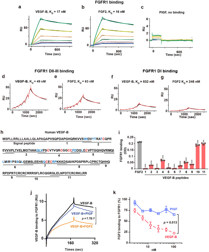
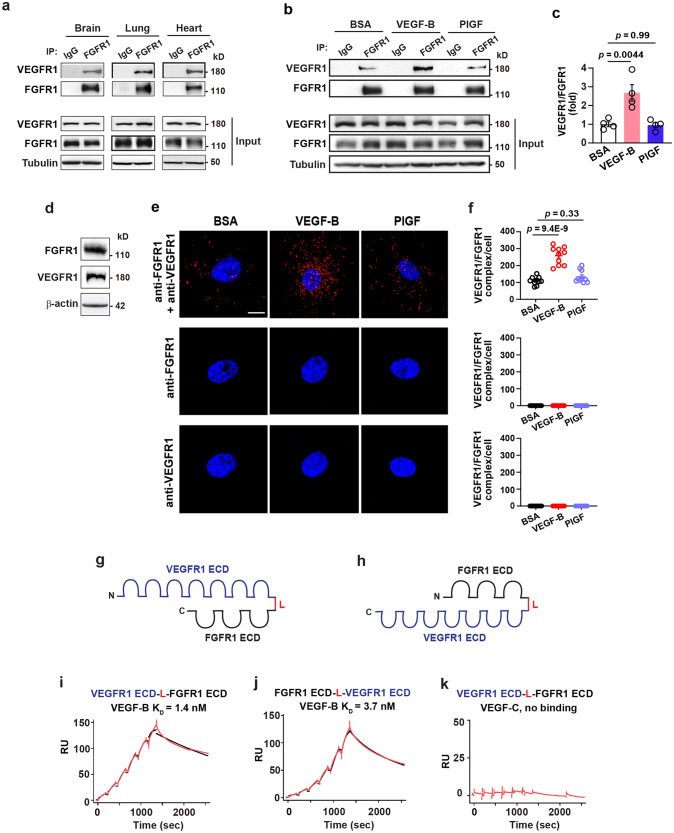
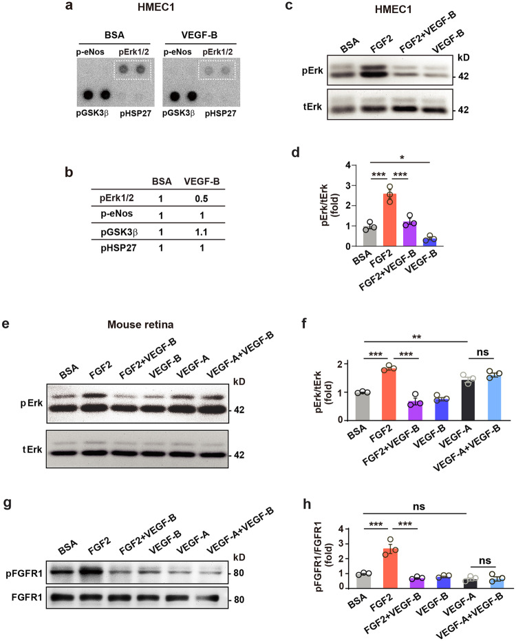
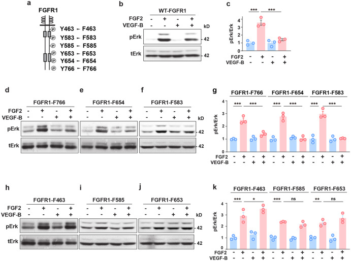
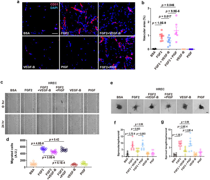
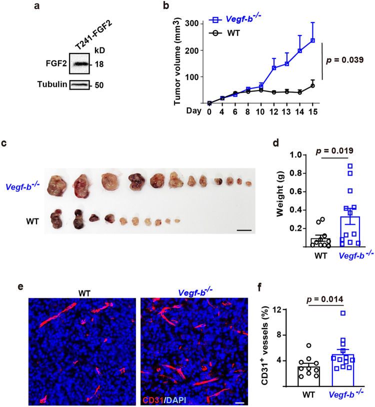
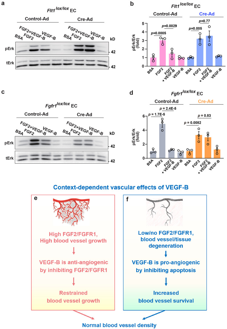
Although VEGF-B was discovered as a VEGF-A homolog a long time ago, the angiogenic effect of VEGF-B remains poorly understood with limited and diverse findings from different groups. Notwithstanding, drugs that inhibit VEGF-B together with other VEGF family members are being used to treat patients with various neovascular diseases. It is therefore critical to have a better understanding of the angiogenic effect of VEGF-B and the underlying mechanisms. Using comprehensive in vitro and in vivo methods and models, we reveal here for the first time an unexpected and surprising function of VEGF-B as an endogenous inhibitor of angiogenesis by inhibiting the FGF2/FGFR1 pathway when the latter is abundantly expressed. Mechanistically, we unveil that VEGF-B binds to FGFR1, induces FGFR1/VEGFR1 complex formation, and suppresses FGF2-induced Erk activation, and inhibits FGF2-driven angiogenesis and tumor growth. Our work uncovers a previously unrecognized novel function of VEGF-B in tethering the FGF2/FGFR1 pathway. Given the anti-angiogenic nature of VEGF-B under conditions of high FGF2/FGFR1 levels, caution is warranted when modulating VEGF-B activity to treat neovascular diseases.


Conflict of interest statement

The authors declare no competing interests.

Figures

Fig. 1
Fig. 1
VEGF-B binds to FGFR1 and competes with FGF2 for FGFR1 binding. ac Surface plasmon resonance (SPR) results showing VEGF-B binding to FGFR1 with a KD value (17 nM, a) similar to that for FGF2 (16 nM, b), whereas PlGF, another VEGFR1-binding member of the VEGF family, does not bind to FGFR1 (c). The lines from bottom to the top represent different concentrations of FGFR1-Fc: 12.5, 25, 50, 100, 200, and 400 nM, respectively. d, e SPR results showing VEGF-B binding to FGFR1 DII-III with a KD value (49 nM, d) similar to that of FGF2 (63 nM, e). The red lines are the resonance unit (RU) values at different concentrations of FGFR1 DII-III (25, 50, 100, 200, 400, 800 nM). The black lines are the fitted curves. f, g SPR results showing a very low binding affinity of VEGF-B (532 nM, f) and FGF2 (245 nM, g) to FGFR1 DI. The red lines are the RU values at different concentrations of FGFR1 DI (18.75, 37.5, 75, 150, 300, and 600 nM). The black lines are the fitted curves. h Scheme of the synthetic VEGF-B peptides. Blue: amino acids important for VEGFR1 binding. Red: eight cysteines forming the cysteine knots. i ELISA results showing the binding of VEGF-B peptides to FGFR1. n = 3. One-way ANOVA followed by Sidak post hoc analysis (number of comparisons against FGF2, 11) was used. Adjusted p values are <1.0E−10 for peptides 1–9, 1.0E−6 and 5.6E−3 for peptides 10 and 11. The experiment was repeated three times. j SPR results showing FGF2 competing with VEGF-B for FGFR1 binding, while PlGF does not. Kruskal-Wallis test with Dunn post hoc analysis (number of comparisons, 3) was used. k ELISA results showing VEGF-B dose-dependently competing with FGF2 for FGFR1 binding, while PlGF does not. Data are mean ± s.e.m. The experiment was repeated three times. Two-way ANOVA was used. **p < 0.01, ***p < 0.001
Fig. 2
Fig. 2
VEGF-B induces VEGFR1/FGFR1 complex formation and binds to VEGFR1/FGFR1 heterodimer with a high affinity. a Immunoprecipitation (IP) followed by Western blot showing VEGFR1 co-precipitated with FGFR1 in mouse brain, lung and heart. b, c Immunoprecipitation followed by Western blot showing that VEGF-B, but not PlGF, induced VEGFR1/FGFR1 complex formation in mouse retinae. d Western blot showing the expression of VEGFR1 and FGFR1 in HRECs. e, f Representative images (e) and corresponding quantification (f, n = 10 per group. The experiment was repeated three times) of in situ proximity ligation assays showing that 30 min treatment of VEGF-B (50 ng/ml), but not PlGF (50 ng/ml), induced VEGFR1/FGFR1 complex formation in HRECs (top panel). Adding one antibody alone at a time did not induce any complex formation (anti-FGFR1: middle panel; anti-VEGFR1: lower panel). Blue: DAPI; red: VEGFR1/FGFR1 complex. Data are mean ± s.e.m., n = 4 for (c) and 10 for (f). For (c) and (f), adjusted p values are from one-way ANOVA followed by Sidak post hoc analysis (number of comparisons, 2). Scale bar: 10 µm. The experiment was repeated three times. g, h Schemes of recombinant VEGFR1/FGFR1 (g) and FGFR1/VEGFR1 (h) heterodimers, each containing the extracellular domain (ECD) of VEGFR1 and FGFR1 connected by a linker (L). ik SPR results showing VEGF-B binding to the VEGFR1/FGFR1 heterodimer (i) and FGFR1/VEGFR1 heterodimer (j) with similar KD values, while VEGF-C shows no binding (k). The red lines are the RU values at different concentrations of VEGFR1/FGFR1 heterodimers (1.875, 3.75, 7.5, 15, 30, and 60 nM). The black lines are the fitted curves
Fig. 3
Fig. 3
VEGF-B inhibits FGF2-induced Erk activation in vitro and in vivo. a, b Images of phospho-kinase antibody array screening (a) using HMEC1s and corresponding quantifications (b). c, d Western blots showing that VEGF-B (100 ng/ml, 10 min treatment) reduced FGF2 (50 ng/ml)-induced Erk phosphorylation in HMEC1. One-way ANOVA followed by Holm-Sidak post hoc analysis was used (number of comparisons, 3). Adjusted p values are 2.1E−4 for BSA vs FGF2; 4.0E−4 for FGF2 vs FGF2 + VEGF-B, and 0.021 for VEGF-B vs BSA. e, f Western blots showing that in mouse retinae, intravitreal injection of VEGF-B inhibited FGF2-, but not VEGF-A-induced Erk phosphorylation (30 min after injection). Adjusted p values are 1.1E−5 for BSA vs FGF2; 1.5E−7 for FGF2 vs FGF2 + VEGF-B; 2.7E−3 for VEGF-A vs BSA and 0.14 for VEGF-A vs VEGF-A + VEGF-B. g, h Western blots showing that in mouse retinae, intravitreal injection of VEGF-B inhibited FGF2-induced FGFR1 phosphorylation (30 min after injection). Adjusted p values are 8.2E−6 for BSA vs FGF2; 5.5E−7 for FGF2 vs FGF2 + VEGF-B and 0.85 for VEGF-A vs VEGF-A + VEGF-B. For (f) and (h), two-way ANOVA followed by Sidak post hoc analysis was used (number of comparisons, 9). n = 3 each group. *p < 0.05, **p < 0.01, ***p < 0.001, ns: p > 0.05. The experiments were repeated three times
Fig. 4
Fig. 4
FGFR1 tyrosine residues important for the inhibitory effect of VEGF-B on FGF2-induced Erk activation. a Scheme showing the tyrosine (Y) residues of FGFR1 replaced by phenylalanine (F). bg Western blots showing that VEGF-B (100 ng/ml, 30 min treatment) inhibits FGF2 (50 ng/ml)-induced Erk phosphorylation in cells expressing wild-type FGFR1 (WT-FGFR1, b, c) or the FGFR1-F766 (d, g), FGFR1-F654 (e, g), FGFR1-F583 (f, g) mutants. n = 3 each group. For (c), adjusted p values are 9.7E−5 for FGF2 vs. BSA and 3.4E−4 for FGF2 vs FGF2 + VEGF-B. For FGFR1-F766, FGFR1-F654, and FGFR1-F583 in (g), adjusted p values are 4.0E−8 for BSA vs FGF2; 7.4E−6 for FGF2 vs FGF2 + VEGF-B; 1.2E−5 for FGF2 vs. BSA; 1.4E−5 for FGF2 vs FGF2 + VEGF-B; 2.9E−5 for FGF2 vs. BSA and 3.6E−5 for FGF2 vs FGF2 + VEGF-B. hk Western blots showing that in cells expressing the FGFR1-F463 (h, k), FGFR1-F585 (i, k), or FGFR1-F653 (j, k) mutants, VEGF-B failed to inhibit FGF2-induced Erk phosphorylation. Adjusted p values are 6.0E−8 for FGFR1-F463; 1.2E−5 for FGFR1-F585 and 1.3E−3 for FGFR1-F653. For (c), (g), and (k), one-way ANOVA followed by Sidak post hoc analysis was used (number of comparisons is 2 for c, g, and k). All data are mean ± s.e.m. *p < 0.05, **p < 0.01, ***p < 0.001, ns: p > 0.05. The experiments were repeated three times
Fig. 5
Fig. 5
VEGF-B inhibits FGF2-induced angiogenesis. a, b Representative images (a) and corresponding quantification (b) of in vivo Matrigel assay showing that VEGF-B inhibits FGF2-induced angiogenesis. n = 8 for FGF2 and FGF2 + VEGF-B, n = 6 for BSA, VEGF-B, FGF2 + PlGF and PlGF. p values are from two-way ANOVA followed by LSD test using the logarithmically transformed data. c, d Representative images (c) of cell migration assays using HRECs and corresponding quantifications of migrated cells (d, n = 12 per group). eg Representative images (e) of HREC spheroid sprouting assays and corresponding quantifications of the number of sprouts/spheroid (f) and sprout length (g). n = 6, 9, and 10 per group for BSA, VEGF-B (100 ng/ml) and FGF2 (50 ng/ml); n = 8 per group for FGF2 (50 ng/ml) + VEGF-B (100 ng/ml), FGF2 (50 ng/ml) + PlGF (100 ng/ml) and PlGF (100 ng/ml). The experiment was repeated three times. For (d), (f), and (g), adjusted p values are from two-way ANOVA followed by Sidak post hoc analysis (number of comparisons, 9). Scale bars: 50 µm for (a) and (c), 100 µm for (e). A.U.: arbitrary unit. All data are mean ± s.e.m. The experiments were repeated three times
Fig. 6
Fig. 6
VEGF-B inhibits FGF2-overexpressing tumor growth and tumor angiogenesis. a Western blot showing the abundant expression of FGF2 in murine T241 fibrosarcoma cells (T241-FGF2). b Tumor growth curves showing that the T241-FGF2 cells formed bigger tumors in Vegf-b−/− mice than in wild-type littermates. P value was from two-way ANOVA followed by LSD multiple comparisons test. c Images showing that the T241-FGF2 cells formed bigger tumors in Vegf-b−/− than in wild-type mice. Scale bar: 1 cm. d Shown are weights of the tumors from (c). P value was from Mann-Whitney test. e Representative images showing that T241-FGF2 tumor angiogenesis was higher in Vegf-b−/− mice than in wild-type littermates. Scale bar: 20 µm. f Quantifications of tumor blood vessel densities in (c). P value was from Mann-Whitney test. All data are mean ± s.e.m. The experiment was repeated twice
Fig. 7
Fig. 7
VEGFR1 and FGFR1 are required for the inhibitory effect of VEGF-B. a, b Western blot showing that VEGF-B (10 min treatment) inhibits FGF2-induced Erk activation in WT ECs (Control-Ad). This inhibition is lost upon Flt1 deletion (Cre-Ad). c, d Western blot showing that VEGF-B (100 ng/ml, 10 min treatment) inhibits FGF2 (50 ng/ml)-induced Erk activation in WT ECs (Control-Ad). This inhibition is lost upon Fgfr1 deletion (Cre-Ad). For (b) and (d), adjusted p values were from one-way ANOVA followed by Sidak post hoc analysis (number of comparisons, 2). The experiments were repeated three times. e, f Scheme illustrating the context-dependent effects of VEGF-B. Under conditions of high FGF2/FGFR1 levels, VEGF-B can be anti-angiogenic by inhibiting the FGF2/FGFR1 pathway, such as in tumors characterized by abundant FGF2/FGFR1 expression (e). Under conditions of low/no FGF2/FGFR1 expression, such as in tissue/blood vessel disintegration (e.g., myocardial infarction or blood vessel regression after FGF2 withdrawal), VEGF-B can be pro-angiogenic due to its known anti-apoptotic and survival effects (f). Therefore, depending on FGFR1/FGF2 levels, VEGF-B can be anti- or pro-angiogenic depending on the specific condition

References

    1. Nag S, Eskandarian MR, Davis J, Eubanks JH. Differential expression of vascular endothelial growth factor-A (VEGF-A) and VEGF-B after brain injury. J. Neuropathol. Exp. Neurol. 2002;61:778–788. - PubMed
    1. Zhang F, et al. VEGF-B is dispensable for blood vessel growth but critical for their survival, and VEGF-B targeting inhibits pathological angiogenesis. Proc. Natl Acad. Sci. USA. 2009;106:6152–6157. - PMC - PubMed
    1. Robciuc MR, et al. VEGFB/VEGFR1-induced expansion of adipose vasculature counteracts obesity and related metabolic complications. Cell Metab. 2016;23:712–724. - PMC - PubMed
    1. Kivela R, et al. Endothelial cells regulate physiological cardiomyocyte growth via VEGFR2-mediated paracrine signaling. Circulation. 2019;139:2570–2584. - PMC - PubMed
    1. Li X, et al. Reevaluation of the role of VEGF-B suggests a restricted role in the revascularization of the ischemic myocardium. Arterioscler Thromb. Vasc. Biol. 2008;28:1614–1620. - PMC - PubMed

Publication types

MeSH terms

Substances