. 2023 Sep;24(9):1511-1526.
doi: 10.1038/s41590-023-01590-2. Epub 2023 Aug 17.

Baseline innate and T cell populations are correlates of protection against symptomatic influenza virus infection independent of serology

Collaborators, Affiliations

Baseline innate and T cell populations are correlates of protection against symptomatic influenza virus infection independent of serology

Robert C Mettelman et al. Nat Immunol. 2023 Sep.

Abstract

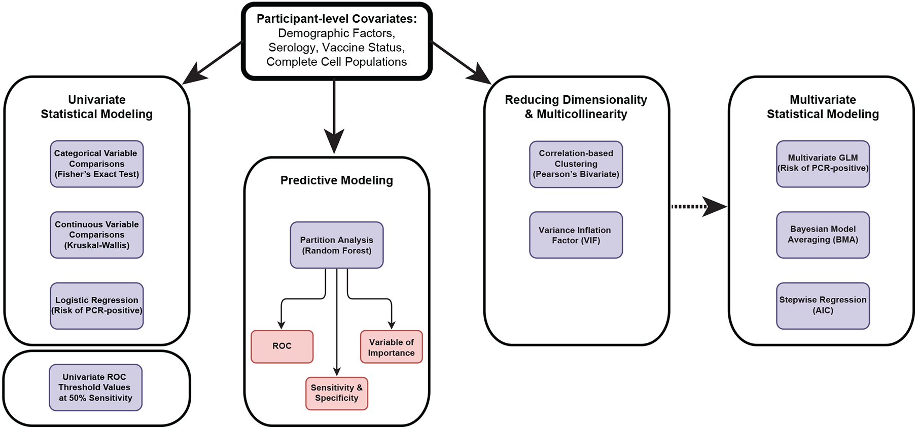
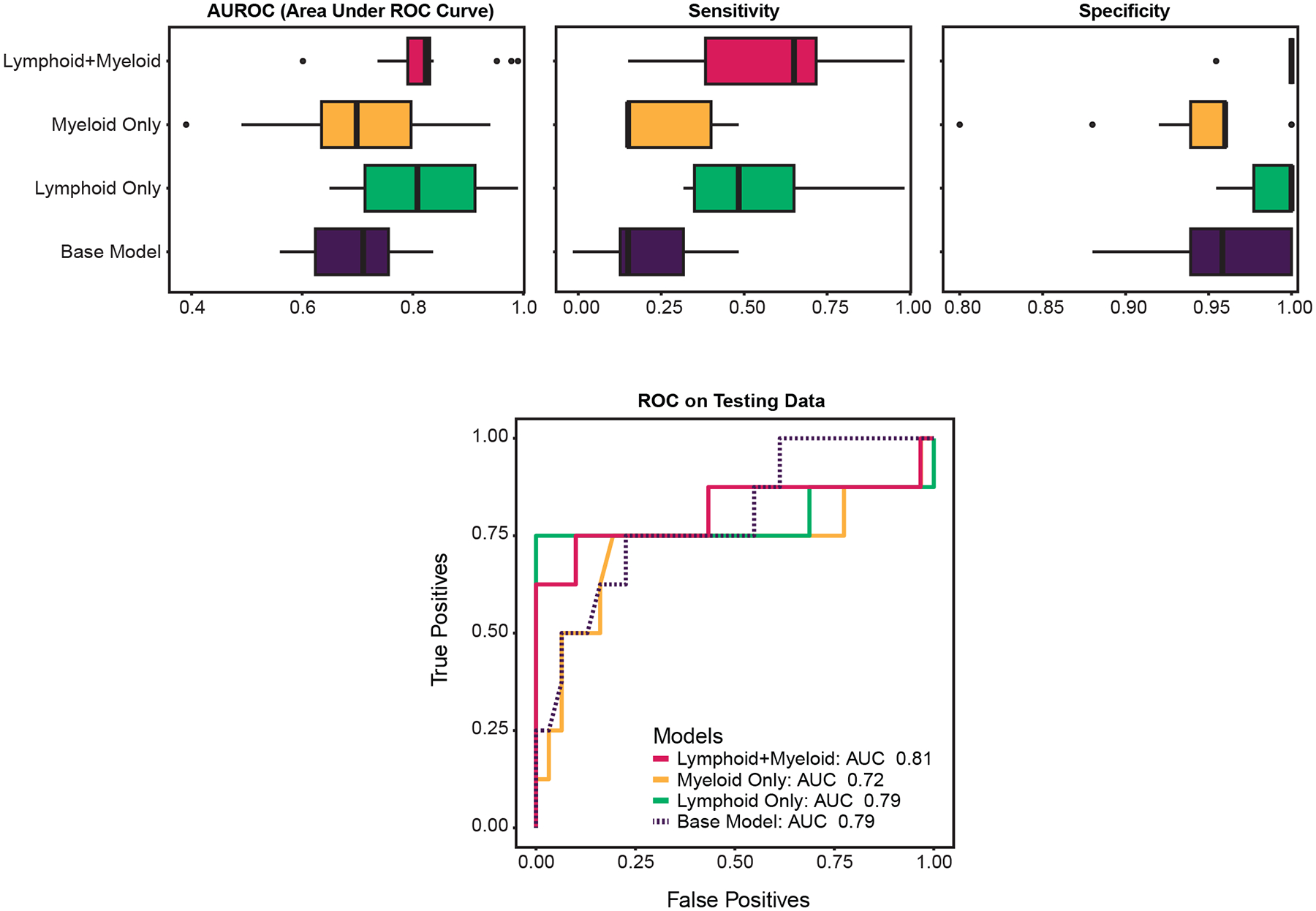
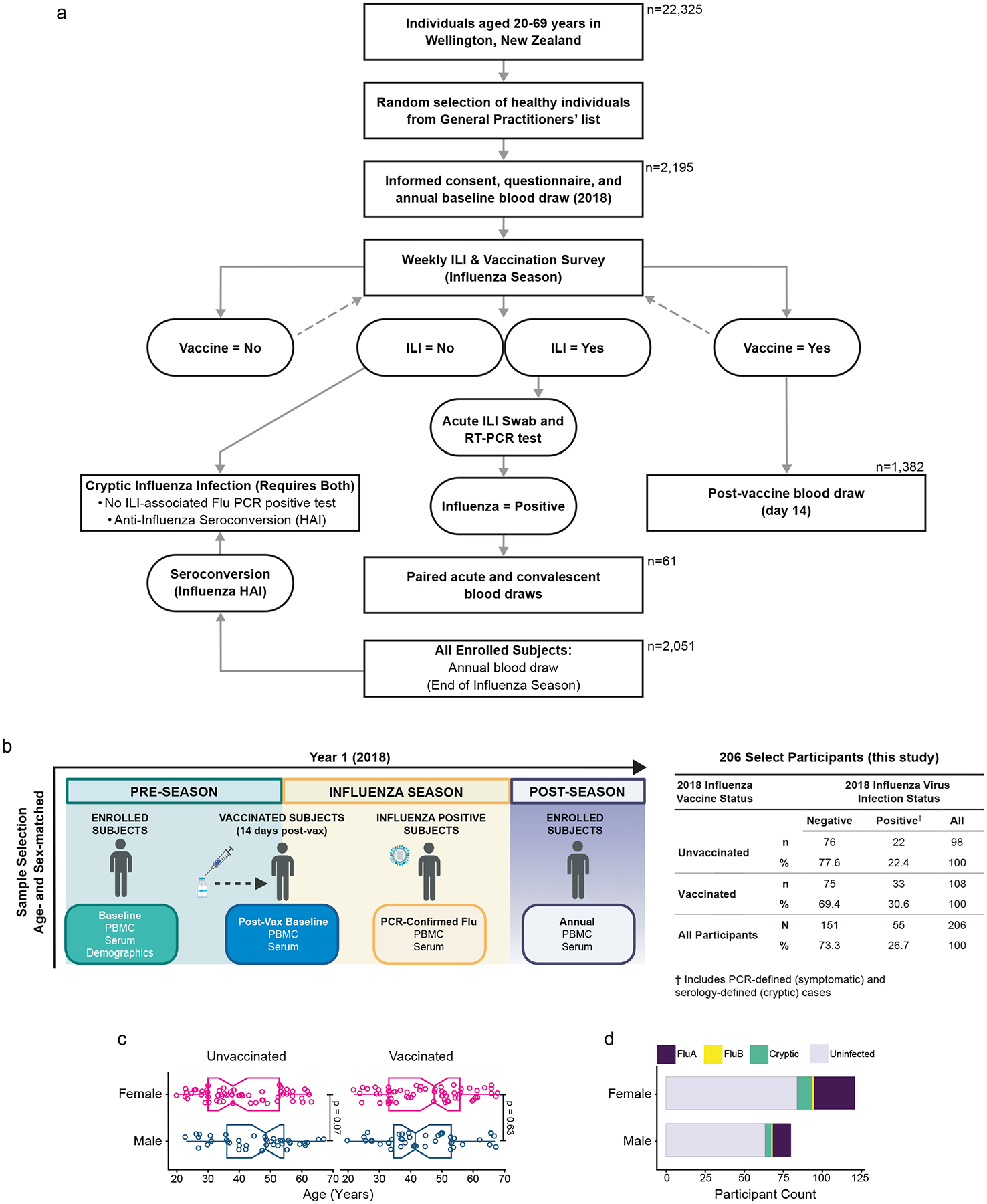
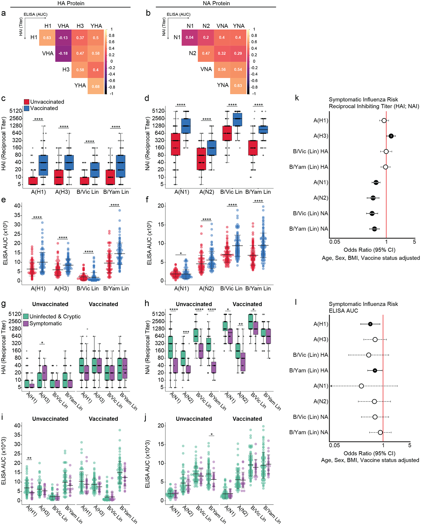
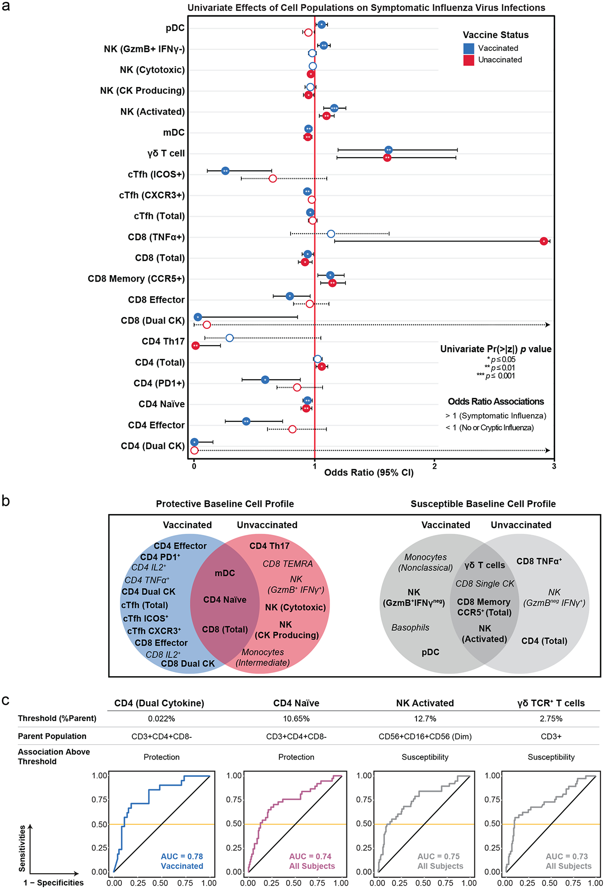
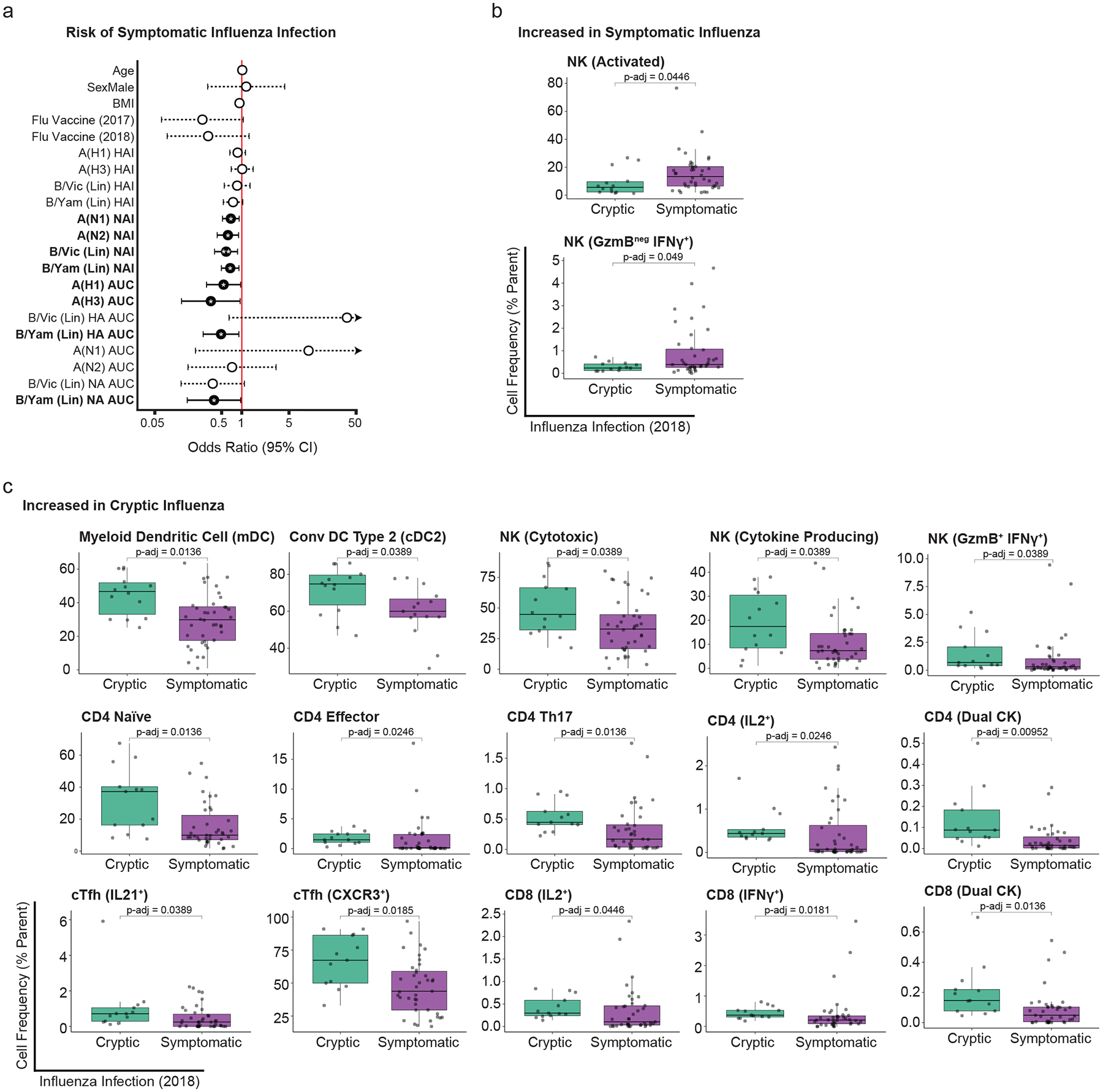
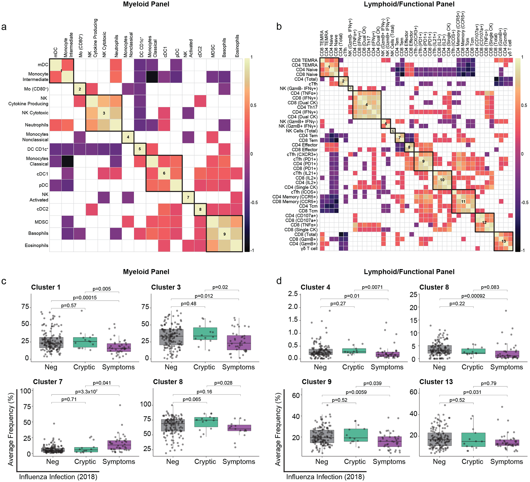
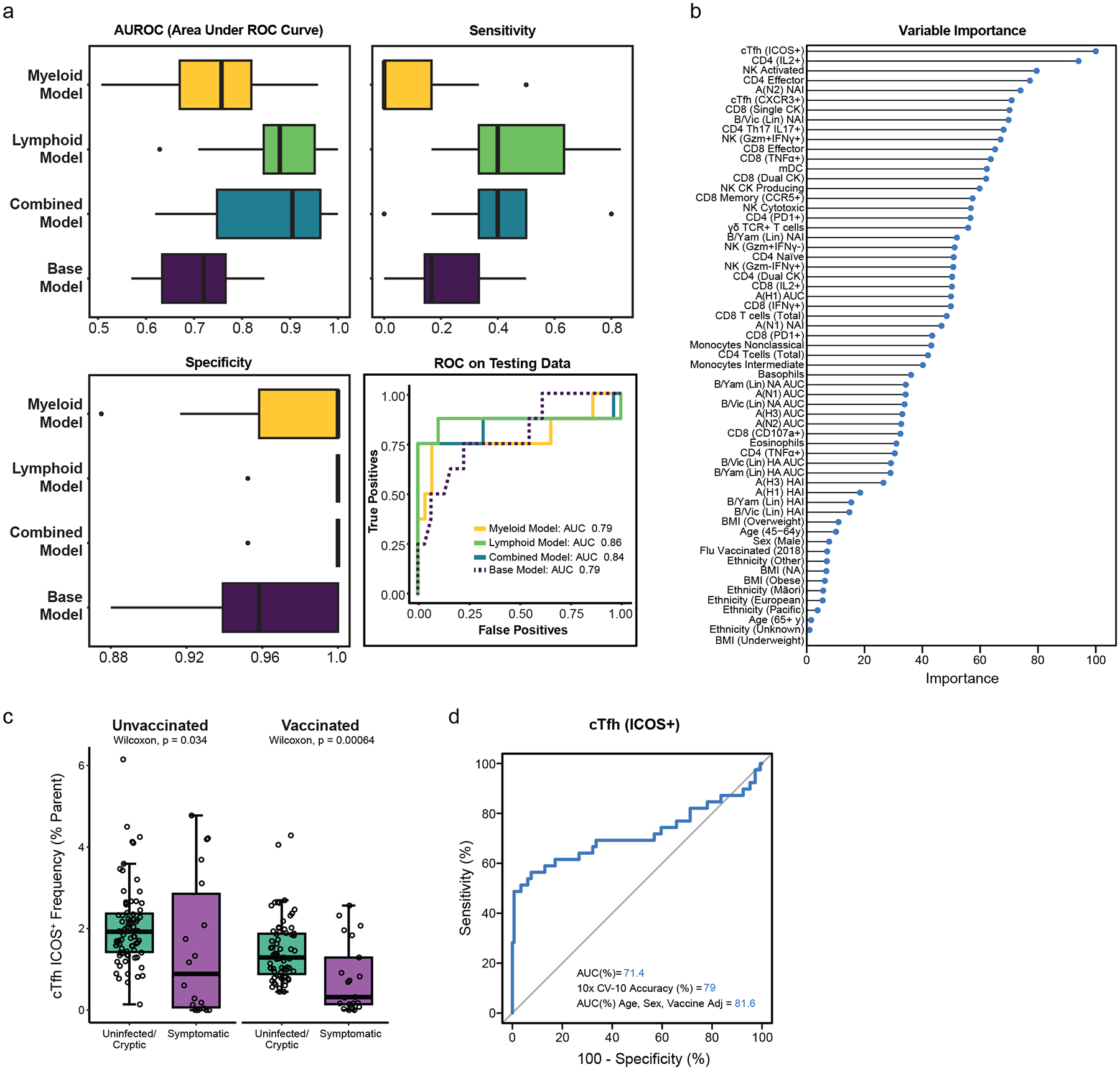
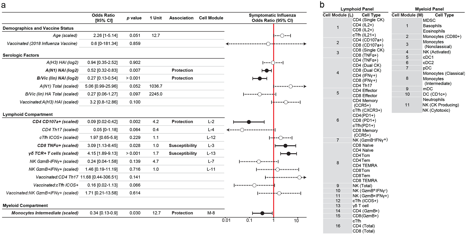
Evidence suggests that innate and adaptive cellular responses mediate resistance to the influenza virus and confer protection after vaccination. However, few studies have resolved the contribution of cellular responses within the context of preexisting antibody titers. Here, we measured the peripheral immune profiles of 206 vaccinated or unvaccinated adults to determine how baseline variations in the cellular and humoral immune compartments contribute independently or synergistically to the risk of developing symptomatic influenza. Protection correlated with diverse and polyfunctional CD4+ and CD8+ T, circulating T follicular helper, T helper type 17, myeloid dendritic and CD16+ natural killer (NK) cell subsets. Conversely, increased susceptibility was predominantly attributed to nonspecific inflammatory populations, including γδ T cells and activated CD16- NK cells, as well as TNFα+ single-cytokine-producing CD8+ T cells. Multivariate and predictive modeling indicated that cellular subsets (1) work synergistically with humoral immunity to confer protection, (2) improve model performance over demographic and serologic factors alone and (3) comprise the most important predictive covariates. Together, these results demonstrate that preinfection peripheral cell composition improves the prediction of symptomatic influenza susceptibility over vaccination, demographics or serology alone.


Conflict of interest statement

P.G.T. has consulted or received honorarium and/or travel support from Illumina, JNJ, Pfizer, and 10X. P.G.T. serves on the Scientific Advisory Board of ImmunoScape and CytoAgents. The remaining authors declare no competing interests.

Figures

Extended Data Fig. 1.
Data analysis pipeline for predictive and statistical modeling
Extended Data Fig. 2:
Participant demographic and serologic correlations
Extended Data Fig. 3:
Myeloid panel gating strategy
Extended Data Fig. 4
Lymphoid and ICS panel gating strategy
Extended Data Fig. 5:
Co-regulated immune cell clusters by vaccine status
Extended Data Fig. 6:
Decision tree model comparison from cellular covariates
Figure 1:
Figure 1:. SHIVERS-II study design, subject enrollment, sample collection, and demographics.
a) Schematic depiction of the Southern Hemisphere Influenza and Vaccine Effectiveness Research and Surveillance 2 (SHIVERS-II) study design and participant numbers for Year 1 (2018). b) Following consented enrollment, demographic information and whole blood draws were collected from all vaccinated and unvaccinated subjects pre-season (non-vaccinated baseline) and 14 days post-vaccination (vaccinated baseline). Subjects meeting WHO-defined criteria for influenza-like illness (ILI) were tested for influenza viruses by PCR and confirmed cases were sampled further during acute infection. All enrolled subjects were sampled post-season. Cryptic infections were adjudicated post-season from ILI- and PCR-negative participants with a 4-fold or greater increase in HAI antibody titers without post-vaccination HAI seroconversion. (Right) 206 enrolled subjects were selected for study inclusion from four baseline comparator groups (unvaccinated-uninfected; unvaccinated-infected; vaccinated-uninfected; vaccinated-infected) based on age- and sex-matching. c) Sex (assigned at birth) of n=206 participants stratified by vaccine status and age (years) compared by two-sided Wilcoxon rank sum test (unvaccinated-female (n=58) vs. unvaccinated-male (n=42) p= 0.07; vaccinated-female (n=66) vs. vaccinated-male (n=42) p= 0.63). Boxes represent the median and 25th to 75th percentiles; whiskers indicate the minimum (left) and maximum (right) values no further than 1.5 times the interquartile (IQR); notches extend 1.58*IQR / sqrt(n) providing 95% CI. P< 0.05 achieving significance. d) Participant sex stratified by influenza virus infection status and strain. Influenza A (FluA) virus category includes A(H1N1) and A(H3N2); influenza B (FluB) viruses include B/Victoria (lineage) and B/Yamagata (lineage) strains. PBMC (peripheral blood mononuclear cells).
Figure 2:
Figure 2:. Individual serology measures correlate with protection from symptomatic influenza disease.
a-b) Spearman Rank correlations between (a) anti-HA (HAI; ELISA) or (b) anti-NA (NAI; ELISA) serology measures. c-f) Serology measures against (c,e) HA and (d,f) NA by influenza virus source comparing vaccinated (n=108) to unvaccinated (n=98) participants by two-sided Wilcoxon rank sum test. For (c) A(H1) p< 2×10⁻¹⁶, A(H3) p= 4.5×10⁻¹¹, B/Vic (lin) p= 1.6×10⁻¹⁵, B/Yam (lin) p= 4.1×10⁻¹²; for (d) A(N1) p< 2×10⁻¹⁶, A(N2) p= 1.5×10⁻⁸, B/Vic (lin) p= 6×10⁻¹², B/Yam (lin) p= 9.4×10⁻¹⁶; for (e) A(H1) p= 5.3×10⁻⁹, A(H3) p= 2×10⁻⁹, B/Vic (lin) p= 4.5×10⁻⁵, B/Yam (lin) p= 4.8×10⁻¹¹; for (f) A(N1) p= 0.033, A(N2) p= 7.4×10⁻⁵, B/Vic (lin) p= 9.7×10⁻¹¹, B/Yam (lin) p= 1.1×10⁻⁹. g-j) Serology measures of vaccinated and unvaccinated participants against (g,i) HA and (h,j) NA by influenza virus source comparing uninfected and cryptic (n=165) to symptomatic (n=41) influenza virus infection using two-sided Wilcoxon rank sum test. For (g) unvaccinated: A(H1) p= 0.280, A(H3) p= 0.024, B/Vic (lin) p= 1.0, B/Yam (lin) p= 0.922; vaccinated A(H1) p= 0.32, A(H3) p= 0.57, B/Vic (lin) p= 0.33, B/Yam (lin) p= 0.65; for (h) unvaccinated: A(N1) p= 1.6×10⁻⁶, A(N2) p= 3.8×10⁻⁴, B/Vic (lin) p= 4.4×10⁻⁷, B/Yam (lin) p= 2.7×10⁻⁸; vaccinated A(N1) p= 0.012, A(N2) p= 0.0013, B/Vic (lin) p= 0.033, B/Yam (lin) p= 0.212; for (i) unvaccinated: A(H1) p= 0.0037, A(H3) p= 0.450, B/Vic (lin) p= 0.713, B/Yam (lin) p= 0.239; vaccinated A(H1) p= 0.103, A(H3) p= 0.155, B/Vic (lin) p= 0.646, B/Yam (lin) p= 0.053; for (j) unvaccinated: A(N1) p= 0.781, A(N2) p= 0.393, B/Vic (lin) p= 0.161, B/Yam (lin) p= 0.028; vaccinated A(N1) p= 0.90, A(N2) p= 0.95, B/Vic (lin) p= 0.40, B/Yam (lin) p= 0.48. Boxes represent the median and 25th to 75th percentiles; whiskers indicate the minimum and maximum values no further than 1.5 times the interquartile (IQR); dot plots presented as mean± SD. Inhibiting titers presented as reciprocal endpoint dilutions calculated from HAI or NAI assays using A(H1N1), A(H3N2), B/Victoria (lineage), and B/Yamagata (lineage) viruses. Total binding antibody titers reported as AUC values calculated from ELISA assay against purified, full-length HA or NA proteins derived from influenza A(H1N1), A(H3N2), B/Victoria (lineage), and B/Yamagata (lineage) viruses. Means compared using two-sided Wilcoxon rank sum test. k-l) Relative risk of symptomatic infection among all participants (n=206) given individual anti-HA and anti-NA serology measures by (k) HAI and NAI, or (l) ELISA adjusted for participant age (years), sex, BMI (kg/m2), and influenza vaccine status (2018) from individual GLMs. Odds Ratio (OR; center circle) with 95% CI (bars) derived from exponential transformation of GLM estimate (logit) values. HAI and NAI reciprocal inhibition endpoint values depicted at 1=log2 interval; ELISA AUC values depicted at 1=5000 interval. Positive effects on symptomatic influenza have OR > 1 while negative effects have OR < 1. Individual HAI or NAI models generated from 185 degrees of freedom (DOF) (symptomatic n=35, uninfected/cryptic n=151) except A(H3) HAI and B/Vic (lin) HAI (DOF = 184; symptomatic n=35, uninfected/cryptic n=150). Significance determined from GLM Pr(>|z|) output where z=estimate/SE. Resulting two-tailed p values: A(H1) HAI p= 0.368, A(H3) HAI p= 0.033, B/Vic (lin) HAI p= 0.983, B/Yam (lin) HAI p= 0.775, A(N1) NAI p= 2.4×10⁻⁶, A(N2) NAI p= 3.0 ×10⁻⁵, B/Vic (lin) NAI p= 1.9 ×10⁻⁶, B/Yam (lin) NAI p= 4.1 ×10⁻⁶. Individual HA and NA ELISA models generated from 187 DOF (symptomatic n=35, uninfected/cryptic n=153); Significance determined from GLM Pr(>|z|) output where z=estimate/SE. Resulting two-tailed p values: A(H1) AUC p= 0.011, A(H3) AUC p= 0.172, B/Vic (lin) AUC p= 0.163, B/Yam (lin) AUC p= 0.033, A(N1) AUC p= 0.232, A(N2) AUC p= 0.3, B/Vic (lin) AUC p= 0.252, B/Yam (lin) AUC p= 0.63. Not significant (blank); * p≤ 0.05; ** p≤ 0.01; *** p≤ 0.001.
Figure 3:
Figure 3:. Univariate effects of cell populations on symptomatic influenza by vaccination status.
A) Univariate generalized logistic regression model (GLM) to determine individual effect of cell population frequency (% parent) effects on symptomatic influenza by vaccine status. Individual GLMs of unvaccinated participants generated from 98 DOF (uninfected/cryptic n=79, symptomatic n=19); GLMs of vaccinated participants generated from 108 DOF (uninfected/cryptic n=86, symptomatic n=22). GLM estimate values (logit) were transformed to Odds Ratio (OR; center circle) using the e-function and presented with associated 95% CI (bars). Cell populations with positive effects on symptomatic influenza have OR > 1 while cell populations with negative effects have OR < 1. Univariate significance determined from GLM Pr(>|z|) output (closed significant; open not significant) where z=estimate/SE. Significance defined as * p≤ 0.05; ** p≤ 0.01; *** p≤ 0.001. Exact Pr(>|z|) p values and FDR-adjusted q values reported in Supplementary Table 4. b) Baseline cell profiles associated with (left) protection from and (right) susceptibility to developing symptomatic influenza by 2018 influenza virus vaccination status. Subjects with significant cell frequency outliers were determined by Grubbs’ test (p<0.05) and removed from this analysis. All cell populations presented significant by one-sided Kruskal-Wallis (italicized) or both Kruskal-Wallis and univariate GLM (bold) evaluations. Exact p values reported in Supplementary Tables 3 and 4. c) Representative thresholds depicted as cell frequency (% parent) with associated ROC curves. A threshold defines the cell frequency at which the ROC curve sensitivity (true positives) equals 0.5 and represents the cutoff above which an individual factor correctly associates 50% of cases as protected or susceptible. AUC values indicate the overall quality (true vs false positives) of the individual measure in discerning protection or susceptibility.
Figure 4:
Figure 4:. Cryptic infections are associated with unique cellular responses.
a) Relative risk of symptomatic infection among influenza virus-infected participants given demographic, serologic, or vaccine history covariates. GLM estimate values (logit) were transformed to Odds Ratio (OR; center circle) using the e-function and presented with associated 95% CI (bars). Univariate GLMs generated from 54 DOF (symptomatic n=41, cryptic n=14) except BMI (DOF = 46; symptomatic n=35, cryptic n=12). Significance determined from GLM Pr(>|z|) output where z=estimate/SE. Resulting two-tailed p values: Age p= 0.528, SexMale p= 0.827, BMI p= 0.24, Flu Vaccine 2017 p= 0.062, Flu Vaccine 2018 p= 0.111, A(H1) HAI p= 0.321, A(H3) HAI p= 0.945, B/Vic (lin) HAI p= 0.495, B/Yam (lin) HAI p= 0.091, A(N1) NAI p= 0.014, A(N2) NAI p= 0.015, B/Vic (lin) NAI p= 0.01, B/Yam (lin) p= 0.014, A(H1) AUC p= 0.041, A(H3) AUC p= 0.041, B/Vic (lin) AUC p= 0.082, B/Yam (lin) AUC p= 0.026, A(N1) NAI p= 0.249, A(N2) NAI p= 0.666, B/Vic NAI p= 0.076, B/Yam NAI p= 0.045. Not significant (blank); *p≤ 0.05. b-c) Comparison of individual cell population frequencies (% parent) across cryptic and symptomatic participants. Cell populations are grouped based on increased frequencies in (b) symptomatic or (c) cryptic influenza cases. In (b) NK Activated (cryptic n= 14 vs. symptomatic n= 40); NK GzmBnegIFNγ+ (cryptic n= 13 vs. symptomatic n= 39) and (c) mDC, NK Cytotoxic, NK Cytokine Producing (cryptic n= 14 vs. symptomatic n= 40); cDC2 (cryptic n= 14 vs. symptomatic n= 15); NK GzmB+IFNγ+, CD4 Naïve, CD4 Effector, CD4 Th17, CD4 IL2+, CD4 Dual CK, cTfh IL21+, cTfh CXCR3+, CD8 IL2+, CD8 IFNγ+, CD8 Dual CK (cryptic n= 13 vs. symptomatic n= 39). Lymphoid panel frequencies represent the average frequency (% parent) across virus (MOI = 4 A/Michigan/45/2015 H1N1pdm09 or A/Singapore/INFIMH-16-019/2016 H3N2) and peptide (1–5 μM /peptide pools containing M1, NP, PB1) stimulation groups. Boxes represent the median and 25th to 75th percentiles; whiskers indicate the minimum and maximum values no further than 1.5 times the interquartile (IQR). Means compared using two-sided Wilcoxon rank sum test with FDR adjustment with q≤ 0.01 achieving significance.
Figure 5:
Figure 5:. Co-regulated CMI and innate immune cell modules.
a-b) Co-regulated cell modules (“Clusters”) determined by average frequency (% parent) of individual cell populations from (a) myeloid or (b) lymphoid compartments with significant positive correlation (Pearson’s bivariate correlation). Lymphoid population frequencies calculated as average frequency across ICS stimulation conditions including peptide pools (M1, NP, PB1) and virus (A(H1N1), A(H3N2). P values were adjusted using false discovery rate (FDR; q) correction for multiple comparisons. Significance *q <0.1; **q<0.05; not significant (blank). c-d) Mean cell cluster frequencies from (c) myeloid or (d) lymphoid correlation matrices compared by influenza virus infection status (myeloid Cluster 1, 3, 7: uninfected (Neg) n=145 vs. cryptic n=14 vs. symptomatic n= 40; myeloid Cluster 8: Neg n=129 vs. cryptic n=14 vs. symptomatic n= 15; lymphoid Cluster 4, 8, 9, 13: Neg n=133 vs. cryptic n=13 vs. symptomatic n= 39) using two-sided Wilcoxon rank sum test. Boxes represent the median and 25th to 75th percentiles; whiskers indicate the minimum and maximum values no further than 1.5 times the interquartile (IQR).
Figure 6:
Figure 6:. Decision tree model comparison.
a) Comparison of the Base (demographic factors + serology + vaccination status), Myeloid (base + myeloid panel cell populations), Lymphoid (base + lymphoid panel cell populations), and Combined (base + myeloid + lymphoid) random forest models built to categorize symptomatic and uninfected/cryptic influenza. Participants were split 80:20 into a training set (symptomatic cases n=31, uninfected/cryptic controls n=128) and testing set (symptomatic cases n=8, uninfected/cryptic controls n=33) ensuring equal proportions of cases and controls. Models were trained, tested, and cross-validated using 10-times cross-validation (10x CV-10). Sensitivity (true positive rate), Specificity (false positive rate) and AUROC (area under the receiver-operating characteristic curve) metrics and an out-of-sample evaluation of the models (bottom right) are provided. Boxes represent the median and 25th to 75th percentiles; whiskers indicate the minimum and maximum values no further than 1.5 times the interquartile (IQR). b) Relative importance of each baseline covariate in the Combined random forest Model; baseline covariates with high importance best categorize symptomatic and uninfected/cryptic influenza cases. Subjects with significant cell frequency outliers were determined by Grubbs’ test (p<0.05) and removed from this analysis. c) Mean cTfh (ICOS+) cell frequency comparison between uninfected/cryptic and symptomatic influenza infection by 2018 influenza vaccine status using two-sided Wilcoxon rank sum test (unvaccinated symptomatic n=18 vs. unvaccinated uninfected/cryptic n=75; vaccinated symptomatic n=21 vs. uninfected/cryptic n=71). Boxes represent the median and 25th to 75th percentiles; whiskers indicate the minimum and maximum values no further than 1.5 times the interquartile (IQR). d) ROC curve generated from GLM predicting symptomatic influenza infection from cTfh (ICOS+) cell frequency. AUC values from cTfh ICOS+ GLM as univariate model, following 10x CV-10, and after age (years), sex, and 2018 influenza vaccine status adjustment.
Figure 7:
Figure 7:. Baseline predictors of influenza virus infection susceptibility accounting for demographic, vaccination, serologic covariates, and cellular.
Multivariate generalized logistic regression model (GLM) predicting symptomatic influenza virus infection accounting for baseline demographics, vaccination status, serology, myeloid, and lymphoid covariates. a) Final logistic regression model with covariates by category predicting risk of symptomatic influenza (symptomatic n=39 and uninfected/cryptic n=161 participants). For covariate selection in the final model, baseline covariates with strong multicollinearity by variance inflation factor (VIF) assessment were excluded; single representative cell populations from each co-regulated cell cluster were included. Models comprising these covariates were compared by stepwise Akaike Information Criterion (AIC; forward and reverse) and Bayesian model averaging (BMA) to account for inherent model uncertainty arising from variable selection. Interaction terms denoted with a colon (:). Odds Ratio [95% CI] derived from exponential transformation of GLM estimate (logit) value; two-tailed p value significance determined from GLM Pr(>|z|) output (closed significant; open not significant) where z=estimate/SE. Variables were scaled as indicated based on their median and standard deviation values. The value for 1 Unit of change is indicated for each scaled value. Units for scaled values are years (Age), AUC (total antibody ELISA measures), log2 reciprocal endpoint titer (HAI; NAI), %parent frequency (cell populations). For example, for every increase of 12.7 years of age, there is an increase in the odds of susceptible influenza infection by 2.26, suggesting younger individuals are better protected. Association of significant baseline covariate predictors with symptomatic (Susceptibility) or uninfected/cryptic (Protection) influenza indicated. Subjects with significant cell frequency outliers were determined by Grubbs’ test (p<0.05) and removed from this analysis. Cell Module column corresponds to (b) strongly correlated cell modules clustered based on absolute value of correlation. Significance *p≤ 0.05; **p≤ 0.01; ***p≤ 0.001.

Similar articles

Cited by

References

    1. CDC. Estimated Influenza Illnesses, Medical visits, Hospitalizations, and Deaths in the United States — 2017–2018 influenza season. Influenza (Flu) https://www.cdc.gov/flu/about/burden/2017–2018.htm#Table1 (2018).
    1. Iuliano AD et al. Estimates of global seasonal influenza-associated respiratory mortality: a modelling study. The Lancet 391, 1285–1300 (2018). - PMC - PubMed
    1. Flannery B et al. Interim estimates of 2016–17 seasonal influenza vaccine effectiveness — United States, February 2017. Morb. Mortal. Wkly. Rep 66, 167–171 (2017). - PMC - PubMed
    1. Flannery B et al. Interim estimates of 2017–18 seasonal influenza vaccine effectiveness - United States, February 2018. Morb. Mortal. Wkly. Rep 67, 180–185 (2018). - PMC - PubMed
    1. Jackson ML et al. Influenza vaccine effectiveness in the United States during the 2015–2016 season. N. Engl. J. Med 377, 534–543 (2017). - PMC - PubMed

Publication types