. 2023 Aug 1:14:1230174.
doi: 10.3389/fphar.2023.1230174. eCollection 2023.

Salvianolic acid B attenuates inflammation and prevent pathologic fibrosis by inhibiting CD36-mediated activation of the PI3K-Akt signaling pathway in frozen shoulder

Affiliations

Salvianolic acid B attenuates inflammation and prevent pathologic fibrosis by inhibiting CD36-mediated activation of the PI3K-Akt signaling pathway in frozen shoulder

Yan Yan et al. Front Pharmacol. .

Abstract

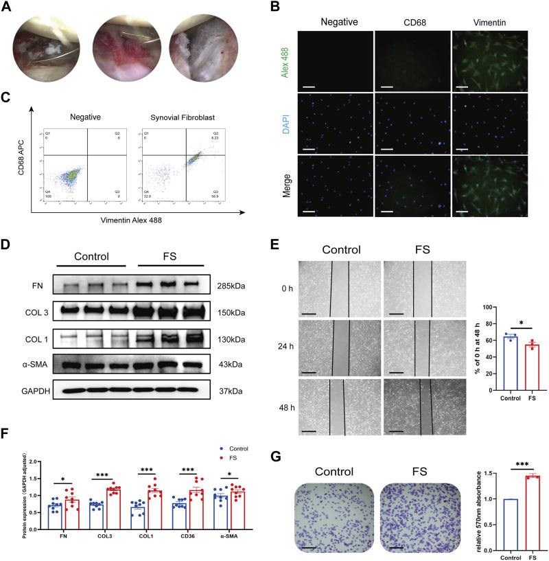
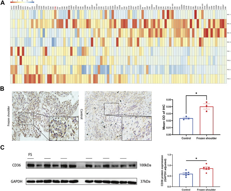
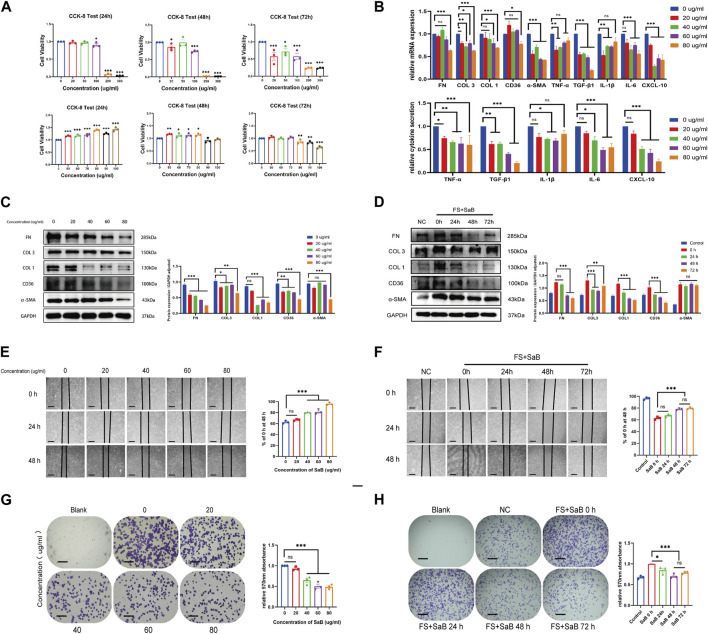
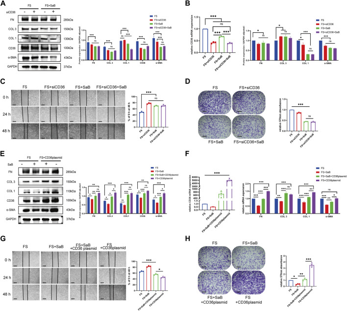
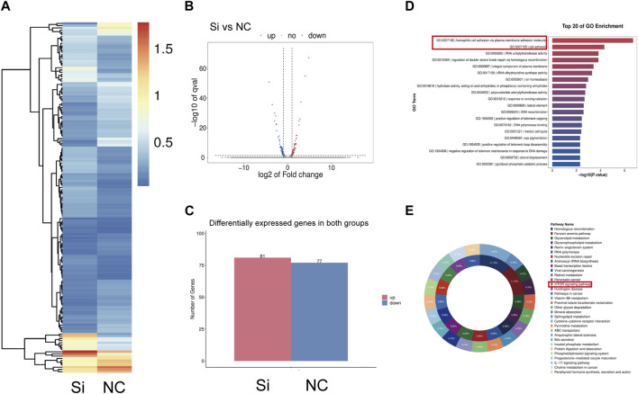
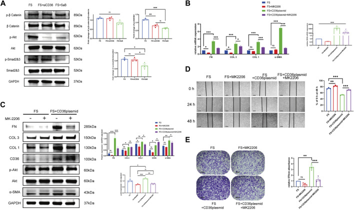
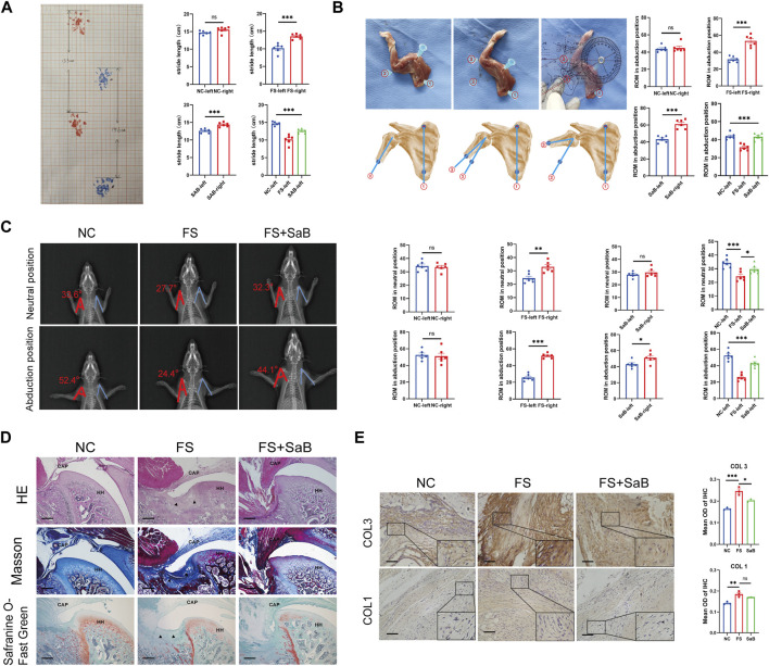
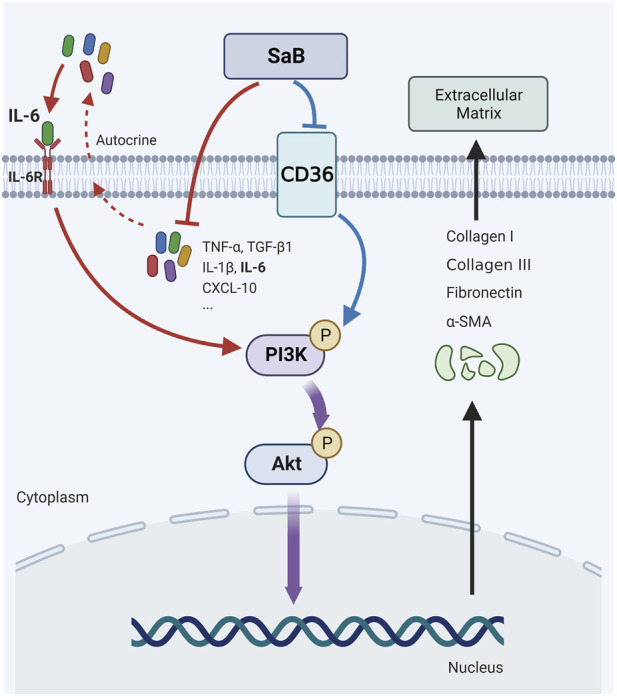
Frozen shoulder (FS) is characterized by pain and limited range of motion (ROM). Inflammation and fibrosis are accepted as main pathologic processes associated with the development of FS. However, the intrinsic mechanisms underlying pathologic fibrosis remain unclear. We aimed to elucidate the key molecules involved in pathologic fibrosis and explore new therapeutic targets for FS. Synovial fibroblasts isolated from patient biopsies were identified using immunofluorescence. Western blotting, RT-qPCR, cell adhesion tests, and would-healing assays were used to evaluate the fibrosis-related functions of synovial fibroblasts. Elevated cluster of differentiation 36 (CD36) expression was detected in FS using Western blotting and immunohistochemistry. Salvianolic acid b (SaB) inhibited CD36, blocking synovial fibroblast-induced inflammation and fibrosis. Our RNA-seq data showed that knocking down CD36 dramatically impaired the capacity of synovial fibroblasts for cell adhesion and that the PI3K-Akt signaling pathway may be crucial to the fibrotic process of FS. By up-regulating CD36 and inhibiting the phosphorylation of Akt, we demonstrated that CD36 promotes pathologic fibrosis by activating the PI3k-Akt pathway. Finally, rats treated with SaB had improved ROM and less collagen fiber deposition than the FS model group. Conclusion: SaB attenuates inflammation and inhibited the CD36-mediated activation of the PI3K-Akt signaling pathway to block pathologic fibrosis of FS in vitro and in vivo models.

Keywords: CD36; PI3K-Akt signaling pathway; fibrosis; frozen shoulder; inflammatory cytokines; salvianolic acid B.


Conflict of interest statement

The authors declare that the research was conducted in the absence of any commercial or financial relationships that could be construed as a potential conflict of interest.

Figures

FIGURE 1
FIGURE 1
Extraction and identification of synovial fibroblasts and evaluation of fibrosis-related abilities. (A) Arthroscopic view of the right shoulder in a 68-year-old woman with primary frozen shoulder. Biopsies were obtained from inflammatory synovium and thickened capsule with a punch forceps. (B,C) Cells extracted from synovium tissue expressed the specific markers for fibroblasts with positive vimentin while negative CD68. Scale bar: 25 µm. (D,F) Western blotting results showed that synovial fibroblasts in FS expressed more COL 1, COL 3, FN, and α-SMA than that in the control group. (E,G) Cell adhesion test and Wound-healing showed that synovial fibroblasts from FS were associated with the enhanced fibrosis-related ability. Scale bar: 50 μm *p < 0.05; **p < 0.01; ***p < 0.001.
FIGURE 2
FIGURE 2
Different expression of CD36 at both tissue and cellular level between FS and control group. (A) RNA-seq analysis suggested that CD36 was one of the most up-regulated molecules on surface of synovial fibroblasts. (B) Western blotting results showed that synovial fibroblasts in FS expressed more CD36 than that in the control group. Scale bar: 50 µm. (C) IHC results suggested that more CD36 was expressed in the synovium tissue derived from FS than that from the control group. *p < 0.05; **p < 0.01; ***p < 0.001.
FIGURE 3
FIGURE 3
SaB inhibited inflammation response and fibrosis-related functions of synovial fibroblasts. (A) The CCK-8 result showed that with the increase of SaB concentration, the number of living synovial fibroblasts decreased gradually. SaB treatment less than 80 μg/mL have no statistically significant effect on the growth and proliferation of synovial fibroblast within 72 h. (B) qRT-PCR results showed a decreasing mRNA trend of inflammatory cytokines and fibrosis-related molecules with increasing SaB concentrations. ELISA results suggested that less inflammatory cytokines were secreted in the supernatant of synovial fibroblast after the administration of SaB. (C) Western blotting results showed that as the concentration of SaB increased, synovial fibroblast expressed less fibrosis-related proteins including COL 1, COL 3, FN, and α-SMA as well as CD36. (D) Synovial fibroblasts expressed less fibrosis-related molecules as the administration time of SaB increased within 72 h. Besides, administration time of 48 h showed no significantly different therapeutic effect than that of 72 h, or even better. (E–H) Cell adhesion test and Wound-healing suggested that SaB concentration of 80 μg/mL and administration time of 48 h may be the appropriate condition for inhibiting the fibrosis-related functions of synovial fibroblasts. Scale bar: 50 μm *p < 0.05; **p < 0.01; ***p < 0.001.
FIGURE 4
FIGURE 4
SaB inhibited pathologic fibrosis at the cellular level via CD36. (A–D) After knocking down CD36, fibrosis-related molecules (COL 1, COL 3, FN, and α-SMA) were down-regulated at both mRNA and protein levels in synovial fibroblast, the effect of which was similar to that seen after SaB treatment. Besides, the healing and adhesion abilities of synovial fibroblast were also impaired in a manner comparable to SaB treatment. Scale bar: 50 µm. (E–H) Synovial fibroblasts secreted more fibrosis-related molecules and showed considerably greater capacity for healing and adhesion after being transfected with CD36 overexpression plasmid. When transfecting plasmid into synovial fibroblast cultured with SaB, the anti-fibrotic effect of which was partially reversed. Scale bar: 50 μm *p < 0.05; **p < 0.01; ***p < 0.001.
FIGURE 5
FIGURE 5
Gene expression change of synovium fibroblasts after CD36 knockdown. (A–C) A total of 158 mRNA were differently expressed (Fold Change ≥2 and Adjusted p-value ≤0.001) between siRNA group and the control group, among which 81 were upregulated and 77 were down-regulated. (D) KEGG pathway analysis was performed to determine the pivotal signaling pathways within the differentially expressed genes. (E) GO analysis was performed to investigate the vital change of cell function after knocking down the CD36 gene in synovial fibroblast.
FIGURE 6
FIGURE 6
CD36 promotes pathologic synovial fibroblast-induced fibrosis in FS through the PI3k-Akt pathway. (A) The expression of p-Akt/Akt, as well as several key molecules in other classic fibrosis-related pathways including the TGF-β1/SMAD pathway and the WNT/β-catenin pathway, was examined in synovial fibroblasts treated with siRNA or SaB. (B,C) Western blotting and qRT-PCR results showed that COL 1, COL 3, FN and α-SMA along with Akt phosphorylation was significantly upregulated in FS synovial fibroblasts after the overexpression of CD36, and this trend was reversed by Akt inhibitor, MK2206. (D,E) Cell adhesion test and Wound-healing suggested that MK2206 inhibited the enhanced fibrosis-related functions in synovial fibroblasts caused by the overexpression of CD36. Scale bar: 50 μm *p < 0.05; **p < 0.01; ***p < 0.001.
FIGURE 7
FIGURE 7
SaB blocks the progression of pathologic fibrosis of frozen shoulder in vivo. (A) Gait analysis by measuring the stride length on a grid paper. Stride length is defined as the longest distance between the front and rear paws. (B) Evaluation of the shoulder ROM. The angle between the scapular medial border (line 1) and humerus shaft (line 2 or 3) was measured, and the ROM was calculated by subtracting the angle in a maximally adducted position from the angle in a maximally abducted position. (C) The neutral and abduction positions’ X-ray films were collected using a flatbed scanner. Consider the axis of the humerus and the lateral edge of the scapula as the two sides of an angle with the humeral head’s center as the vertex. (D) The capsular areas were increased in the left immobilized shoulder in groups B and C, while group C showed a significantly smaller capsular area than that of group B. Besides, the amount of inflammatory cells observed in group C was significantly lower than that in group B, but higher than in group A, although not statistically significant. Black arrow: Deposition of collagen. HH: humeral head; CAP: capsule of joint. Scale bar: 100 µm. (E) Higher expression of COL 1 and COL 3 in capsule was observed in group B than the other 2 groups, while no significant difference was found between group A and C. Scale bar: 50 μm *p < 0.05; **p < 0.01; ***p < 0.001.
FIGURE 8
FIGURE 8
CD36 were upregulated in synovial fibroblasts of FS and promote pathologic fibrosis in FS through the PI3k-Akt signaling pathway; SaB inhibited inflammation response and CD36-mediated pathologic fibrosis of FS. Created by Biorender.com.

References

    1. Akbar M., Crowe L., McLean M., Garcia-Melchor E., MacDonald L., Carter K., et al. (2021). Translational targeting of inflammation and fibrosis in frozen shoulder: Molecular dissection of the T cell/IL-17A axis. Proc. Natl. Acad. Sci. U. S. A. 118 (39), e2102715118. 10.1073/pnas.2102715118 - DOI - PMC - PubMed
    1. Akbar M., McLean M., Garcia-Melchor E., Crowe L. A., McMillan P., Fazzi U. G., et al. (2019). Fibroblast activation and inflammation in frozen shoulder. PloS one 14 (4), e0215301. 10.1371/journal.pone.0215301 - DOI - PMC - PubMed
    1. Bao Y., Wang L., Xu Y., Yang Y., Wang L., Si S., et al. (2012). Salvianolic acid B inhibits macrophage uptake of modified low density lipoprotein (mLDL) in a scavenger receptor CD36-dependent manner. Atherosclerosis 223 (1), 152–159. 10.1016/j.atherosclerosis.2012.05.006 - DOI - PMC - PubMed
    1. Blessing W. A., Okajima S. M., Cubria M. B., Villa-Camacho J. C., Perez-Viloria M., Williamson P. M., et al. (2019). Intraarticular injection of relaxin-2 alleviates shoulder arthrofibrosis. Proc. Natl. Acad. Sci. U. S. A. 116 (25), 12183–12192. 10.1073/pnas.1900355116 - DOI - PMC - PubMed
    1. Bunker T. D., Anthony P. P. (1995). The pathology of frozen shoulder. A Dupuytren-like disease. J. bone Jt. Surg. 77 (5), 677–683. 10.1302/0301-620x.77b5.7559688 - DOI - PubMed

LinkOut - more resources