

Association between carotid and coronary atherosclerotic plaque morphology: a virtual histology intravascular ultrasound study
- PMID: 37593244
- PMCID: PMC10431194
Association between carotid and coronary atherosclerotic plaque morphology: a virtual histology intravascular ultrasound study
Abstract
Background and aim: Atherosclerosis is considered to be a systemic disease; however, evidence exists on the heterogeneous nature of atherosclerotic disease. To date, continuous research seeks to determine the morphological differences between carotid and coronary artery disease. This study aimed to evaluate the relationship of morphological characteristics assessed by virtual histology intravascular ultrasound (VH-IVUS) between carotid and coronary plaque composition among patients with and without a history of cerebrovascular events.
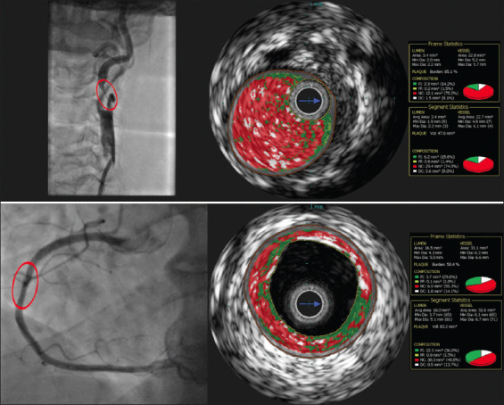
Methods: This study was a single-center prospective study (n = 100; age 69.6 ± 8.4). All patients were scheduled for carotid or coronary artery stenting and underwent VH-IVUS examination of the carotid and coronary arteries before intervention.
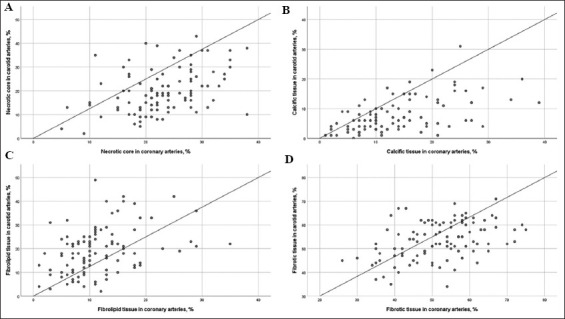
Results: There was a modest, but statistically significant correlation between the carotid and coronary necrotic core ([NC] r = 0.46, P < 0.01), fibrofatty ([FF] r = 0.38, P < 0.01), dense calcium (r = 0.56, P < 0.01), and fibrous (r = 0.42, P < 0.01) plaque composition. The high amount of NC was detected in both arteries of the carotid artery stenting (CAS) group with higher proportion in the coronary artery (20.2% ± 9.4 % vs. 22.7% ± 6.8%, P = 0.02). More fibrolipid content was observed in carotid plaque compared to coronary (19.6% ± 9.9% vs. 12.2% ± 8.1%, P < 0.01). Patients with a history of cerebrovascular events had a numerically greater proportion of necrotic tissue in the carotid artery compared to asymptomatic and symptomatic CAS group patients (23.5% ± 10.7% vs. 18.9% ± 8.2% and 18.7% ± 9.5%, P = 0.11).
Conclusion: The percentage of all analyzed plaque components was moderately correlated between coronary and carotid artery plaques. Nevertheless, the proportion of NC plaque tissue was greater in the coronary arteries, while the carotid arteries showed more %FF atherosclerotic lesions. CAS group patients with a history of cerebrovascular events had a tendency of greater proportion of necrotic tissue in analyzed carotid plaques compared to others in the CAS group.
Relevance for patients: In this study, we found that patients with a history of cerebrovascular event had a tendency of increased NC content in culprit lesion of carotid artery. Complementary use of non-invasive and invasive imaging modalities allows to detect high-risk atherosclerotic plaques and adjust treatment strategy.
Keywords: Atherosclerosis; Carotid artery stenting; Percutaneous coronary intervention; Virtual histology.
Copyright: © 2023 Author(s).
Conflict of interest statement
None.
Figures
References
-
- Shenouda R, Vancheri S, Bassi EM, Nicoll R, Sobhi M, El Sharkawy E, et al. The Relationship between Carotid and Coronary Calcification in Patients with Coronary Artery Disease. Clin Physiol Funct Imaging. 2021;41:271–80. - PubMed
-
- Cohen GI, Aboufakher R, Bess R, Frank J, Othman M, Doan D, et al. Relationship between Carotid Disease on Ultrasound and Coronary Disease on CT Angiography. JACC Cardiovasc Imaging. 2013;6:1160–7. - PubMed
-
- Den Ruijter HM, Peters SA, Anderson TJ, Britton AR, Dekker JM, Eijkemans MJ, et al. Common Carotid Intima-Media Thickness Measurements in Cardiovascular Risk Prediction:A Meta-Analysis. JAMA. 2012;308:796–803. - PubMed
-
- Amato M, Montorsi P, Ravani A, Oldani E, Galli S, Ravagnani PM, et al. Carotid Intima-Media Thickness by B-Mode Ultrasound as Surrogate of Coronary Atherosclerosis:Correlation with Quantitative Coronary Angiography and Coronary Intravascular Ultrasound Findings. Eur Heart J. 2007;28:2094–101. - PubMed
-
- Nasu K, Tsuchikane E, Katoh O, Vince DG, Virmani R, Surmely JF, et al. Accuracy of In Vivo Coronary Plaque Morphology Assessment:A Validation Study of In Vivo Virtual Histology Compared with In Vitro Histopathology. J Am Coll Cardiol. 2006;47:2405–12. - PubMed
LinkOut - more resources
Full Text Sources