

Prevalence of immune mediated vesiculobullous lesions among patients visiting a private dental hospital with special emphasis on gingival manifestation
- PMID: 37593559
- PMCID: PMC10431229
- DOI: 10.4103/jisp.jisp_646_21
Prevalence of immune mediated vesiculobullous lesions among patients visiting a private dental hospital with special emphasis on gingival manifestation
Abstract
Background and aim: Vesiculobullous lesions are a group of mucocutaneous lesions that are predominantly immune-mediated but may also have a genetic or viral origin. The most common site of occurrence is buccal mucosa, whereas the number of cases involving gingiva is comparatively low. Based on the literature, although numerous studies have reported the prevalence of vesiculobullous lesions in the nonkeratinized epithelium, there is a dearth of knowledge about its occurrence in keratinized oral mucosa, especially gingiva. The objective of the study was to assess the prevalence of immune-mediated oral vesiculobullous lesions emphasizing the occurrence in keratinized mucosa, especially the gingiva, among patients visiting a private dental hospital.
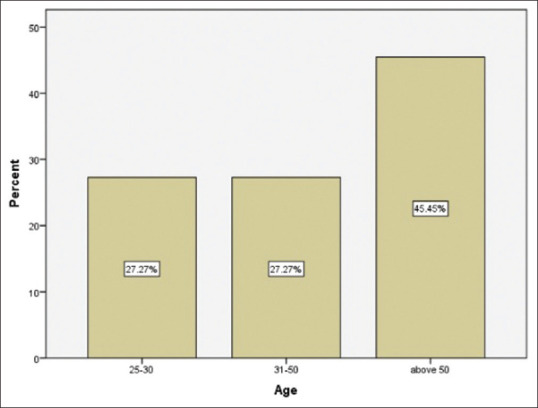
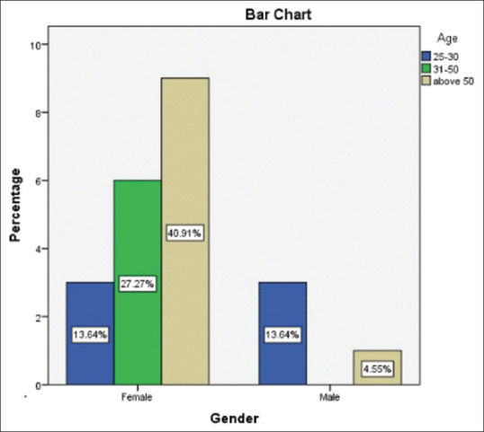
Materials and methods: The study was conducted in a private teaching dental institute and hospital setting. Out of 615 incisional biopsies received in the department of oral pathology, between June 2019 and April 2021, n = 22 samples were immune-mediated vesiculobullous lesions confirmed by clinical and histopathological diagnosis after eliminating lesions of viral origin. Patient details including age, gender, site, duration, and systemic illness were collected from the digital information archiving software and analyzed by appropriate statistics using SPSS software.
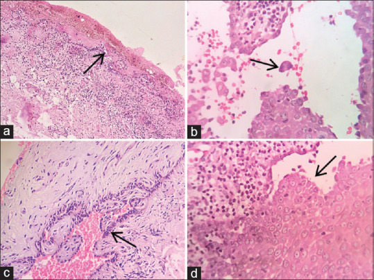
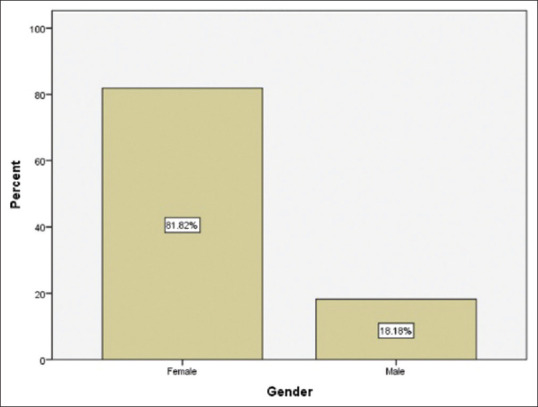
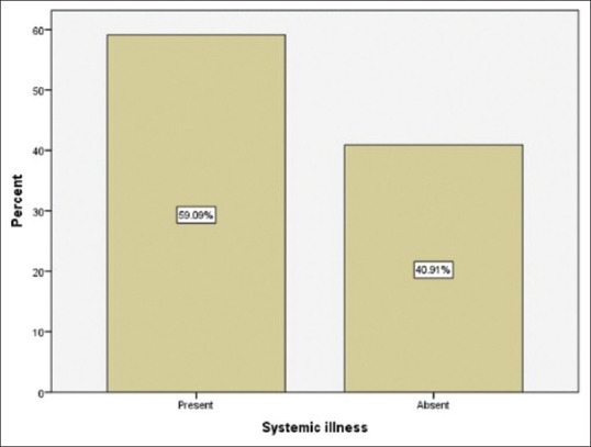
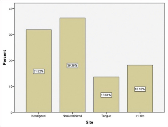
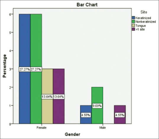
Results: Based on the results, 95.5% of the patients had histopathological features of intraepithelial clefting and only 4.5% of them showed subepithelial clefting. Female predilection was 6.3:1. The most common site of involvement was nonkeratinized mucosa (36.36%) and 59.09% of the patients presented with systemic illness.
Conclusion: The study shows most of the features of pemphigus is consistent in gingiva and other parts of oral mucosa. The dental practitioners should be aware of the various oral manifestations of such lesions to ensure accurate diagnosis and adequate treatment.
Keywords: Autoimmune; gingiva; immune mediated; vesiculobullous.
Copyright: © 2023 Indian Society of Periodontology.
Conflict of interest statement
There are no conflicts of interest.
Figures

Similar articles
-
Prevalence of Oral Mucosal Lesions in a Tertiary Care Dental Hospital of Kathmandu.JNMA J Nepal Med Assoc. 2017 Jul-Sep;56(207):362-6. JNMA J Nepal Med Assoc. 2017. PMID: 29255321
-
Gingival Manifestations in Oral Chronic Autoimmune Bullous Diseases: A Retrospective Study.Medicina (Kaunas). 2024 Jan 17;60(1):167. doi: 10.3390/medicina60010167. Medicina (Kaunas). 2024. PMID: 38256427 Free PMC article.
-
Oral mucocutaneous lesions - a comparative clinicopathological and immunofluorescence study.J Int Oral Health. 2015 Mar;7(3):59-63. J Int Oral Health. 2015. PMID: 25878481 Free PMC article.
-
Dental care of patients with autoimmune vesiculobullous diseases: case reports and literature review.Quintessence Int. 2006 Nov-Dec;37(10):777-87. Quintessence Int. 2006. PMID: 17078276 Review.
-
Diagnostic procedures for autoimmune vesiculobullous diseases: A review.J Oral Maxillofac Pathol. 2014 Sep-Dec;18(3):390-7. doi: 10.4103/0973-029X.151324. J Oral Maxillofac Pathol. 2014. PMID: 25948994 Free PMC article. Review.
Cited by
-
Effects of Probiotics on Inflammatory Biomarkers and Its Associations With Cardiac Autonomic Function in Women With Arterial Hypertension: A Secondary Analysis of a Randomized Clinical Trial.Probiotics Antimicrob Proteins. 2024 Jun 6. doi: 10.1007/s12602-024-10303-6. Online ahead of print. Probiotics Antimicrob Proteins. 2024. PMID: 38842655
-
Screening of the Lipid-Lowering Probiotic Lactiplantibacillus Plantarum SDJ09 and its Anti-Obesity Mechanism.Appl Biochem Biotechnol. 2025 Jan;197(1):35-54. doi: 10.1007/s12010-024-05034-x. Epub 2024 Aug 2. Appl Biochem Biotechnol. 2025. PMID: 39093349
References
-
- Bickle K, Roark TR, Hsu S. Autoimmune bullous dermatoses: A review. Am Fam Physician. 2002;65:1861–70. - PubMed
-
- Regezi JA, Sciubba JJ, Jordan RC. Oral Pathology: Clinical-Pathologic Correlations. 7th ed. St. Louis, Mo: Elsevier; 2017.
-
- Dagistan S, Goregen M, Miloglu O, Cakur B. Oral pemphigus vulgaris: A case report with review of the literature. J Oral Sci. 2008;50:359–62. - PubMed
LinkOut - more resources
Full Text Sources
Miscellaneous