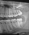
Paradental cyst with hyaline ring granuloma masquerading as pericoronitis
- PMID: 37593562
- PMCID: PMC10431234
- DOI: 10.4103/jisp.jisp_292_22
Paradental cyst with hyaline ring granuloma masquerading as pericoronitis
Abstract
Paradental cyst is an odontogenic cyst associated with pericoronitis in partly erupted mandibular third molars. It is an inflammatory cyst common among the mandibular molars. The cyst is most commonly seen on the distal or distobuccal aspect of the third molars. The angle of tooth and food impaction has been postulated to be responsible for the development of the cyst in third molars. The source of the epithelium has been reported as reduced enamel epithelium. The paradental cyst is frequently misdiagnosed as a radicular cyst or dentigerous cyst. We report a case of paradental cyst in a patient with partially erupted mandibular third molar with food impaction and resulting hyaline ring granuloma.
Keywords: Food impaction; hyaline ring granuloma; mandibular molar; paradental cyst; pericoronitis.
Copyright: © 2023 Indian Society of Periodontology.
Conflict of interest statement
There are no conflicts of interest.
Figures


References
-
- Friedrich RE, Scheuer HA, Zustin J. Inflammatory paradental cyst of the first molar (buccal bifurcation cyst) in a 6-year-old boy: Case report with respect to immunohistochemical findings. In Vivo. 2014;28:333–9. - PubMed
-
- Mohan A, Sivakumar TT, Joseph AP, BR Varun, Mony V, LK Kumar S. Inflammatory paradental cyst on the distobuccal aspect of an impacted mandibular third molar: A case report. Int J Case Rep Imag. 2017;8:592–6.
-
- Kimura TC, Carneiro MC, Coelho YF, de Sousa SC, Veltrini VC. Hyaline ring granuloma of the mouth-A foreign-body reaction that dentists should be aware of: Critical review of literature and histochemical/immunohistochemical study of a new case. Oral Dis. 2021;27:391–403. - PubMed
-
- Colgan CM, Henry J, Napier SS, Cowan CG. Paradental cysts: A role for food impaction in the pathogenesis?A review of cases from Northern Ireland. Br J Oral Maxillofac Surg. 2002;40:163–8. - PubMed
Publication types
LinkOut - more resources
Full Text Sources
