Review
. 2023 Jul 24:21:3933-3945.
doi: 10.1016/j.csbj.2023.07.024. eCollection 2023.

Structural aspects of enzymes involved in prokaryotic Gram-positive heme biosynthesis

Affiliations
Review

Structural aspects of enzymes involved in prokaryotic Gram-positive heme biosynthesis

Nikolaus Falb et al. Comput Struct Biotechnol J. .

Abstract

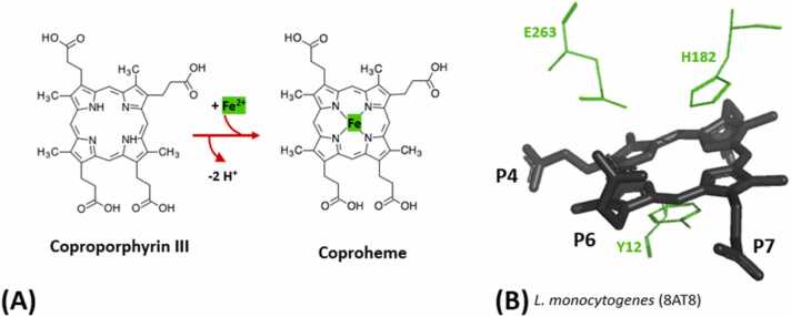
The coproporphyrin dependent heme biosynthesis pathway is almost exclusively utilized by Gram-positive bacteria. This fact makes it a worthwhile topic for basic research, since a fundamental understanding of a metabolic pathway is necessary to translate the focus towards medical biotechnology, which is very relevant in this specific case, considering the need for new antibiotic targets to counteract the pathogenicity of Gram-positive superbugs. Over the years a lot of structural data on the set of enzymes acting in Gram-positive heme biosynthesis has accumulated in the Protein Database (www.pdb.org). One major challenge is to filter and analyze all available structural information in sufficient detail in order to be helpful and to draw conclusions. Here we pursued to give a holistic overview of structural information on enzymes involved in the coproporphyrin dependent heme biosynthesis pathway. There are many aspects to be extracted from experimentally determined structures regarding the reaction mechanisms, where the smallest variation of the position of an amino acid residue might be important, but also on a larger level regarding protein-protein interactions, where the focus has to be on surface characteristics and subunit (secondary) structural elements and oligomerization. This review delivers a status quo, highlights still missing information, and formulates future research endeavors in order to better understand prokaryotic heme biosynthesis.

Keywords: Coproheme decarboxylase; Coproporphyrin ferrochelatase; Coproporphyrinogen oxidase; Frataxin; Molecular enzymology; Structure determination; Uroporphyrinogen decarboxylase.


Conflict of interest statement

The authors of the submitted manuscript “Structural aspects of enzymes involved in prokaryotic Gram-positive heme biosynthesis” declare no conflict of interests.

Figures

ga1
Graphical abstract
Fig. 1
Fig. 1
Steps of the Gram-positive CPD pathway (A) and Gram-negative/eukaryotic PPD pathway (B) from the common precursor uroporphyrinogen III and a common decarboxylation step, performed by UroD. To highlight the reaction sites on the substrate molecule, green shades mark the respective area. Chemical structures were downloaded from the KEGG compound database (https://www.genome.jp/kegg/compound/), provided by the Bioinformatics Center, Institute for Chemical Research, Kyoto University and the Human Genome Center, Institute of Medical Science, University of Tokyo. The respective identification codes are: C01051 (Uroporphyrinogen III), C03263 (Coproporphyrinogen III), C05770 (Coproporphyrin III), C21284 (Coproheme), C01079 (Protoporphyrinogen IX), C02191 (Protoporphyrin IX) and C00032 (Heme b).
Fig. 2
Fig. 2
Available crystal structures of the CPD pathway in the PDB, found by keyword search. Protein structures shown are (A) UroD, (B) CgoX, (D) CpfC, (D) ChdC and (E) frataxin orthologues, which are possible iron transporters for the pathway. The figure depicts a surface overview and for (A) UroD and (D) ChdC an orientation view of the total structure/oligomer. The initial two letters of the structure titles in bold correspond to the organism of origin and are explained in the text and the glossary. PDB-IDs are depicted below. The cofactors if present are presented as sticks. Additionally, a CAVER calculation (Minimum probe radius = 0.9, Shell depth = 4, Shell radius = 3, Clustering threshold = 3.5) was performed originating from the presumed active site/cofactor binding site to further highlight its location on the protein monomer. CAVER calculations from porphyrin binding sites are depicted in yellow, for CgoX the calculations originated from the bound FAD cofactor in the structure and is depicted in red.
Fig. 3
Fig. 3
(A) General reaction catalysed by UroD, with labeled propionate (P) residues. Remaining methyl groups in the product are labeled (M), side chains changing during reaction are highlighted in green. (B & C) Cofactor binding site overlay of UroD from B. subtilis (2INF) (green), N. tabacum (1J93) (yellow) and H. sapiens (2Q71) (magenta). 2Q71 is a crystal structure with a coproporphyrin III molecule (black) bound, and the hydrogen bonds it forms with nearby residues are highlighted in orange. (B) shows distal site of the cofactor with tyrosine and aspartate residues visible, (C) shows the proximal site of the cofactor with two arginine residues visible.
Fig. 4
Fig. 4
Reaction catalysed by CgoX. The formed methyl groups which connect the macrocyclic tetrapyrrole ring are labeled by Greek letters (α,β,γ,δ) and marked in green to highlight them as reaction sites.
Fig. 5
Fig. 5
(A) Reaction catalysed by CpfC. Inserted Fe2+ is highlighted in green. (B) Active site of CpfC from Listeria monocytogenes (8AT8). The cofactor coproporphyrin III is shown in black with visible propionates labeled (P) for orientation. Catalytically important residues are shown as green lines. On the proximal side a tyrosine residue is located and on the distal side a glutamate and a histidine residue are visible.
Fig. 6
Fig. 6
(A) Reaction catalysed by ChdC. Original propionate groups and emerging vinyl groups are labeled (P,V). Sidechains which are reaction sites are marked in green. Included in the reaction is also the three-propionate reaction intermediate MMD. Chemical structure was downloaded from the KEGG compound database (https://www.genome.jp/kegg/compound/), provided by the Bioinformatics Center, Institute for Chemical Research, Kyoto University and the Human Genome Center, Institute of Medical Science, University of Tokyo. The identification code is C22173. (B) Monomeric subunit of ChdC from Corynebacterium diphtheriae (6XUC) with black coproheme cofactor marking the active site. Flexible linker is distinctly colored in cyan (C) Cofactor in the active site with propionates labeled for orientation (P). Visible residues are a histidine and a tyrosine on the distal site and a histidine on the proximal site.
Fig. 7
Fig. 7
(A) Crystal structure of BsFrat (2OC6), with highlighted conserved residues in red, conserved antiparallel beta sheet (wheat) and alpha helices (gray). (B) Close Up of conserved antiparallel sheet region (cartoon in wheat), with conserved residues presented as red lines. An additional scheme shows their structural positions in the sheet, according to 2OC6 and the sequence alignment in (C). (C) Results of the multiple sequence alignment of the relevant sequence range, depicted by Berkeley WebLogo. The conserved leucine residue is at alignment position 299, the conserved tyrosine/tryptophan residue at 305, proline at 308 and glycine at 331.
Fig. 8
Fig. 8
Known frataxin and CyaY crystal structures in cartoon depiction and from the same perspective to allow comparison of metal binding sites. Structures include: Apo-frataxin from Homo sapiens (splitpea) with possible residues involved in metal binding highlighted in red (1EKG), apo-frataxin from Bacillus subtilis (black) (2OC6), Co2+ bound frataxin from Saccharomyces cerevisiae (lightpink) and Co2+ in cyan (3OER), Fe2+ bound frataxin from Saccharomyces cerevisiae in (lightpink) and Fe2+ in orange (4EC2), Co2+ bound CyaY from Escherichia coli (marine) and Co2+ in cyan (2EFF), Eu3+ bound CyaY from Escherichia coli (marine) and Eu3+ in black (2P1X), Co2+ bound CyaY from Psychchromonas ingrahamii (lightorange) and Co2+ in cyan (4LK8), Eu3+ bound CyaY from Psychchromonas ingrahamii (lightorange) and Eu3+ in black (4LP1).
Fig. 9
Fig. 9
(A) Active sites of coproporphyrin ferrochelatases and overall monomeric structure, represented for LmCpfC (blue). (B) Active sites and orientation of flexible loop regions for various coproheme decarboxylases and overall pentameric structure with highlighted subunit, represented for LmChdC (magenta).

Similar articles

Cited by

References

    1. Poulos T.L. Heme enzyme structure and function. Chem Rev. 2014;114(7):3919–3962. doi: 10.1021/cr400415k. - DOI - PMC - PubMed
    1. Shi Y. A glimpse of structural biology through X-Ray crystallography. Cell. 2014;159(5):995–1014. doi: 10.1016/j.cell.2014.10.051. - DOI - PubMed
    1. Lobo S.A.L., Scott A., Videira M.A.M., Winpenny D., Gardner M., Palmer M.J., et al. Staphylococcus aureus Haem Biosynthesis: characterisation of the enzymes involved in final steps of the pathway: S. Aureus Haem Biosynthesis: from uroporphyrinogen III to haem. Mol Microbiol. 2015;97(3):472–487. doi: 10.1111/mmi.13041. - DOI - PubMed
    1. Celis A.I., DuBois J.L. Making and breaking heme. Curr Opin Struct Biol. 2019;59:19–28. doi: 10.1016/j.sbi.2019.01.006. - DOI - PMC - PubMed
    1. A.I. Celis J.E. Choby J. Kentro E.P. Skaar J.L. DuBois Control of Metabolite Flux during the Final Steps of Heme b Biosynthesis in Gram-Positive Bacteria 2020. - PMC - PubMed

LinkOut - more resources