. 2023 Oct;80(10):2072-2087.
doi: 10.1161/HYPERTENSIONAHA.123.21392. Epub 2023 Aug 18.

Cellular Senescence Contributes to Large Elastic Artery Stiffening and Endothelial Dysfunction With Aging: Amelioration With Senolytic Treatment

Affiliations

Cellular Senescence Contributes to Large Elastic Artery Stiffening and Endothelial Dysfunction With Aging: Amelioration With Senolytic Treatment

Zachary S Clayton et al. Hypertension. 2023 Oct.

Abstract

Background: Here, we assessed the role of cellular senescence and the senescence associated secretory phenotype (SASP) in age-related aortic stiffening and endothelial dysfunction.

Methods: We studied young (6-8 mo) and old (27-29 mo) p16-3MR mice, which allows for genetic-based clearance of senescent cells with ganciclovir (GCV). We also treated old C57BL/6N mice with the senolytic ABT-263.

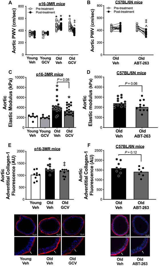
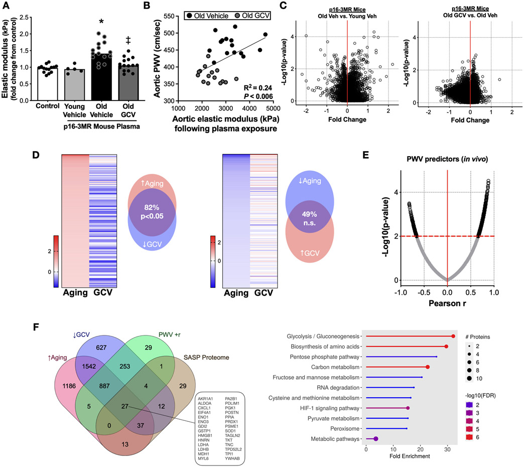
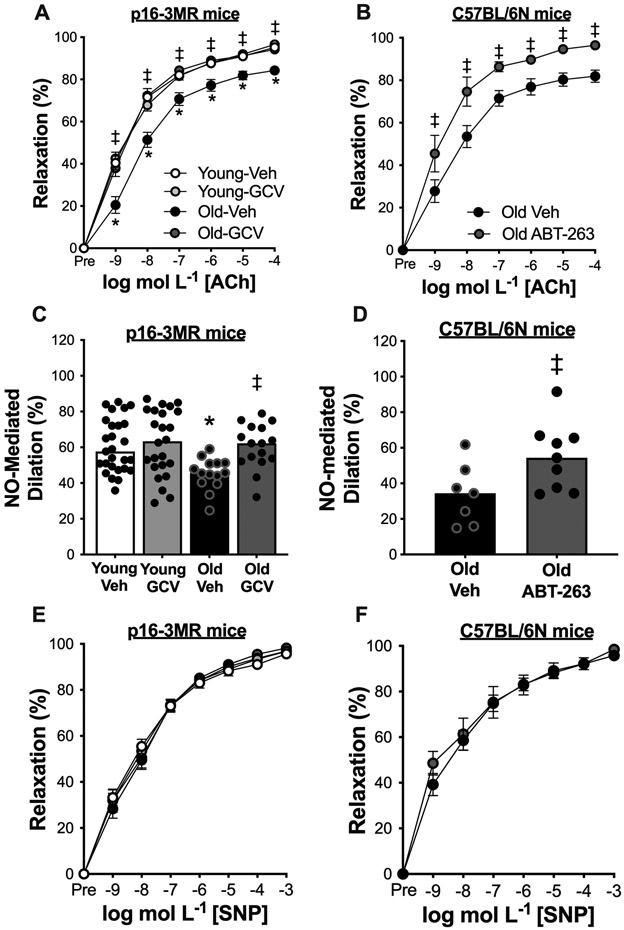
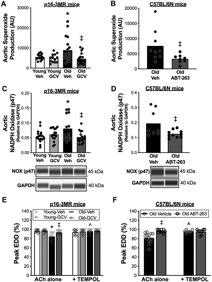
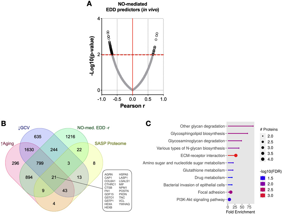
Results: In old mice, GCV reduced aortic stiffness assessed by aortic pulse wave velocity (PWV; 477±10 vs. 382±7 cm/s, P<0.05) to young levels (old-GCV vs. young-vehicle, P=0.35); ABT-263 also reduced aortic PWV in old mice (446±9 to 356±11 cm/s, P<0.05). Aortic adventitial collagen was reduced by GCV (P<0.05) and ABT-263 (P=0.12) in old mice. To show an effect of the circulating SASP, we demonstrated that plasma exposure from Old-vehicle p16-3MR mice, but not from Old-GCV mice, induced aortic stiffening assessed ex vivo (elastic modulus; P<0.05). Plasma proteomics implicated glycolysis in circulating SASP-mediated aortic stiffening. In old p16-3MR mice, GCV increased endothelial function assessed via peak carotid artery endothelium-dependent dilation (EDD; Old-GCV, 94±1% vs. Old-vehicle, 84±2%, P<0.05) to young levels (Old-GCV vs. young-vehicle, P=0.98), and EDD was higher in old C57BL/6N mice treated with ABT-263 vs. vehicle (96±1% vs. 82±3%, P<0.05). Improvements in endothelial function were mediated by increased nitric oxide (NO) bioavailability (P<0.05) and reduced oxidative stress (P<0.05). Circulating SASP factors related to NO signaling were associated with greater NO-mediated EDD following senescent cell clearance.

Conclusions: Cellular senescence and the SASP contribute to vascular aging and senolytics hold promise for improving age-related vascular function.

Keywords: aging; aortic stiffness; cellular senescence; endothelial function; senescence-associated secretory phenotype.


Conflict of interest statement

Disclosures None.

Figures

Figure 1.
Figure 1.. Cellular senescence mediates aortic stiffening with aging and senolytic treatment lowers aortic stiffness in advanced age.
In young (6-8 months) and old (27-29 months) male and female (sexes combined) p16-3MR mice treated with vehicle (Veh; sterile saline) or ganciclovir (GCV, in sterile saline) at a dose of 25 mg/kg/day for five consecutive days via intraperitoneal injection: (A) Aortic pulse wave velocity (PWV) before and after treatment. (C) Aortic elastic modulus at time of sacrifice. (E) Immunofluorescence staining for Type-1 Collagen in the adventitial layer of aortic rings collected at time of sacrifice. In old male C57BL/6N mice treated with vehicle (10% ethanol; 30% PEG400; 60% Phosal 50PG) or ABT-263 (in the vehicle) at a dose of 50 mg/kg/day via oral gavage following a one week on – two weeks off – one week on dosing regimen: (B) Aortic pulse wave velocity before and after treatment. (D) Aortic intrinsic mechanical wall stiffness at time of sacrifice. (F) Immunofluorescence staining for type-1 collagen in the adventitial layer of aortic rings collected at time of sacrifice. All data are mean ± SEM. N = 16-23/group, p16-3MR study aortic PWV. N = 15-20/group, p16-3MR study aortic intrinsic mechanical wall stiffness. N = 10/group, p16-3MR study aortic adventitial type-1 collagen fluorescence. N = 14-16/group, ABT-263 study aortic PWV. N = 11-14/group, ABT-263 study aortic intrinsic mechanical wall stiffness. N = 9/group, ABT-263 study aortic adventitial type-1 collagen fluorescence. * P < 0.05, effect of aging within group; P < 0.05, effect of treatment within age group; ^ P < 0.05, effect of time.
Figure 2.
Figure 2.. The age-related plasma proteome is mediated, in part, by cellular senescence and related to changes in aortic stiffness.
(A) Changes in elastic modulus of aorta rings from young intervention naïve p16-3MR mice induced by plasma from Old vehicle (Veh; sterile saline) and ganciclovir (GCV, in sterile saline) treated old (27-29 months) p16-3MR mice. (B) Aortic elastic modulus following plasma exposure relative to the post-intervention aortic pulse wave velocity (PWV) value of the plasma donor mouse. Targeted plasma proteomic analyses of young (6-8 months) male and female vehicle-treated p16-3MR mice and old (27-29 months) male and female p16-3MR mice: (C) Volcano plots of differentially expressed plasma proteins in old vehicle (sterile saline)-treated mice (Old Veh) relative to young vehicle-treated (Young Veh) mice (left-side panel); Volcano plots of differentially expressed plasma proteins in old GCV relative to Old Veh-treated animals (right-side panel). (D) Left side: Top 200 proteins that were elevated with aging (Old Veh vs. Young Veh) and their relative changes with GCV treatment (Old GCV vs. Old Veh); Right side: Top 200 proteins that were lower with aging (Old Veh vs. Young Veh) and their relative changes with GCV treatment (Old GCV vs. Old Veh). (E) Correlation among individual plasma proteins and post-intervention aortic PWV values. (F) Left side: Plasma proteins that were: i) higher with aging; ii) lower with GCV treatment; iii) positively related to post-intervention aortic PWV; and iv) accepted as senescence-associated secretory phenotype proteins; Right side: KEGG pathway analyses of the 27 proteins that met the criteria presented in Figure 2H. Data are mean ± SEM. n = 15/group (panels A-B); n = 10/group for all proteomics results. * P < 0.05 vs. control; P < 0.05 vs. Old Vehicle plasma.
Figure 3.
Figure 3.. Cellular senescence impairs endothelial function with aging by reducing nitric oxide (NO) bioavailability not by influencing vascular smooth muscle sensitivity to NO, and senolytic treatment increases vascular endothelial function in advanced age by increasing NO bioavailability, not by altering vascular smooth muscle sensitivity to NO.
In young (6-8 months) and old (27-29 months) male and female (sexes combined) p16-3MR mice treated with vehicle (Veh; sterile saline) or ganciclovir (GCV, in sterile saline) at a dose of 25 mg/kg/day for five consecutive days via intraperitoneal injection: (A) Carotid artery endothelium-dependent dilation (EDD) to increasing doses of acetylcholine (ACh). (C) NO-mediated EDD calculated as peak EDD alone (−) peak EDD in the presence of L-NAME. (E) Carotid artery endothelium-independent dilation to increasing doses of the NO donor sodium nitroprusside (SNP). In old male C57BL/6N mice treated with vehicle (10% ethanol; 30% PEG400; 60% Phosal 50PG) or ABT-263 (in the vehicle) at a dose of 50 mg/kg/day via oral gavage following a one week on – two weeks off – one week on dosing regimen: (B) Carotid artery EDD to increasing doses of ACh. (D) NO-mediated EDD. (F) Carotid artery endothelium-independent dilation (EID) to increasing doses of SNP. All data are mean ± SEM. N = 15-20/group, p16-3MR study EDD, NO-mediated EDD, and EID. N = 10-14/group, ABT-263 study EDD, NO-mediated EDD, and EID. * P < 0.05, effect of aging within group; P < 0.05, effect of treatment within age group.
Figure 4.
Figure 4.. Cellular senescence promotes vascular superoxide-related oxidative stress with aging and senolytic treatment lowers vascular superoxide-related oxidative stress in advanced age.
In young (6-8 months) and old (27-29 months) male and female (sexes combined) p16-3MR mice treated with vehicle (Veh; sterile saline) or ganciclovir (GCV, in sterile saline) at a dose of 25 mg/kg/day for five consecutive days via intraperitoneal injection: (A) Aortic superoxide production (electron paramagnetic resonance spectroscopy amplitude units [AU]). (C) Aortic abundance of nicotinamide adenine dinucleotide phosphate (NAPDH) oxidase p47 normalized to glyceraldehyde-3-phosphate dehydrogenase (GAPDH). (E) Peak carotid artery endothelium dependent dilation (EDD) to acetylcholine (ACh) with vs. without the presence of the superoxide dismutase mimetic TEMPOL. In old male C57BL/6N mice treated with vehicle (10% ethanol; 30% PEG400; 60% Phosal 50PG) or ABT-263 (in the vehicle) at a dose of 50 mg/kg/day via oral gavage following a one week on – two weeks off – one week on dosing regimen: (B) Aortic superoxide production (electron paramagnetic resonance spectroscopy, AU). (D) Aortic abundance of NAPDH oxidase p47 normalized to GAPDH. (F) Peak carotid artery EDD to ACh with vs. without the presence of TEMPOL. All data are mean ± SEM. N = 15-20/group, p16-3MR mice aortic superoxide production, and aortic NAPDH oxidase p47 and CuZn SOD abundance. N = 10-15/group, p16-3MR mice peak EDD with TEMPOL. N = 8-9/group, ABT-263 study aortic superoxide production. N = 8-10/group, ABT-263 study aortic NADPH oxidase p47 abundance. N = 10-12/group, ABT-263 study aortic CuZn SOD abundance. N = 7-9/group, ABT-263 study peak EDD with TEMPOL. * P < 0.05, effect of aging within group; P < 0.05, effect of treatment within age group; ^ P < 0.05, ACh alone vs. ACh + TEMPOL.
Figure 5.
Figure 5.. The age-related plasma proteome is mediated, in part, by cellular senescence and related to changes in nitric oxide (NO)-mediated endothelium-dependent dilation (EDD).
(A) Relative plasma protein changes that were related to post-intervention NO-mediated EDD. (B) Plasma proteins that were relatively: i) higher with aging; ii) lower with GCV treatment; iii) positively related to post-intervention NO-mediated EDD; and iv) accepted as senescence-associated secretory phenotype (SASP) proteins. (C) KEGG pathway analyses of the 21 proteins that met the criteria presented in Figure 5B. Data are mean ± SEM. n = 10/group (Young Vehicle; Old Vehicle; Old GCV).

References

    1. Lakatta EG, Levy D. Arterial and cardiac aging: major shareholders in cardiovascular disease enterprises: Part I: aging arteries: a "set up" for vascular disease. Circulation. Jan 2003;107(1):139–46. - PubMed
    1. Najjar SS, Scuteri A, Lakatta EG. Arterial aging: is it an immutable cardiovascular risk factor? Hypertension. Sep 2005;46(3):454–62. doi: 10.1161/01.HYP.0000177474.06749.98 - DOI - PubMed
    1. Lakatta EG. Arterial and cardiac aging: major shareholders in cardiovascular disease enterprises: Part III: cellular and molecular clues to heart and arterial aging. Circulation. Jan 2003;107(3):490–7. doi: 10.1161/01.cir.0000048894.99865.02 - DOI - PubMed
    1. Ungvari Z, Tarantini S, Donato AJ, Galvan V, Csiszar A. Mechanisms of Vascular Aging. Circ Res. Sep 2018;123(7):849–867. doi: 10.1161/CIRCRESAHA.118.311378 - DOI - PMC - PubMed
    1. Campisi J, d'Adda di Fagagna F. Cellular senescence: when bad things happen to good cells. Nat Rev Mol Cell Biol. Sep 2007;8(9):729–40. doi: 10.1038/nrm2233 - DOI - PubMed

Publication types