. 2023 Dec;36(9):2499-2506.
doi: 10.1007/s40620-023-01725-6. Epub 2023 Aug 18.

Human parietal epithelial cells (PECs) and proteinuria in lupus nephritis: a role for ClC-5, megalin, and cubilin?

Affiliations

Human parietal epithelial cells (PECs) and proteinuria in lupus nephritis: a role for ClC-5, megalin, and cubilin?

Monica Ceol et al. J Nephrol. 2023 Dec.

Abstract

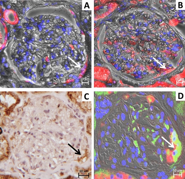
Background: Parietal epithelial cells are a heterogeneous population of cells located on Bowman's capsule. These cells are known to internalize albumin with a still undetermined mechanism, although albumin has been shown to induce phenotypic changes in parietal epithelial cells. Proximal tubular cells are the main actors in albumin handling via the macromolecular complex composed by ClC-5, megalin, and cubilin. This study investigated the role of ClC-5, megalin, and cubilin in the parietal epithelial cells of kidney biopsies from proteinuric lupus nephritis patients and control subjects and identified phenotypical changes occurring in the pathological milieu.

Methods: Immunohistochemistry and immunofluorescence analyses for ClC-5, megalin, cubilin, ANXA3, podocalyxin, CD24, CD44, HSA, and LTA marker were performed on 23 kidney biopsies from patients with Lupus Nephritis and 9 control biopsies (obtained from nephrectomies for renal cancer).

Results: Two sub-populations of hypertrophic parietal epithelial cells ANXA3+/Podocalyxin-/CD44-, both expressing ClC-5, megalin, and cubilin and located at the tubular pole, were identified and characterized: the first one, CD24+/HSA-/LTA- had characteristics of human adult parietal epithelial multipotent progenitors, the second one, CD24-/LTA+/HSA+ committed to become phenotypically proximal tubular cells. The number of glomeruli presenting hypertrophic parietal epithelial cells positive for ClC-5, megalin, and cubilin were significantly higher in lupus nephritis patients than in controls.

Conclusions: Our results may provide further insight into the role of hypertrophic parietal epithelial cells located at the tubular pole and their possible involvement in protein endocytosis in lupus nephritis patients. These data also suggest that the presence of hypertrophic parietal epithelial cells in Bowman's capsule represents a potential resource for responding to protein overload observed in other glomerulonephritis.

Keywords: ClC-5; Cubilin; Hypertrophic PECs; Kidney biopsy; Megalin.


Conflict of interest statement

All the authors declare no competing interests.

Figures

Fig. 1
Fig. 1
Phenotypic characterization of PECs in kidney biopsies from LN patients: identification of the CD24+ subpopulation expressing the protein uptake machinery. Representative images of serial sections of glomerulus with hypertrophic PECs identified by arrows. PECs A ANXA3+ (red: CFL647) showing positivity also for B megalin (red: CFL647), C cubilin (green: fluorescein isothiocyanate [FITC], D ClC-5 (brown: DAB), G CD24 (green: Alexa Fluor 488). Representative immunofluorescence images showing PECs negative for E podocalyxin (green: Alexa Fluor 488), F CD44 (green: Alexa Fluor 488) and H HSA (red: CFL647). Nuclei were counterstained with DAPI (IF images blue), and Mayer hematoxylin (IHC image blue). Immunofluorescence magnification 40X/0.6, IHC magnification 20X/0.45. Scale bar 10 µm
Fig. 2
Fig. 2
Phenotypic characterization of PECs in control kidney biopsies: identification of the CD24+ subpopulation expressing the protein uptake machinery. Representative images of serial sections of glomerulus with hypertrophic PECs identified by arrows. Immunofluorescence microscopy demonstrating positivity for A ANXA3+ (red: CFL647) and B megalin (red: CFL647), magnification 40X/0.6. C Immunohistochemistry staining showing positive signal for ClC-5 (brown: DAB), magnification 20X/0.45. D Confocal image showing co-localization between megalin (red: CFL647) and CD24 (green: Alexa Fluor 488) in yellow/orange PECs, magnification 63X/1.4. Nuclei were counterstained with DAPI (IF images blue), and Mayer hematoxylin (IHC image blue). Scale bar 10 µm
Fig. 3
Fig. 3
Phenotypic characterization of PECs in kidney biopsies from proteinuric patients: identification of a subpopulation of CD24 PECs committed to PTC phenotype expressing the protein uptake machinery. Representative images of serial sections of glomerulus with hypertrophic PECs identified by arrows. A Immunofluorescence image showing co-localization between megalin (red: CFL647) and cubilin (green: FITC) in yellow/orange PECs, magnification 40X/0.6. B Confocal image showing co-localization between megalin (red: CFL647) and LTA (green: FITC) in yellow/orange PECs, magnification 63X/1.4. C Immunofluorescence microscopy showing the presence of HSA (red: CFL647) and D absence of CD24 signal (green: Alexa Fluor 488) in the same PECs, magnification 40X/0.6. Nuclei were counterstained with DAPI (Blue). Scale bar 10 µm

References

    1. Ronconi E, Sagrinati C, Angelotti ML, et al. Regeneration of glomerular podocytes by human renal progenitors. J Am Soc Nephrol JASN. 2009;20:322–332. doi: 10.1681/ASN.2008070709. - DOI - PMC - PubMed
    1. Smeets B, Angelotti ML, Rizzo P, et al. Renal progenitor cells contribute to hyperplastic lesions of podocytopathies and crescentic glomerulonephritis. J Am Soc Nephrol JASN. 2009;20:2593–2603. doi: 10.1681/ASN.2009020132. - DOI - PMC - PubMed
    1. Kuppe C, Leuchtle K, Wagner A, et al. Novel parietal epithelial cell subpopulations contribute to focal segmental glomerulosclerosis and glomerular tip lesions. Kidney Int. 2019;96:80–93. doi: 10.1016/j.kint.2019.01.037. - DOI - PMC - PubMed
    1. D’Agati VD, Shankland SJ. Recognizing diversity in parietal epithelial cells. Kidney Int. 2019;96:16–19. doi: 10.1016/j.kint.2019.02.036. - DOI - PubMed
    1. Sagrinati C, Netti GS, Mazzinghi B, et al. Isolation and characterization of multipotent progenitor cells from the Bowman’s capsule of adult human kidneys. J Am Soc Nephrol JASN. 2006;17:2443–2456. doi: 10.1681/ASN.2006010089. - DOI - PubMed

Publication types

MeSH terms

Substances

LinkOut - more resources