. 2023 Aug 18;9(33):eadg6262.
doi: 10.1126/sciadv.adg6262. Epub 2023 Aug 18.

Commensal Cutibacterium acnes induce epidermal lipid synthesis important for skin barrier function

Affiliations

Commensal Cutibacterium acnes induce epidermal lipid synthesis important for skin barrier function

Samia Almoughrabie et al. Sci Adv. .

Abstract

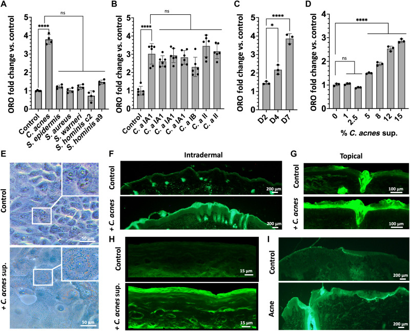
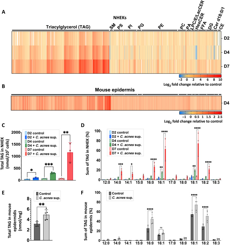
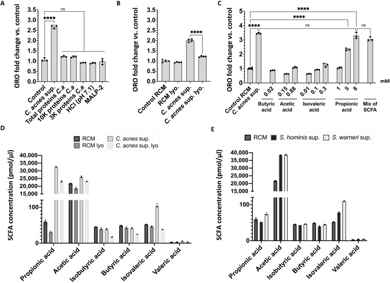
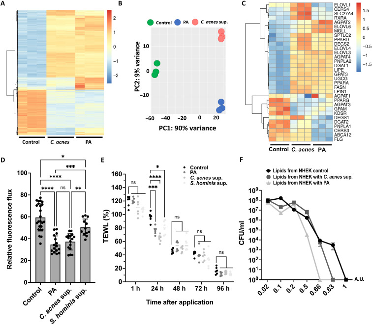
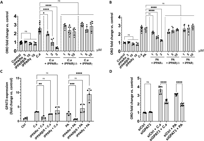
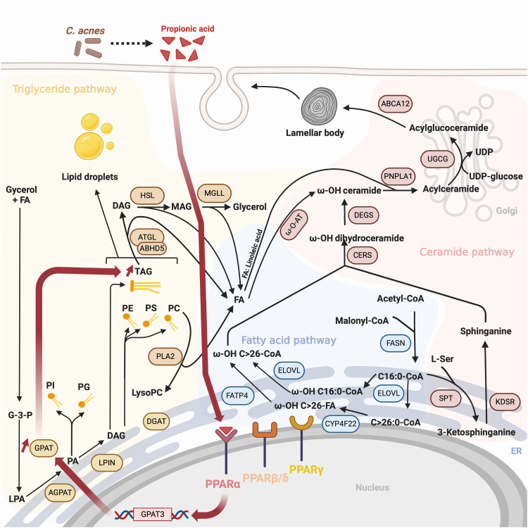
Lipid synthesis is necessary for formation of epithelial barriers and homeostasis with external microbes. An analysis of the response of human keratinocytes to several different commensal bacteria on the skin revealed that Cutibacterium acnes induced a large increase in essential lipids including triglycerides, ceramides, cholesterol, and free fatty acids. A similar response occurred in mouse epidermis and in human skin affected with acne. Further analysis showed that this increase in lipids was mediated by short-chain fatty acids produced by Cutibacterium acnes and was dependent on increased expression of several lipid synthesis genes including glycerol-3-phosphate-acyltransferase-3. Inhibition or RNA silencing of peroxisome proliferator-activated receptor-α (PPARα), but not PPARβ and PPARγ, blocked this response. The increase in keratinocyte lipid content improved innate barrier functions including antimicrobial activity, paracellular diffusion, and transepidermal water loss. These results reveal that metabolites from a common commensal bacterium have a previously unappreciated influence on the composition of epidermal lipids.


Figures

Fig. 1.
Fig. 1.. C. acnes induces increased lipid staining in mouse and human skin.
(A) Oil Red O staining (ORO) of total lipids in NHEKs after treatment with sterile supernatants from media conditioned by the growth of different species of bacteria commonly found on human skin or (B) different C. acnes strains (C. a). (C) Oil Red O quantification of total lipids in NHEKs at days 2 (D2), 4 (D4), and 7 (D7) after exposure to 15% of sterile supernatant from C. acnes CM. (D) Dose-response at day 4 Oil Red O staining of total lipids in NHEK after treatment with different concentrations of sterile supernatant from C. acnes conditioned culture media. (E) Oil Red O lipid staining of NHEKs with and without sterile supernatant from C. acnes conditioned culture media. (F) Bodipy lipid staining of mouse skin after intradermal injection of 107 CFU/ml of C. acnes or (G) topical application of 107 CFU/cm2 of C. acnes on mice. (H) Bodipy lipid staining of reconstructed human epidermis treated with or without sterile supernatant from C. acnes CM. (I) Bodipy lipid staining of an acne lesion and nonlesional human skin. Experiments conducted were performed at least in triplicate. Not significant (ns) = P > 0.05, *P < 0.05, ****P < 0.0001.
Fig. 2.
Fig. 2.. C. acnes induces an increase of triglycerides in mice and human keratinocytes.
(A) DMS-based shotgun lipidomic analysis of extracted lipids from normal NHEKs after exposure of C. acnes CM for 2, 4, and 7 days (n = 3) and (B) from mouse epidermis exposed to live C. acnes at day 4 (n = 6). (C) Total TAG content in NHEKs and (E) in mouse skin treated as in (A) and (B). (D) Fatty acid composition analysis of TAG from NHEK and (F) from mouse epidermis treated as in (A) and (B). CE, cholesterol esters; Cer d18:1, ceramides; DG, diacylglycerols; Cer d18:0, dihydroceramides; FFA, free fatty acids; HexCER, hexosyl ceramides; LacCER, lactosyl ceramides; LPC, lysophosphatidylcholine; LPE, lysophosphatidylethanolamine; PA, phosphatidic acid; PC, phosphatidylcholine; PE, phosphatidylethanolamine; PG, phosphatidylglycerol; PI, phosphatidylinositol; PS, phosphatidylserine; SM, sphingomyelin. Lipidomic analysis has been performed in triplicate for each time point in NHEKs and in six replicates in mouse epidermis. *P < 0.05, **P < 0.01, ***P < 0.001, ****P < 0.0001.
Fig. 3.
Fig. 3.. Propionic acid produced by C. acnes induces lipid accumulation in keratinocytes.
(A) Oil Red O quantification at day 4 in NHEKs after adding sterile CM from C. acnes supernatant (C.a) or proteins from C. acnes media precipitated with ammonium sulfate (85% saturation) or ultrafiltrates from C. acnes media at less than 10 and 3 kDa or media adjusted to pH 7.1 with hydrochloric acid (HCl) or Malp-2 (100 ng/ml). (B) Oil Red O quantification of NHEK at day 4 after adding 15% of C. acnes conditioned culture media supernatant or media control without C. acnes [reinforced clostridial medium (RCM)] or C. acnes CM after lyophilization (lyo.) or nonlyophilized. (C) Oil Red O quantification of NHEK at day 4 after adding different concentration of SCFA alone or in a mix containing 8 mM propionic acid, 0.6 mM acetic acid, 0.01 mM isovaleric acid, and 0.02 mM butyric acid. (D and E) SCFA composition of bacterial supernatants by gas chromatography–mass spectrometry. Experiments conducted were performed at least in triplicate. ****P < 0.0001.
Fig. 4.
Fig. 4.. C. acnes and propionic acid increase expression of lipid synthesis genes and enhance the barrier and the antimicrobial function of keratinocytes.
(A) RNA-seq heatmap showing the top 2000 genes differentially expressed in NHEKs in response to exposure to 15% of C. acnes CM or 8 mM propionic acid for 4 days. (B) PCA of the normalized RNA-seq data. (C) Heatmap showing effect of C. acnes CM or propionic acid (PA) on genes involved in the lipid. (D) Paracellular diffusion of FD-4 solution (1.25 mg/ml) after exposure of confluent NHEK to 8 mM propionic acid or 15% of C. acnes supernatant or S. hominis supernatant. (E) Transepidermal water loss (TEWL) on tape-stripped mouse skin after applying C. acnes supernatant or 8 mM propionic acid topically. (F) Growth curves of C. acnes treated with different concentrations of lipids extracted from NHEK after exposing to 15% C. acnes supernatant or 8 mM propionic acid for 4 days. Note that the heatmaps were scaled by row. Experiments conducted were performed at least in triplicate. A.U., arbitrary unit. *P < 0.05, **P < 0.01, ***P < 0.001, and ****P < 0.0001.
Fig. 5.
Fig. 5.. Lipid induction by propionic acid involves PPARs and the activity of GPAT3.
(A) Oil Red O lipid quantification in NHEKs after exposure to sterile conditioned C. acnes supernatant (C.a) or (B) of 8 mM propionic acid (PA) with different concentrations of inhibitors for PPARα (GW6471), PPARβ/δ (GSK3787), and PPARγ (T0070907). (C) Expression of GPAT3 measured by qPCR in NHEKs after adding 15% C. acnes supernatant (C.a) or 8 mM PA and PPARα inhibitor at 3 μM or PPARβ/δ and PPARγ inhibitors at 10 μM (D) Oil Red O lipid quantification in NHEK after exposure to sterile conditioned 15% C. acnes supernatant or 8 mM PA after silencing GPAT3. Experiments conducted were performed at least in four replicates. *P < 0.05, **P < 0.01, ***P < 0.001, ****P < 0.0001.
Fig. 6.
Fig. 6.. Proposed model of the influence of C. acnes on triglyceride synthesis in NHEKs and relationship to other lipids that form the epidermal barrier.
Illustration of lipid synthesis response to C. acnes SCFAs such as propionic acid. Dark red bold arrows show enzymatic steps and products induced by activation of PPARα. ER (Endoplasmic Reticulum), LPA (Lysophosphatidic acid), UDP (Uridine Diphosphate).

References

    1. P. M. Elias, Structure and function of the stratum corneum permeability barrier. Drug Dev. Res. 13, 97–105 (1988).
    1. G. K. Menon, G. W. Cleary, M. E. Lane, The structure and function of the stratum corneum. Int. J. Pharm. 435, 3–9 (2012). - PubMed
    1. T. A. Harris-Tryon, E. A. Grice, Microbiota and maintenance of skin barrier function. Science 376, 940–945 (2022). - PubMed
    1. K. R. Feingold, Y. J. Jiang, The mechanisms by which lipids coordinately regulate the formation of the protein and lipid domains of the stratum corneum. Dermatoendocrinol. 3, 113–118 (2011). - PMC - PubMed
    1. B. MacLean, D. M. Tomazela, N. Shulman, M. Chambers, G. L. Finney, B. Frewen, R. Kern, D. L. Tabb, D. C. Liebler, M. J. MacCoss, Skyline: An open source document editor for creating and analyzing targeted proteomics experiments. Bioinformatics 26, 966–968 (2010). - PMC - PubMed