Review
. 2023 Oct:123:110719.
doi: 10.1016/j.intimp.2023.110719. Epub 2023 Aug 16.

Etiology of lipid-laden macrophages in the lung

Affiliations
Review

Etiology of lipid-laden macrophages in the lung

E R Stevenson et al. Int Immunopharmacol. 2023 Oct.

Abstract

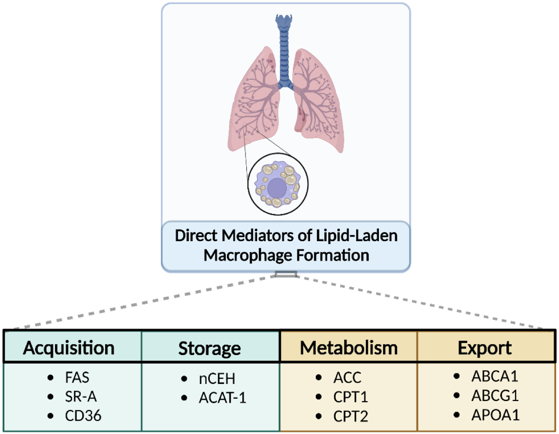
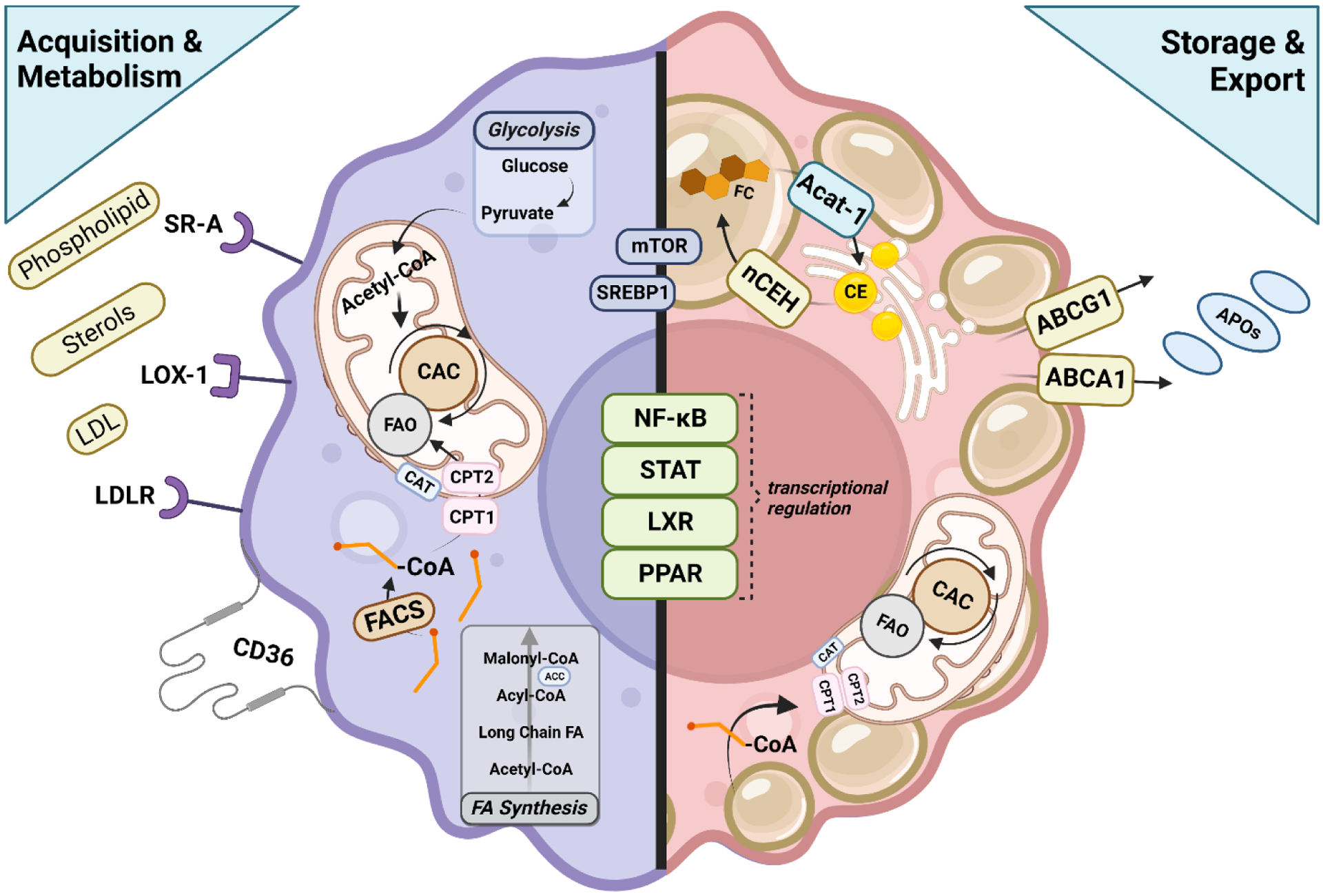
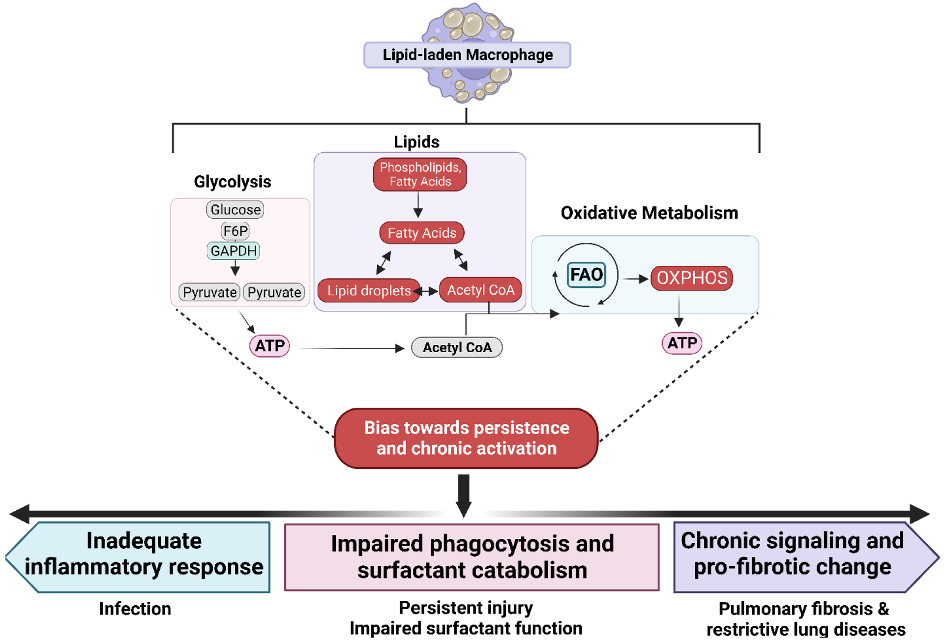
Uniquely positioned as sentinel cells constantly exposed to the environment, pulmonary macrophages are vital for the maintenance of the lung lining. These cells are responsible for the clearance of xenobiotics, pathogen detection and clearance, and homeostatic functions such as surfactant recycling. Among the spectrum of phenotypes that may be expressed by macrophages in the lung, the pulmonary lipid-laden phenotype is less commonly studied in comparison to its circulatory counterpart, the atherosclerotic lesion-associated foam cell, or the acutely activated inflammatory macrophage. Herein, we propose that lipid-laden macrophage formation in the lung is governed by lipid acquisition, storage, metabolism, and export processes. The cellular balance of these four processes is critical to the maintenance of homeostasis and the prevention of aberrant signaling that may contribute to lung pathologies. This review aims to examine mechanisms and signaling pathways that are involved in lipid-laden macrophage formation and the potential consequences of this phenotype in the lung.

Keywords: Foam cell; Immunometabolism; Inflammation; Lipid; Lung; Macrophage.


Conflict of interest statement

Declaration of Competing Interest The authors declare that they have no known competing financial interests or personal relationships that could have appeared to influence the work reported in this paper.

Figures

Figure 1.
Figure 1.
Balance between lipid acquisition, export, metabolism, and storage in pulmonary macrophages are the primary processes that prevent the development of a lipid-laden phenotype. Dysregulation or imbalance of these processes leading to the excess storage of lipid are the most significant when considering the development of this cell phenotype and potential therapeutic intervention in the lung.
Figure 2.
Figure 2.. Cellular processes governing lipid accumulation in alveolar macrophages.
Macrophages acquire lipids and cholesterol through a variety of receptors including SR-A, LOX-1, LDLR and CD36. These cells also acquire lipid through de novo fatty acid synthesis. FACS conjugates free Co-A to the molecule, allowing for the conversion of the fatty acyl CoA to acyl carnitine, which can then be transported across the mitochondria membrane by CAT. CPT2 converts acyl carnitine back to fatty acyl CoA which can then be oxidized and feed into the CAC. NADH, FADH2, and GTP produced from glycolysis/CAC are oxidized and the resulting electrons flow through the complexes of the electron transport chain, creating a protein gradient that drives ATP synthase to produce usable energy for the cell in the form of ATP. Upregulated storage of lipids in the macrophage drive sterol-sensitive signaling pathways, some of which oppose lipid accumulation. LXR signaling induces the transcription of cholesterol efflux transporters like ABCG1 and ABCA1 to promote lipid homeostasis within the cell, similar to PPARα-mediated reduction in triglyceride levels. PPARγ signaling increases glucose metabolism and upregulates the expression of CD36, promoting the uptake of lipids in the macrophage and, along with STAT6, also induces FAO. Contributing to increased substrate availability for FAO, NFκB controls the transcription of pro-inflammatory genes and contributes to macrophage signaling, including activation of SREBP1 which promotes lipogenesis, a process also controlled by mTOR signaling. Importantly, the cell must also have mechanisms to store lipid to be oxidized at a later time for energy and to counteract free cholesterol-induced cytotoxicity, thus storage of these molecules is vitally important. Free cholesterol is esterified by ACAT-1, forming CE, the critical reaction leading to lipid droplet formation in the macrophage. This action is opposed by nCEH which releases free cholesterol for export for loading onto various apolipoprotein carriers.
Figure 3.
Figure 3.
Persistence of lipid laden macrophages in the lung may lead to dampened inflammation and fibrotic change. Due to excess lipid accumulation in macrophages of this phenotype, it is proposed that there is significant reliance on fatty acid oxidation (FAO) rather than glycolysis which is typical of an acute inflammatory response. Increased reliance on these processes is represented by red boxes. The downstream effects of these changes are proposed to bias towards chronic activation, which may ultimately lead to an inadequate inflammatory response upon stimulation, priming the lung for susceptibility to infection and persistent injury. Furthermore, this may impair the phagocytic capacity of the macrophage, leading to both ineffective inflammation and poor surfactant catabolism (pale blue box), further negatively impacting lung function. Persistence of this phenotype may also contribute to persistence of this traditionally “anti-inflammatory phenotype,” leading to pro-fibrotic signaling and collagen deposition in the lung (purple box), leading to fibrosis or other restrictive lung diseases.

References

    1. Bain CC & MacDonald AS The impact of the lung environment on macrophage development, activation and function: diversity in the face of adversity. Mucosal Immunol 15, 223–234 (2022). 10.1038/s41385-021-00480-w - DOI - PMC - PubMed
    1. Laskin DL et al. Macrophages, reactive nitrogen species, and lung injury. Ann N Y Acad Sci 1203, 60–65 (2010). 10.1111/j.1749-6632.2010.05607.x - DOI - PMC - PubMed
    1. Shibata N & Glass CK Macrophages, oxysterols and atherosclerosis. Circ J 74, 2045–2051 (2010). 10.1253/circj.cj-10-0860 - DOI - PubMed
    1. Aluganti Narasimhulu C et al. Atherosclerosis--do we know enough already to prevent it? Curr Opin Pharmacol 27, 92–102 (2016). 10.1016/j.coph.2016.02.006 - DOI - PubMed
    1. Gowdy KM & Fessler MB Emerging roles for cholesterol and lipoproteins in lung disease. Pulm Pharmacol Ther 26, 430–437 (2013). 10.1016/j.pupt.2012.06.002 - DOI - PMC - PubMed

LinkOut - more resources