. 2023 Sep;64(9):100427.
doi: 10.1016/j.jlr.2023.100427. Epub 2023 Aug 16.

Metabolic changes and propensity for inflammation, fibrosis, and cancer in livers of mice lacking lysosomal acid lipase

Affiliations

Metabolic changes and propensity for inflammation, fibrosis, and cancer in livers of mice lacking lysosomal acid lipase

Ivan Bradić et al. J Lipid Res. 2023 Sep.

Abstract

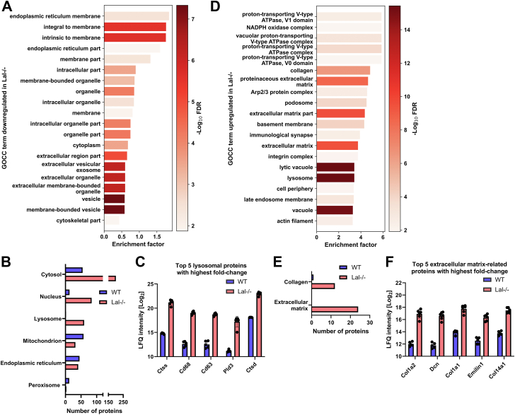
Lysosomal acid lipase (LAL) is the sole lysosomal enzyme responsible for the degradation of cholesteryl esters and triacylglycerols at acidic pH. Impaired LAL activity leads to LAL deficiency (LAL-D), a severe and fatal disease characterized by ectopic lysosomal lipid accumulation. Reduced LAL activity also contributes to the development and progression of non-alcoholic fatty liver disease (NAFLD). To advance our understanding of LAL-related liver pathologies, we performed comprehensive proteomic profiling of livers from mice with systemic genetic loss of LAL (Lal-/-) and from mice with hepatocyte-specific LAL-D (hepLal-/-). Lal-/- mice exhibited drastic proteome alterations, including dysregulation of multiple proteins related to metabolism, inflammation, liver fibrosis, and cancer. Global loss of LAL activity impaired both acidic and neutral lipase activities and resulted in hepatic lipid accumulation, indicating a complete metabolic shift in Lal-/- livers. Hepatic inflammation and immune cell infiltration were evident, with numerous upregulated inflammation-related gene ontology biological process terms. In contrast, both young and mature hepLal-/- mice displayed only minor changes in the liver proteome, suggesting that loss of LAL solely in hepatocytes does not phenocopy metabolic alterations observed in mice globally lacking LAL. These findings provide valuable insights into the mechanisms underlying liver dysfunction in LAL-D and may help in understanding why decreased LAL activity contributes to NAFLD. Our study highlights the importance of LAL in maintaining liver homeostasis and demonstrates the drastic consequences of its global deficiency on the liver proteome and liver function.

Keywords: cholesterol; cholesterol ester storage disease; lipase/lysosomal; lipid metabolism; lipids; liver; lysosomal storage disorder; non-alcoholic fatty liver disease; proteomics.


Conflict of interest statement

Conflict of interest The authors declare that they have no known competing financial interests or personal relationships that could have appeared to influence the work reported in this paper.

Figures

None
Graphical abstract
Fig. 1
Fig. 1
Significant proteome alterations in livers of Lal-deficient (Lal−/−) mice. A: Experimental setup of 4 h fasting of wild-type (WT) and Lal−/− mice followed by an oral oil bolus and sacrifice 2 h post-gavage. B: Quantified total proteome depth in wild-type (WT) and Lal−/− mice livers. C: Dynamic range of the liver proteome from WT and Lal−/− mice based on log10 of the mean intensity of relative label-free quantification (LFQ), ordered by rank of abundance. D: Volcano plot of the liver proteome showing 480 significantly upregulated and 234 significantly downregulated proteins in Lal−/− mice (FDR < 0.01, S0 = 2). Figures represent data from 5 WT and 6 Lal−/− mice. Data are presented as mean ± SD.
Fig. 2
Fig. 2
LAL deficiency impairs hepatic glucose and lipid metabolism. A: Top 20 gene ontology biological process (GOBP) terms downregulated in livers of Lal−/− mice. B: Significantly changed proteins from WT and Lal−/− livers annotated to selected carbohydrate metabolism-related UniProt keyword terms. C: Top five proteins with highest fold change annotated to Glycolysis UniProt keyword. D: Significantly changed proteins from WT and Lal−/− livers annotated to selected lipid metabolism-related UniProt keyword terms. Top five proteins with the highest fold change annotated to (E) Cholesterol metabolism, (F) Fatty acid metabolism, and (G) Hydrolase UniProt keywords. All figures represent data from 5 WT and 6 Lal−/− mice. Data are presented as mean or as mean ± SD.
Fig. 3
Fig. 3
Impaired lipid hydrolysis in livers of Lal−/− mice. Liver tissue was isolated from WT and Lal−/− mice after 4 h of fasting and after an oral lipid bolus. Acid (pH 4.5) (A) cholesteryl ester hydrolase (CEH) and (B) triacylglycerol (TG) hydrolase (TGH) activities (n = 3–4), neutral (pH 7) (C) CEH and (D) TGH activities (n = 3–4), hepatic (E) total cholesterol (TC) and (F) TG concentrations (n = 3–4), and plasma (G) TC, and (H) TG concentrations (n = 3–10). Statistically significant differences were calculated by 2-way ANOVA followed by Tukey’s post hoc test. ∗P < 0.05, ∗∗P ≤ 0.01, ∗∗∗P ≤ 0.001, and ∗∗∗∗P ≤ 0.0001 for comparison between different genotypes within the same group (WT vs. Lal−/−, WT fasted vs. Lal−/− fasted). ###P ≤ 0.001 for comparison between the different conditions in the same genotypes (WT fasted vs. WT, Lal−/− fasted vs. Lal−/−). Data are presented as mean ± SD.
Fig. 4
Fig. 4
LAL deficiency is associated with pronounced liver inflammation. A: Top 20 gene ontology biological process (GOBP) terms upregulated in the liver of Lal−/− mice. B: Significantly changed proteins from WT and Lal−/− livers annotated to selected UniProt keyword terms. C: Top five proteins with the highest fold change annotated to the UniProt keyword Inflammatory Response. D: Top five proteins with the highest fold change annotated to the UniProt keyword Immunity. All figures represent data from 5 WT and 6 Lal−/− mice. Data are presented as mean or as mean ± SD.
Fig. 5
Fig. 5
LAL deficiency causes major changes in liver cellular components. A: Top 20 gene ontology cellular component (GOCC) terms downregulated in livers of Lal−/− mice. B: Significantly changed proteins from WT and Lal−/− livers annotated to selected UniProt keyword terms representing various cellular compartments. C: Top five proteins with the highest fold change annotated to the UniProt keyword Lysosome. D: Top 20 GOCC terms upregulated in livers of Lal−/− mice. E: Significantly changed proteins from WT and Lal−/− livers annotated to selected UniProt keyword terms representing extracellular matrix. F: Top five proteins with the highest fold change annotated to the UniProt keyword Extracellular Matrix. All figures represent data from 5 WT and 6 Lal−/− mice. Data are presented as mean or as mean ± SD.
Fig. 6
Fig. 6
Minor proteome alterations in livers of young and mature hepatocyte-specific Lal−/− (hepLal−/−) mice. A: Body weight and (B) liver-to-body weight ratio of young (9–11 weeks of age) and mature (50–60 weeks of age) WT and hepLal−/− mice (n = 4–6). C: Quantified total proteome depth in livers from young (n = 5) and mature (n = 5–6) WT and hepLal−/− mice. D: Volcano plot of the liver proteome showing 11 significantly upregulated and four significantly downregulated proteins in young hepLal−/− mice (n = 5, FDR < 0.05, S0 = 0.1). E: Volcano plot of the liver proteome showing three significantly upregulated and 18 significantly downregulated proteins in mature hepLal−/− mice (n = 5–6, FDR < 0.05, S0 = 0.1). Kyoto encyclopedia of genes and genomes (KEGG) pathways enriched in livers from (F) young (n = 5) and (G) mature (n = 5–6) WT and hepLal−/− mice. Statistically significant differences for (A–C) were calculated by 2-way ANOVA followed by Tukey’s post-hoc test. ∗P < 0.05 for comparison between different genotypes within the same group (young WT vs. young hepLal−/−, mature WT vs. mature hepLal−/−). ####P ≤ 0.001 for comparison between the different conditions in the same genotypes (young WT vs. mature WT, young hepLal−/− vs. mature hepLal−/−). Data are presented as mean ± SD.

Comment in

Similar articles

Cited by

References

    1. Goldstein J.L., Dana S.E., Faust J.R., Beaudet A.L., Brown M.S. Role of lysosomal acid lipase in the metabolism of plasma low density lipoprotein. Observations in cultured fibroblasts from a patient with cholesteryl ester storage disease. J. Biol. Chem. 1975;250:8487–8495. - PubMed
    1. Ouimet M., Franklin V., Mak E., Liao X., Tabas I., Marcel Y.L. Autophagy regulates cholesterol efflux from macrophage foam cells via lysosomal acid lipase. Cell Metab. 2011;13:655–667. - PMC - PubMed
    1. Bernstein D.L., Hulkova H., Bialer M.G., Desnick R.J. Cholesteryl ester storage disease: review of the findings in 135 reported patients with an underdiagnosed disease. J. Hepatol. 2013;58:1230–1243. - PubMed
    1. Carter A., Brackley S.M., Gao J., Mann J.P. The global prevalence and genetic spectrum of lysosomal acid lipase deficiency: a rare condition that mimics NAFLD. J. Hepatol. 2019;70:142–150. - PubMed
    1. Reiner Z., Guardamagna O., Nair D., Soran H., Hovingh K., Bertolini S., et al. Lysosomal acid lipase deficiency--an under-recognized cause of dyslipidaemia and liver dysfunction. Atherosclerosis. 2014;235:21–30. - PubMed

Publication types