Review
. 2023 Oct 10;101(15):e1483-e1494.
doi: 10.1212/WNL.0000000000207701. Epub 2023 Aug 18.

Mapping Essential Tremor to a Common Brain Network Using Functional Connectivity Analysis

Affiliations
Review

Mapping Essential Tremor to a Common Brain Network Using Functional Connectivity Analysis

Ellen Younger et al. Neurology. .

Erratum in

Abstract

Background and objectives: The cerebello-thalamo-cortical circuit plays a critical role in essential tremor (ET). However, abnormalities have been reported in multiple brain regions outside this circuit, leading to inconsistent characterization of ET pathophysiology. Here, we test whether these mixed findings in ET localize to a common functional network and whether this network has therapeutic relevance.

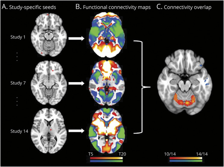
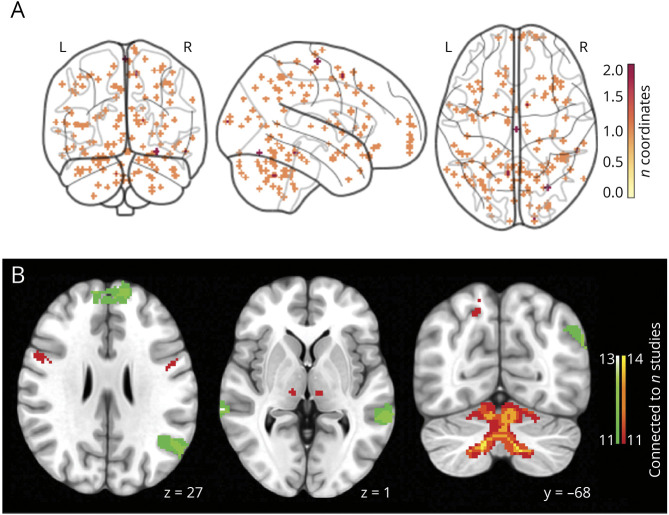
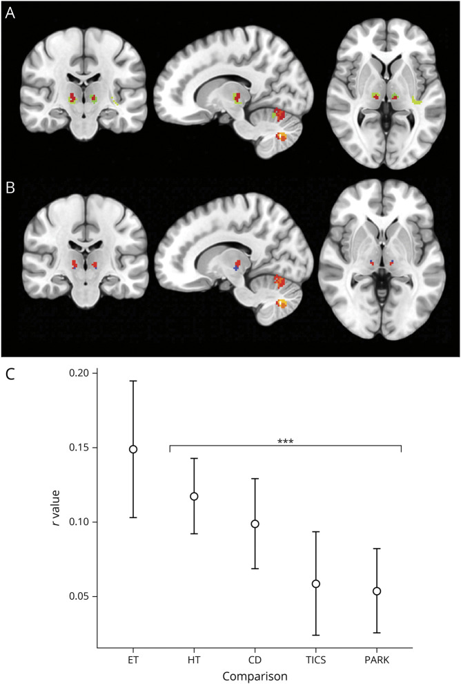
Methods: We conducted a systematic literature search to identify studies reporting structural or metabolic brain abnormalities in ET. We then used 'coordinate network mapping,' which leverages a normative connectome (n = 1,000) of resting-state fMRI data to identify regions commonly connected to findings across all studies. To assess whether these regions may be relevant for the treatment of ET, we compared our network with a therapeutic network derived from lesions that relieved ET. Finally, we investigated whether the functional connectivity of this ET symptom network is abnormal in an independent cohort of patients with ET as compared with healthy controls.

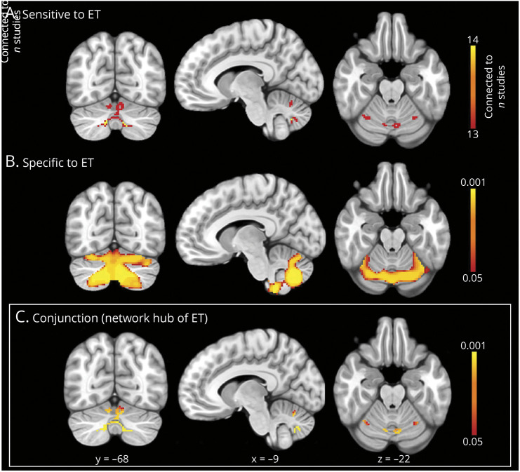
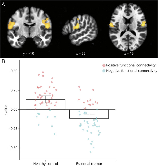
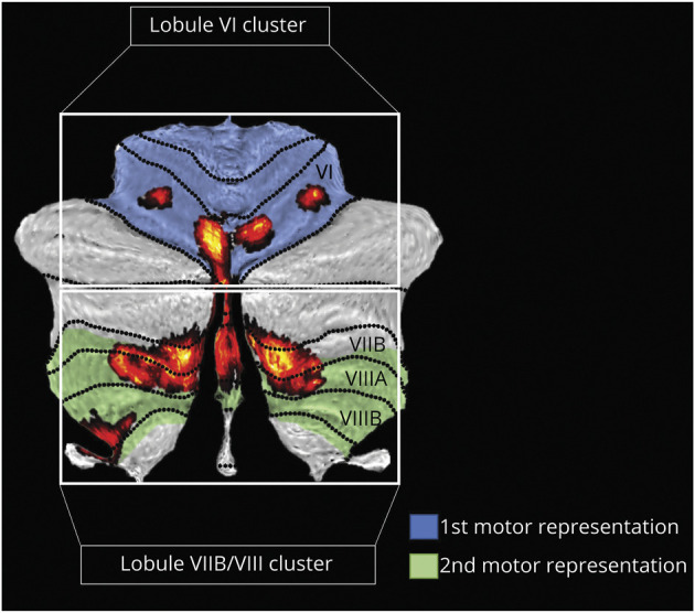
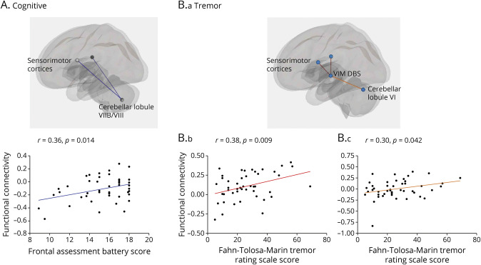
Results: Structural and metabolic brain abnormalities in ET were located in heterogeneous regions throughout the brain. However, these coordinates were connected to a common functional brain network, including the cerebellum, thalamus, motor cortex, precuneus, inferior parietal lobe, and insula. The cerebellum was identified as the hub of this network because it was the only brain region that was both functionally connected to the findings of over 90% of studies and significantly different in connectivity compared with a control data set of other movement disorders. This network was strikingly similar to the therapeutic network derived from lesions improving ET, with key regions aligning in the thalamus and cerebellum. Furthermore, positive functional connectivity between the cerebellar network hub and the sensorimotor cortices was significantly reduced in patients with ET compared with healthy controls, and connectivity within this network was correlated with tremor severity and cognitive functioning.

Discussion: These findings suggest that the cerebellum is the central hub of a network commonly connected to structural and metabolic abnormalities in ET. This network may have therapeutic utility in refining and informing new targets for neuromodulation of ET.


Conflict of interest statement

E.F.P. Younger, E.G. Ellis and N. Parsons are funded by the Deakin University Postgraduate Research Scholarship; P. Pantano has received funding for travel from Novartis, Genzyme, and Bracco and a speaking honorarium from Biogen, research support from Italian Ministry of Foreign Affairs and Fondazione Italiana Sclerosi Multipla; K. Caeyenberghs is supported by a Veski Fellowship, the Victorian Near-miss Award Pilot is administered by Veski for the Victorian Health and Medical Research Workforce Project on behalf of the Victorian Government and the Association of Australian Medical Research Institutes, funding for the Pilot has been provided by the Victorian Department of Jobs, Precincts and Regions; J. Benito-Leon is supported by the National Institutes of Health, Bethesda, MD, USA (NINDS #R01 NS39422), the European Commission (grant ICT-2011-287739, NeuroTREMOR), the Ministry of Economy and Competitiveness (grant RTC-2015-3967-1, NetMD—platform for the tracking of movement disorder), and the Spanish Health Research Agency (grant FIS PI12/01602 and grant FIS PI16/00451); J.P. Romero was funded by the Spanish Ministry of Science and Innovation grant (PID2020-113222RB- C21/AEI/10.13039/501100011033); J. Joutsa has received research grants from the Finnish Medical Foundation, Instrumentarium Research Foundation, Sigrid Juselius Foundation, Finnish Foundation for Alcohol Studies, University of Turku (private donation, Sigrid Juselius), Turku University Hospital (ERVA funds), and conference travel support from Abbvie and Abbott, and lecturer honoraria from Lundbeck; D.T. Corp and S. Tommasin report no disclosures relevant to the manuscript. Go to Neurology.org/N for full disclosures.

Figures

Figure 1
Figure 1. Coordinate Network Mapping Method
(A) Study-specific seeds: 4-mm seeds were created at every coordinate reported as significantly abnormal in essential tremor for each included study. (B) Functional connectivity maps: Maps of voxels that were functionally connected to the study-specific seed were generated using a normative human connectome (n = 1,000). (C) Connectivity overlap: Each study's functional connectivity map was thresholded, binarised, and overlaid to identify the regions that are functionally connected with the most study-specific seeds. Cool colors = negative connectivity; warm colors = positive connectivity.
Figure 2
Figure 2. Coordinate Network Mapping Results
(A) Locations of structural or metabolic abnormality in essential tremor. 150 coordinates of significant structural or metabolic differences between patients with ET and controls, extracted from 14 neuroimaging studies. Reported areas of abnormality were widely dispersed across the brain. (B) The network of essential tremor. The coordinate network mapping analysis revealed a network of regions sensitive to essential tremor (connected to 11/14 study-specific seeds). Warm colors = positive connectivity; cool colors = negative connectivity.
Figure 3
Figure 3. Relevance of the Essential Tremor Network to Tremor Relief
(A) Our essential tremor symptom network (red) aligned closely with the tremor relief network (green; Joutsa et al., 2018) in the thalamus and cerebellum. (B) The established deep brain stimulation target (blue) also overlapped with our essential tremor network (red) in the ventral intermediate nucleus of the thalamus. (C) Spatial correlations revealed that our essential tremor symptom network was more similar to the tremor relief network (Joutsa et al., 2018) than networks of a control group of all control disorders. CD = cervical dystonia; ET = essential tremor; HT = Holmes tremor; PARK = Parkinsonism; TICS = Tics, ***p < 0.001.
Figure 4
Figure 4. Defining the Hub of the Essential Tremor Network
(A) Sensitive to ET: Coordinate network mapping found the cerebellum to be most strongly connected to coordinates of brain abnormality in ET (thresholded at 90%, connected to 13/14 study-specific seeds); (B) Specific to ET: A specificity analysis revealed voxels with significantly greater functional connectivity to coordinates of abnormality in essential tremor than control disorders (p < 0.05). (C) Conjunction (network hub of ET): A conjunction analysis identified a network hub that was both sensitive and specific to essential tremor.
Figure 5
Figure 5. Abnormal Functional Connectivity in Essential Tremor
(A) Functional connectivity between the network hub in the cerebellum and the sensorimotor cortices was significantly different in patients with essential tremor compared with healthy controls. (B) Bar charts (mean ± 95% CI for each group) and individual participant connectivity values show that, on average, these regions were positively connected in healthy controls (left) yet negatively functionally connected in patients with essential tremor (right).
Figure 6
Figure 6. Hub of the Essential Tremor Network: Division of Distinct Functional Representations
The cerebellum can be organized based on functional gradients, which includes 2 functionally distinct motor representations: motor representation 1 (shaded blue) and motor representation 2 (shaded green)., When overlaid onto these functional gradients, our network hub of ET (red) falls largely within the 2 motor representations. Therefore, we split the hub into 2 separate clusters along the fissure between lobule VI and VII to investigate whether these portions of the hub may have differential involvement in ET. These 2 hub clusters (red) are referred to as (1) Lobule VI Cluster, and (2) Lobule VIIB/VIII Cluster.
Figure 7
Figure 7. Correlations Between Functional Connectivity and Clinical Symptoms
(A) Cognitive: Functional connectivity between cerebellar lobule VIIB/VIII cluster and sensorimotor cortices was positively correlated with Frontal Assessment Battery scores in patients with essential tremor. (B) Tremor: Fahn-Tolosa-Marin tremor rating scale scores were positively correlated with functional connectivity between the VIM DBS site and the sensorimotor cortices (B.b, red) and with functional connectivity between the VIM DBS site and cerebellar lobule VI cluster (B.c, orange). Visual inspection of the rightmost scatterplot (Figure 7B.c) indicates the presence of a possible outlier with low functional connectivity between lobule VI and the VIM DBS site. The removal of this data point changed the correlation from r = 0.30; p = 0.042 to r = 0.26; p = 0.089. However, this data point fell well below our objective Cook distance cutoff for data point removal (>0.5) (Cook distance = 0.19) and was therefore retained. DBS = deep brain stimulation; VIM = ventral intermediate nucleus of the thalamus.

References

    1. Louis ED, McCreary M. How common is essential tremor? Update on the worldwide prevalence of essential tremor. Tremor Other Hyperkinet Mov (N Y). 2021;11(1):28. doi:10.5334/tohm.632 - DOI - PMC - PubMed
    1. Welton T, Cardoso F, Carr JA, et al. . Essential tremor. Nat Rev Dis Primers. 2021;7(1):83. doi:10.1038/s41572-021-00314-w - DOI - PubMed
    1. Deuschl G, Bain P, Brin M. Consensus statement of the movement disorder society on tremor. Ad Hoc Scientific Committee. Mov Disord. 2008;13(S3):2-23. doi:10.1002/mds.870131303 - DOI - PubMed
    1. Raethjen J, Deuschl G. The oscillating central network of essential tremor. Clin Neurophysiol. 2012;123(1):61-64. doi:10.1016/j.clinph.2011.09.024 - DOI - PubMed
    1. Louis ED, Faust PL. Essential tremor pathology: neurodegeneration and reorganization of neuronal connections. Nat Rev Neurol. 2020;16(2):69-83. doi:10.1038/s41582-019-0302-1 - DOI - PubMed

Publication types