

Applying deep learning to recognize the properties of vitreous opacity in ophthalmic ultrasound images
- PMID: 37596401
- PMCID: PMC10810903
- DOI: 10.1038/s41433-023-02705-7
Applying deep learning to recognize the properties of vitreous opacity in ophthalmic ultrasound images
Abstract
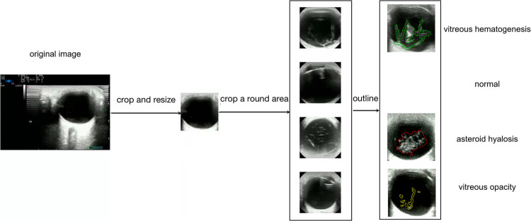
Background: To explore the feasibility of artificial intelligence technology based on deep learning to automatically recognize the properties of vitreous opacities in ophthalmic ultrasound images.
Methods: A total of 2000 greyscale Doppler ultrasound images containing non-pathological eye and three typical vitreous opacities confirmed as physiological vitreous opacity (VO), asteroid hyalosis (AH), and vitreous haemorrhage (VH) were selected and labelled for each lesion type. Five residual networks (ResNet) and two GoogLeNet models were trained to recognize vitreous lesions. Seventy-five percent of the images were randomly selected as the training set, and the remaining 25% were selected as the test set. The accuracy and parameters were recorded and compared among these seven different deep learning (DL) models. The precision, recall, F1 score, and area under the receiver operating characteristic curve (AUC) values for recognizing vitreous lesions were calculated for the most accurate DL model.
Results: These seven DL models had significant differences in terms of their accuracy and parameters. GoogLeNet Inception V1 achieved the highest accuracy (95.5%) and minor parameters (10315580) in vitreous lesion recognition. GoogLeNet Inception V1 achieved precision values of 0.94, 0.94, 0.96, and 0.96, recall values of 0.94, 0.93, 0.97 and 0.98, and F1 scores of 0.94, 0.93, 0.96 and 0.97 for normal, VO, AH, and VH recognition, respectively. The AUC values for these four vitreous lesion types were 0.99, 1.0, 0.99, and 0.99, respectively.
Conclusions: GoogLeNet Inception V1 has shown promising results in ophthalmic ultrasound image recognition. With increasing ultrasound image data, a wide variety of confidential information on eye diseases can be detected automatically by artificial intelligence technology based on deep learning.
© 2023. The Author(s), under exclusive licence to The Royal College of Ophthalmologists.
Conflict of interest statement
The authors declare no competing interests.
Figures


Comment on
-
Clinically applicable deep learning for diagnosis and referral in retinal disease.Nat Med. 2018 Sep;24(9):1342-1350. doi: 10.1038/s41591-018-0107-6. Epub 2018 Aug 13. Nat Med. 2018. PMID: 30104768
References
-
- Duke-Elder S. Diseases of the vitreous body. In: Henry Kimpton SD-E, editors. London. 1969; pp 315–75.
-
- Liu SF, Wang Y, Yang X, Lei BY, Liu L, Li SX, et al. Deep learning in medical ultrasound analysis: a review. Engineering. 2019;5:261–75. doi: 10.1016/j.eng.2018.11.020. - DOI
Publication types
MeSH terms
LinkOut - more resources
Full Text Sources
