. 2023 Aug 18;23(1):582.
doi: 10.1186/s12909-023-04575-9.

An ex vivo model for education and training of bilateral cleft lip surgery

Affiliations

An ex vivo model for education and training of bilateral cleft lip surgery

Rainer Lutz et al. BMC Med Educ. .

Abstract

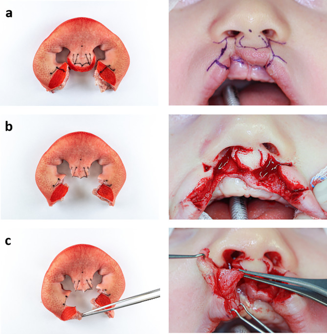
Background: Bilateral cleft lip surgery is very challenging and requires a high level of skill, knowledge and experience. Existing high-fidelity simulation models that can be used by novice cleft surgeons to gain experience and expand their knowledge are rare and expensive. In this study, we developed a bilateral cleft lip model using porcine snout discs, which are available anywhere and inexpensive.

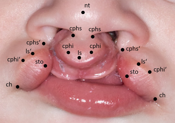
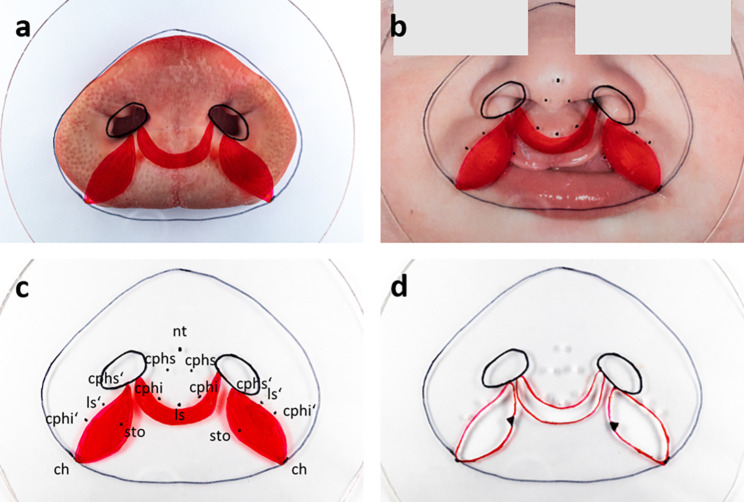
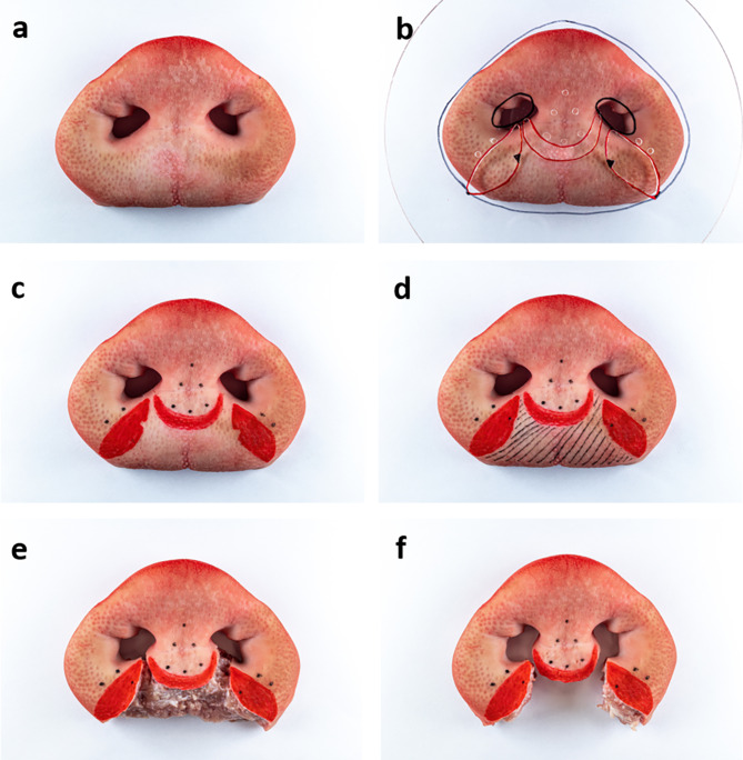
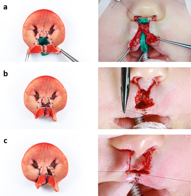
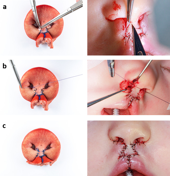
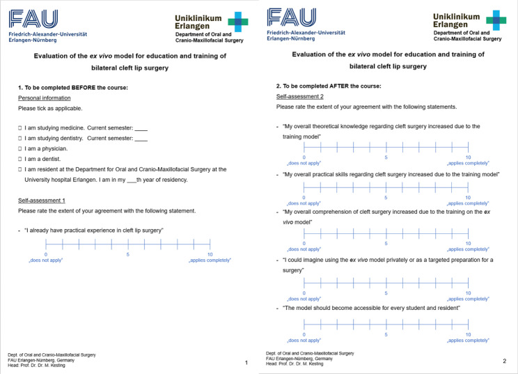
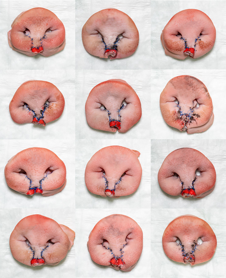
Methods: Anatomic reference points of a patient with a bilateral cleft lip were superimposed with landmarks of the porcine snout disc on a foil template. The template was used to construct an ex vivo bilateral cleft lip model. Surgery was performed on the model according to Millard and the surgical steps were photodocumented analogous to two clinical cases of bilateral cleft lip surgery. The suitability of the model was further tested by twelve participants and evaluated using self-assessment questionnaires.

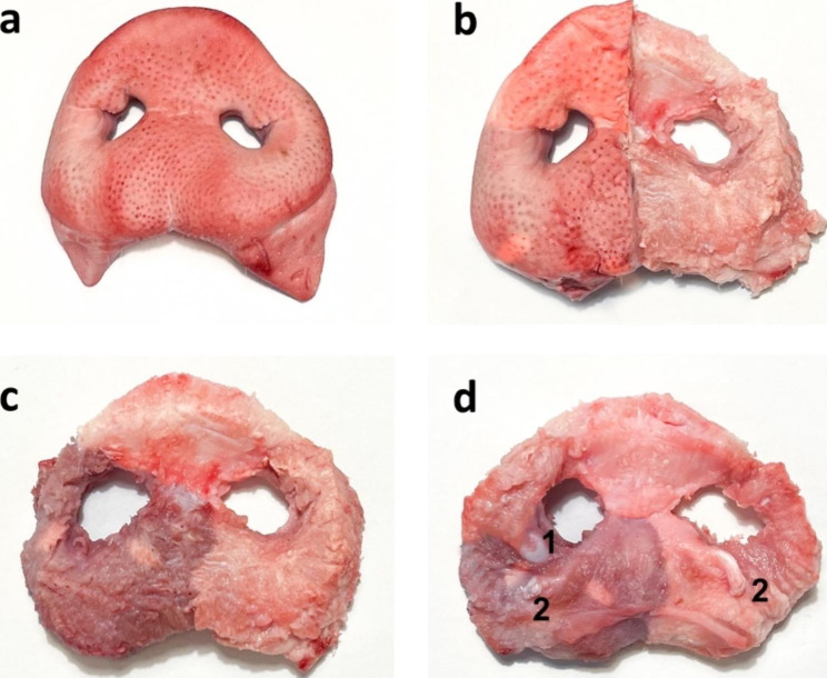
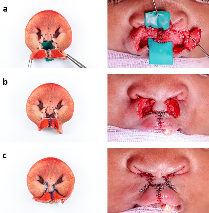
Results: The bilateral cleft lip ex vivo model made of a porcine snout disc proved to be a suitable model with very low cost and ease of fabrication, as the template is reusable on any snout disc. The Millard procedure was successfully performed and the surgical steps of the lip plasty were simulated close to the clinical situation. Regarding the nasal reconstruction, the model lacks three-dimensionality. As a training model, it enhanced the participants comprehension of cleft surgery as well as their surgical skills. All participants rated the model as valuable for teaching and training.

Conclusions: The porcine snout discs can be used as a useful ex vivo model for bilateral cleft lip surgery with limitations in the construction of the nose, which cannot be realistically performed with the model due to anatomical differences with humans. Benefits include a realistic tissue feel, the simulation of a multi-layered lip construction, a wide and rapid availability and low cost. This allows the model to be used by novice surgeons also in low-income countries. It is therefore useful as a training model for gaining experience, but also as a model for refining, testing and evaluating surgical techniques for bilateral lip plasty.

Keywords: Bilateral cleft lip; Cadaver model; Cheiloplasty; Ex vivo model; Lip plasty; Porcine snout disc; Residency training; Skills lab; Surgical simulation; Teaching.


Conflict of interest statement

The authors declare no competing interests.

Figures

Fig. 1
Fig. 1
Anatomic reference points on a picture of a cleft patient with bilateral cleft lip and palate: nasal tip (nt): tip of nose; crista philtri superior (cphs): highest point of philtrum edge; crista philtri inferior (cphi): tip of the cupid’s bow; labiale superius (ls): median (lowest point) of cupid’s bow; stomion (sto): lowest median point of upper lip; cheilion (ch): lateral commissure of the lip
Fig. 2
Fig. 2
Template foil with anatomic landmarks of both snout disc and cleft child: on the porcine snout disc (a), on a double magnified picture of the cleft child with anatomic reference points (b), with labelled reference points (c), final template foil with drill holes (d)
Fig. 3
Fig. 3
Creation of the bilateral cleft lip model: porcine snout disc (a), with template foil (b), after marking the anatomic reference points (c), with sto points marked and cleft area shaded (d), after skin excision (e), after muscle excision: final ex vivo model (f)
Fig. 4
Fig. 4
Anatomy of the porcine snout disc: snout disc before preparation (a), after the skin on the left half of the pig’s snout was removed to show the relationship between skin and muscle (b), after removal of the skin, visualizing the main component of the snout disc: M. nasalis, a muscle which has no attachment to the bony skeleton of the pig’s skull (c), view from the back of the nasal disc: the Cartilago nasi lateralis ventralis (1) and the end tendons of the caninus muscle (2) which is closely interwoven with the levator labii superioris muscle, radiate into the nasal muscle (d)
Fig. 5
Fig. 5
Millard surgery of a bilateral cleft on the ex vivo model (left) and patient A (right), part one: initial situation with drawn incision lines (a), after incisions and vermillion cut out (b), mobilised orbicularis oris muscle (c)
Fig. 6
Fig. 6
Millard surgery of a bilateral cleft on the ex vivo model (left) and patient A (right), part two: suture of the orbicularis oris muscle (a), suture of the vermillion (b), suture fixating the prolabium to the right lip segment on point cphi (c)
Fig. 7
Fig. 7
Millard surgery of a bilateral cleft on the ex vivo model (left) and patient A (right), part three: removing the parking flaps (a), suture of the nasal entrance (b), finished skin suture and final result (c)
Fig. 8
Fig. 8
Millard surgery of a bilateral cleft on the ex vivo model (left) and patient B (right), part one: initial situation with drawn incision lines (a), after incisions and vermillion cut out (b), mobilised orbicularis oris muscle (c)
Fig. 9
Fig. 9
Millard surgery of a bilateral cleft on the ex vivo model (left) and patient B (right), part two: attached orbicularis oris muscle (a), suture of the vermilion, in the patient the prolabium has already been sutured to the vermillion (b), completed suture of the skin (c)
Fig. 10
Fig. 10
Translated version of the applied, self-designed questionnaire
Fig. 11
Fig. 11
Surgical outcomes after Millard surgery on the ex vivo models

References

    1. Khosla RK, McGregor J, Kelley PK, Gruss JS. Contemporary concepts for the bilateral cleft lip and nasal repair. Semin Plast Surg. 2012;26(4):156–63. - PMC - PubMed
    1. Nadjmi N. Primary unilateral cleft lip-nose repair. In: Nadjmi N, editor. Surgical Management of Cleft lip and palate: a Comprehensive Atlas. Cham: Springer International Publishing; 2018. p. 44.
    1. Ghali GE, Ringeman JL. Primary bilateral cleft lip/nose repair using a modified Millard technique. Atlas Oral Maxillofac Surg Clin North Am. 2009;17(2):117–24. doi: 10.1016/j.cxom.2009.05.002. - DOI - PubMed
    1. Millard DR. Jr. Cleft craft: the evolution of its surgery—volume II: bilateral and rare deformities. Boston: Little Brown and Company; 1977.
    1. Mulliken JB, Wu JK, Padwa BL. Repair of bilateral cleft lip: review, revisions, and reflections. J Craniofac Surg. 2003;14(5):609–20. doi: 10.1097/00001665-200309000-00003. - DOI - PubMed