

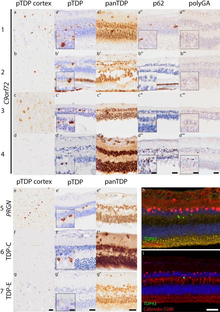
TDP-43 pathology in the retina of patients with frontotemporal lobar degeneration
- PMID: 37597044
- PMCID: PMC10564657
- DOI: 10.1007/s00401-023-02623-8
TDP-43 pathology in the retina of patients with frontotemporal lobar degeneration
Conflict of interest statement
Declarations. Conflict of interest: Anke A. Dijkstra, Tjado H. J. Morrema, Frederique Hart-de Ruijter, Priya Gami-Patel, Frank D. Verbraak, Jurre den Haan, and Annemieke J. Rozemuller report no competing interests; Prof. dr. J. F. de Boer has acquired grant support (for the institution; Department of Physics, VU) from the Dutch Research Council (NWO) and from industry (Thorlabs, ASML, Heidelberg Engineering). He has received royalties related to IP on OCT technologies and semiconductor metrology. He has acted as an expert witness for a UK-based law firm; Dr. F. Bouwman performs contract research for Optina Dx and Optos, she has been an invited speaker at Roche and has been invited for expert testimony at Biogen. All funding is paid to her institution; Dr. J. J. M. Hoozemans received grants from the Dutch Research Council (ZonMW) and, Alzheimer Netherlands, performed contract research or received grants from Merck, ONO Pharmaceuticals, Janssen Prevention Center, DiscovericBio, AxonNeurosciences, Roche, Genentech, Promis, Denali, First Biotherapeutics, and Ensol Biosciences. All payments were made to the institution. Dr. J. J. M. Hoozemans participates in the scientific advisory board of Alzheimer Netherlands and is editor-in-chief for Acta Neuropathologica Communications. Ethical approval: Prior to death donors signed informed consent for brain and eye autopsy and use of brain and eye tissue as well as medical records for research activities. This study was approved by the medical ethical committee of the VUmc (reference# 2009/148).
Figures
References
-
- Goodwill VDC, Chea L, Sigurdson C, Alvarez V, McKee A, Lin J (2020) Chronic traumatic encephalopathy is associated with TDP-43 retinal pathology. https://iovs.arvojournals.org/article.aspx?articleid=2768896
Grants and funding
LinkOut - more resources
Full Text Sources
