. 2023 Aug 19;14(1):5053.
doi: 10.1038/s41467-023-40681-4.

CD98hc is a target for brain delivery of biotherapeutics

Affiliations

CD98hc is a target for brain delivery of biotherapeutics

Kylie S Chew et al. Nat Commun. .

Erratum in

  • Author Correction: CD98hc is a target for brain delivery of biotherapeutics.
    Chew KS, Wells RC, Moshkforoush A, Chan D, Lechtenberg KJ, Tran HL, Chow J, Kim DJ, Robles-Colmenares Y, Srivastava DB, Tong RK, Tong M, Xa K, Yang A, Zhou Y, Akkapeddi P, Annamalai L, Bajc K, Blanchette M, Cherf GM, Earr TK, Gill A, Huynh D, Joy D, Knight KN, Lac D, Leung AW, Lexa KW, Liau NPD, Becerra I, Malfavon M, McInnes J, Nguyen HN, Lozano EI, Pizzo ME, Roche E, Sacayon P, Calvert MEK, Daneman R, Dennis MS, Duque J, Gadkar K, Lewcock JW, Mahon CS, Meisner R, Solanoy H, Thorne RG, Watts RJ, Zuchero YJY, Kariolis MS. Chew KS, et al. Nat Commun. 2023 Sep 7;14(1):5516. doi: 10.1038/s41467-023-41355-x. Nat Commun. 2023. PMID: 37679403 Free PMC article. No abstract available.

Abstract

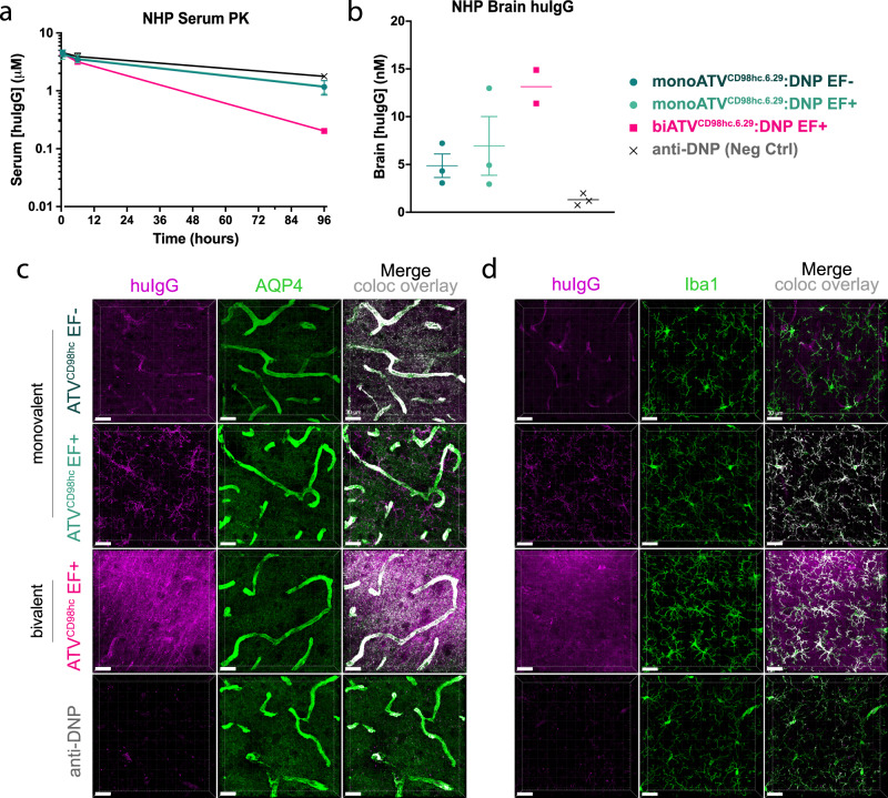
Brain exposure of systemically administered biotherapeutics is highly restricted by the blood-brain barrier (BBB). Here, we report the engineering and characterization of a BBB transport vehicle targeting the CD98 heavy chain (CD98hc or SLC3A2) of heterodimeric amino acid transporters (TVCD98hc). The pharmacokinetic and biodistribution properties of a CD98hc antibody transport vehicle (ATVCD98hc) are assessed in humanized CD98hc knock-in mice and cynomolgus monkeys. Compared to most existing BBB platforms targeting the transferrin receptor, peripherally administered ATVCD98hc demonstrates differentiated brain delivery with markedly slower and more prolonged kinetic properties. Specific biodistribution profiles within the brain parenchyma can be modulated by introducing Fc mutations on ATVCD98hc that impact FcγR engagement, changing the valency of CD98hc binding, and by altering the extent of target engagement with Fabs. Our study establishes TVCD98hc as a modular brain delivery platform with favorable kinetic, biodistribution, and safety properties distinct from previously reported BBB platforms.


Conflict of interest statement

K.S.C., R.C.W., A.M., D.C., K.J.L., H.L.T., J.C., D.J.K., Y.R.C., D.B.S., R.K.T., M.T., K.X., A.Y., Y.Z., P.A., L.A., G.M.C., T.K.E., A.G., D.H., D.J., K.N.K., D.L., A.W.S.L., K.W.L., N.P.D.L., I.B., J.M., H.N.N., E.I.L., M.E.P., E.R., P.S., M.E.K.C., M.S.D., J.D., K.G., J.W.L., C.S.M., R.M., H.S., R.G.T., R.J.W., Y.J.Y.Z., M.S.K. are paid employees of Denali Therapeutics Inc. Denali has filed patent application no. PCT/US2022/053220 related to the subject matter of this paper, which includes the discovery and application of the CD98hc TVs. K.S.C., R.C.W., H.L.T., P.A., G.M.C., M.S.D., Y.J.Y.Z. and M.S.K. are inventors of this patent application. There are no other competing interests.

Figures

Fig. 1
Fig. 1. Transport vehicles engineered at an Fc β-sheet surface bind with a range of affinities to CD98hc.
a Cartoon of the ATVCD98hc bound to the CD98hc-LAT1 complex with the relative position of the membrane highlighted (dotted line). b Outline of the steps used to identify and engineer TVs. Positions in the naïve and maturation libraries are modeled onto the human IgG1 Fc domain (PDB ID: 1HZH) with positions randomized in the original library (orange), randomized expanded positions (green), unaltered original positions (tan), and unaltered peripheral positions (gray) highlighted. An example of one of ten patch libraries is provided. c Representative TV sequences from each stage are shown with corresponding affinities to human and cynomolgus CD98hc of the ATVCD98hc:DNP clone, as measured by surface-plasmon resonance. d A compilation of affinities of ATVCD98hc:DNP variants to human and cynomolgus CD98hc across rounds of maturation. e Immunocytochemistry detection of huIgG 1 h after incubation of ATVCD98hc:DNP (62.5 nM), anti-CD98hc (62.5 nM) and anti-DNP (500 nM) to human (HeLa), cynomolgus CD98hc-expressing cells (CHO: cynomolgus CD98hc) and parental cells (CHO). Representative images are shown from at least 15 images per well, n = 2 independent experiments. Scale bars = 50 µm. Source data are provided as a Source Data file.
Fig. 2
Fig. 2. Crystal structure of the TV6.6 and human CD98hc co-complex provides molecular details of the interaction.
a Model of Fc with TV6.6 (red and blue) in complex with two copies of CD98hc (tan); PDB ID: 8G0M b Zoom in of the TV6.6 surface with engineered residues (blue sticks) and wild-type positions (gray sticks) that contact CD98hc. Surface model of CD98hc indicating residues in contact with the TV (orange) and surface exposed positions that differ between human and cynomolgus (pink). c, d Model of monovalent (c) and bivalent (d) TV binding to the CD98hc-LAT1 complex (PDB ID: 6JMQ) on the cell surface.
Fig. 3
Fig. 3. Monovalent and bivalent ATVCD98hc.6.8:DNP have prolonged brain exposure after a single dose in CD98mu/hu KI mice.
a Cartoons of monovalent and bivalent ATVCD98hc6.8:DNP, with (EF−) and without (EF+) mutations to mitigate FcγR binding. bc Brain and plasma concentrations of ATVCD98hc6.8:DNP variants (170 nM KD) after a single intravenous (IV) 50 mg/kg dose in CD98hcmu/hu KI mice. Plasma clearance values were 19.6–21.9 mL/d/kg and 43–49 mL/d/kg for monoATVCD98hc.6.8:DNP and biATVCD98hc.6.8:DNP, respectively, compared to 7.1 and 8.3 mL/d/kg for mono- or biATVCD98hc.6.8:DNP in WT mice (Supplementary Table 1). All ATVCD98hc variants had higher plasma clearance and brain exposure compared to the anti-DNP control, and biATVCD98hc6.8:DNP variants had higher plasma clearance and brain exposure compared monoATVCD98hc6.8:DNP variants. FcγR binding did not impact plasma clearance and was associated with a small increase in brain exposure. de Concentrations of huIgG in brain parenchymal (d), vascular fractions (e), and cell-associated and non-cell associated fractions (f) obtained by capillary depletion at 7 days post dose. There were increased concentrations of all ATVCD98hc TV variants in the parenchymal and vascular fractions. Consistent with whole-brain exposure, biATVCD98hc6.8:DNP EF+ had the highest concentration in the parenchyma and biATVCD98hc6.8:DNP variants were found at higher concentrations in the vascular fraction. biATVCD98hc6.8:DNP variants were also more cell associated than monoATVCD98hc6.8:DNP variants. be Two-way ANOVA. f One-way ANOVA. bf Graphs display mean ± SEM, n = 4–5, see Source Data for exact n/group. *p < 0.05, ****p < 0.0001. See Supplementary Table 6 for exact p values. g Immunohistochemical localization of ATVCD98hc6.8:DNP variants in brain cortical sections as assessed with anti-huIgG across timepoints. Scale bars = 15 µm. Representative image shown from n = 5/group, n = 2 stained sections per animal. Source data are provided as a Source Data file.
Fig. 4
Fig. 4. Biodistribution of ATVCD98hc6.8:DNP variants in the brain is dependent on valency and FcγR binding in CD98hcmu/hu KI mice.
a Immunohistochemical cell-specific localization of ATVCD98hc6.8:DNP variants (170 nM KD) in brain cortical sections with antibodies against huIgG (purple) and AQP4 (a, green, scale bars = 15 µm) or Iba1 (b, green, scale bars = 20 µm). Overlays are shown with colocalization pseudocolored in white. Representative image shown from n = 5/group, n = 2 stained sections per animal.
Fig. 5
Fig. 5. Pharmacokinetics and distribution of ATVCD98hc are distinct from ATVTfR.
a, b PK of ATVCD98hc.6.8:DNP (170 nM KD) and ATVTfR.35.23.4:DNP (620 nM KD) in plasma (a) and brain (b) following a single 50 mg/kg IV dose. Plasma clearances for both ATVs were increased relative to anti-DNP (5.3 mL/d/kg), and plasma clearance for ATVTfR.35.23.4:DNP (39.0 mL/d/kg) was increased compared to ATVCD98hc.6.8:DNP (17.7 mL/d/kg). Both ATVCD98hc.6.8:DNP and ATVTfR.35.23.4:DNP had increased brain exposure compared to control and had different exposure profiles compared to each other. c, d PK of mono- and biATVCD98hc6.39:BACE1 (94 nM KD) in plasma (c) and brain (d) following a single 50 mg/kg IV dose. Both mono- and biATVCD98hc6.39:BACE1 had higher plasma clearance compared to control, and biATVCD98hc6.39:BACE1 (35.9 mL/d/kg) had higher plasma clearance compared to monovalent (19.9 mL/d/kg). Compare to the plasma clearance measured for mono- and biATVCD98hc.6.8:DNP (19.6 and 43.9 mL/d/kg, respectively Fig. 3b). Both mono- and biATVCD98hc6.39:BACE1 had increased brain exposure compared to control, and were not significantly different from each other. ad Graphs display mean ± SEM, n = 5/group. Two-way ANOVA. Data points not shown were <LLOD. *p < 0.05, ****p < 0.0001. See Supplementary Table 6 for exact p values. e Immunohistochemical localization of huIgG for mono- and biATVCD98hc.6.8:BACE1 (purple) in cortical brain sections, LAMP2 (green), and NeuN (yellow) 7 days after a single 50 mg/kg IV dose. Scale bars = 7 µm. Overlays are shown with colocalization of huIgG and Lamp2 within NeuN positive neurons is pseudocolored in white. Representative images are shown from n = 5 animals/treatment group, n = 2 IHC sections per animal. f, g Simulated brain uptake, clearance, and total net flux for mono- and biATVCD98hc:DNP (f) and ATVCD98hc: BACE1(g). Simulations are performed for a 50 mg/kg IV dose of ATVCD98hc, CD98hc KD = 170 nM. Source data are provided as a Source Data file.
Fig. 6
Fig. 6. Multidose of ATVCD98hc6.8:DNP variants in CD98hcmu/hu KI mice accumulate in brain without impacting circulating cells.
a, c Plasma PK of mono- and biATVCD98hc6.8:DNP (170 nM KD) with (EF−) and without (EF+) mutations to mitigate FcγR binding after 5 weekly IV doses (50 mg/kg). Plasma concentrations were assessed at 30 min after each dose (Cmax) and 24 h before the next dose (Ctrough). Two-way ANOVA. b, d Concentrations of ATVCD98hc:DNP variants in brain 24 h after the final dose. One-way ANOVA ****p < 0.0001. See Supplementary Table 6 for exact p values. ej Circulating cell counts for monocytes (e, h), lymphocytes (f, i) and reticulocytes (g, j) as measured by complete blood count 24 h after the final dose. Cell numbers compared with a one-way ANOVA. aj All graphs display n = 4–5/group (see Source Data for exact n/group), mean ± SEM. Source data are provided as a Source Data file.
Fig. 7
Fig. 7. ATVCD98hc uptake is enhanced in brain and localize to astrocytes and microglia in cynomolgus monkeys.
a, b Plasma and brain exposure of mono- and biATVCD98hc6.29:DNP (205 nM KD to cyno CD98hc) EF+ and monoATVCD98hc6.29:DNP EF- in cynomolgus monkeys 4 days after a single 30 mg/kg IV dose. Graphs display mean ± SEM (for n = 2 or 3 as noted in figure) c, d Localization of ATVCD98hc6.29:DNP variations in cortical brain sections assessed by IHC with antibodies against for huIgG (purple) and AQP4 (c, green) or Iba1 (d, green). Scale bars = 30 µm. Overlays are shown with colocalization pseudocolored in white. Representative image shown from n = 3 animals/treatment group (except for biATVCD98hc.6.29:DNP, where one animal was excluded), n = 3 stained sections per animal. Source data are provided as a Source Data file.

References

    1. Abbott NJ, Patabendige AA, Dolman DE, Yusof SR, Begley DJ. Structure and function of the blood-brain barrier. Neurobiol. Dis. 2010;37:13–25. - PubMed
    1. Chow BW, Gu C. The molecular constituents of the blood-brain barrier. Trends Neurosci. 2015;38:598–608. - PMC - PubMed
    1. Daneman R, Prat A. The blood-brain barrier. Cold Spring Harb. Perspect. Biol. 2015;7:a020412. - PMC - PubMed
    1. Iadecola C. The neurovascular unit coming of age: a journey through neurovascular coupling in health and disease. Neuron. 2017;96:17–42. - PMC - PubMed
    1. Sweeney MD, Sagare AP, Zlokovic BV. Blood-brain barrier breakdown in Alzheimer disease and other neurodegenerative disorders. Nat. Rev. Neurol. 2018;14:133–150. - PMC - PubMed