Review
. 2023 Oct 12;44(38):3827-3844.
doi: 10.1093/eurheartj/ehad484.

Perivascular adipose tissue as a source of therapeutic targets and clinical biomarkers

Affiliations
Review

Perivascular adipose tissue as a source of therapeutic targets and clinical biomarkers

Charalambos Antoniades et al. Eur Heart J. .

Abstract

Obesity is a modifiable cardiovascular risk factor, but adipose tissue (AT) depots in humans are anatomically, histologically, and functionally heterogeneous. For example, visceral AT is a pro-atherogenic secretory AT depot, while subcutaneous AT represents a more classical energy storage depot. Perivascular adipose tissue (PVAT) regulates vascular biology via paracrine cross-talk signals. In this position paper, the state-of-the-art knowledge of various AT depots is reviewed providing a consensus definition of PVAT around the coronary arteries, as the AT surrounding the artery up to a distance from its outer wall equal to the luminal diameter of the artery. Special focus is given to the interactions between PVAT and the vascular wall that render PVAT a potential therapeutic target in cardiovascular diseases. This Clinical Consensus Statement also discusses the role of PVAT as a clinically relevant source of diagnostic and prognostic biomarkers of vascular function, which may guide precision medicine in atherosclerosis, hypertension, heart failure, and other cardiovascular diseases. In this article, its role as a 'biosensor' of vascular inflammation is highlighted with description of recent imaging technologies that visualize PVAT in clinical practice, allowing non-invasive quantification of coronary inflammation and the related residual cardiovascular inflammatory risk, guiding deployment of therapeutic interventions. Finally, the current and future clinical applicability of artificial intelligence and machine learning technologies is reviewed that integrate PVAT information into prognostic models to provide clinically meaningful information in primary and secondary prevention.

Keywords: Atherosclerosis; Coronary computed tomography angiography; Fat attenuation index; Peri-vascular adipose tissue.


Figures

Graphical Abstract
Graphical Abstract
Perivascular adipose tissue (PVAT) as a source of imaging biomarkers and a therapeutic target. FAI, fat attenuation index.
Figure 1
Figure 1
Imaging of human adipose tissue depots by computed tomography. Axial (A), coronal (B), and sagittal (C) views of the chest showing subcutaneous, visceral abdominal, thoracic (including the pericardial and epicardial) adipose tissue. Reconstruction of perivascular adipose tissue (PVAT) around an epicardial coronary artery (D) and thoracic aorta (E). PVAT is defined as the adipose tissue lying within a radial distance from the outer vessel wall equal to the vessel diameter (or at a maximum distance of 2 cm in the case of large vessels with diameter >2 cm, like the aorta).
Figure 2
Figure 2
(A) Routes via which remote and local adipose tissue depots affect the cardiovascular system. (B) Illustration of the major components of the bi-directional interplay between the vascular wall and fat in the peri-vascular space. EC, endothelial cell; eNOS, endothelial nitric oxide synthase; PVAT, perivascular adipose tissue; VSMC, vascular smooth muscle cell. (Reused with permission by Kotanidis and Antoniades.) An animated video web link presents how the cross-talk between PVAT and the vascular wall leads to changes in PVAT texture and composition, visible with computed tomography.
Figure 3
Figure 3
Schematic representation of the biology underlying the detection of coronary inflammation by imaging perivascular adipose tissue (PVAT). Illustration of a healthy artery and surrounding PVAT (bottom) and the resulting PVAT phenotype in states of high vascular wall inflammation (top). Biopsies of PVAT surrounding inflamed vessels demonstrates high macrophage infiltration and contains small adipocytes that when cultured in the presence of inflammatory cytokines, do not store intracellular lipids. Indeed, in the presence of vascular inflammation there is a steep change in adipocyte size and lipid:water content with increasing distance from the vascular wall due to the paracrine effects of vascular inflammation on surrounding peri-vascular adipose tissue (PVAT). These changes in lipid:water balance can be detected by coronary computed tomography angiography (CCTA), visualized by the fat attenuation index (FAI) mapping of PVAT and quantified using FAI-Score. The latter is interpreted clinically by using age and gender nomograms, and when it is used in prognostic models with plaque and clinical risk factors, it provides a powerful was to calculate the patient’s specific risk for cardiovascular events. An example of a patient with high FAI-Score at baseline who reduced vascular inflammation after 1-year treatment with atorvastatin 40 mg once daily. Images obtained with permission from Antonopoulos et al. The animated images on the process by which vascular inflammation drives changes to PVAT visible by CCTA have been obtained with permission from this animated video web link.
Figure 4
Figure 4
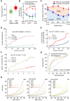
Pericoronary adipose tissue attenuation for detecting vascular inflammation. (A) Peri-coronary fat attenuation index (FAI) is increased around culprit lesions in acute coronary syndrome patients with evidence of inflammation as assessed by 18F-sodium fluoride (18FNaF) by positron emission tomography/computed tomography (adapted from Kwiecinski et al.). (B) Changes in FAI of perivascular adipose tissue (PVAT) around ruptured (culprit) atherosclerotic lesions of acute myocardial infarction patients, non-culprit lesions of the same patients, or lesions in stable CAD patients. (C) Temporal changes in FAI around a culprit lesion in patients with acute ST elevation myocardial infarction (n = 10). (D) Prognostic value of FAI for cardiac mortality and (E) cardiac mortality or non-fatal myocardial infarction in CRISP-CT study in a cohort of 2040 patients undergoing diagnostic CCTA in Cleveland Clinic, US. (F) Prognostic value of unadjusted coronary PVAT attenuation values in SCOT-HEART trial in 1697 evaluable participants. (G) The superior performance of fully weighted FAI vs. unadjusted PVAT density; unadjusted PVAT density had borderline predictive value for cardiac mortality, whereas fully weighted FAI led to improved risk prediction by 13.6% in CRISP-CT study. (H) Standardization of coronary peri-vascular FAI and nomograms for age (FAI-Score) for left anterior descending artery (LAD) right coronary artery (RCA) and left circumflex coronary artery (LCx). FAI-Score informs on the coronary vessel-specific inflammation burden and the associated relative risk for a fatal cardiac event compared to the age group of reference. For instance, a young individual with no traditional risk factors may be at low absolute risk for a fatal cardiac event; however, a high FAI-Score may indicate increased relative risk for cardiac events in the long-term as a result of sub-clinical vascular inflammation. All figure panels were reproduced with permission from the authors/publishers. CAD,coronary artery disease.
Figure 5
Figure 5
Radiomic phenotyping of coronary perivascular adipose tissue (PVAT). (A) Coronary PVAT imaging features can be used to extract a high number of shape-, attenuation-, and texture-related statistics (i.e. radiomics). (B) Heatmap of scaled radiomic features (right) of all 1391 stable radiomic features in the SCOT-HEART population (n = 1575 patients). (C) Extracted radiomic features can be tested against the transcriptome profile of PVAT to identify features informing on distinct biological processes such as adipose tissue inflammation, vascularity, and fibrosis. (D) Selected radiomic features can form distinct radiomic signatures of PVAT, in this case fat radiomic profile adipose tissue inflammation, vascularity, and fibrosis, which had strong independent predictive value for major adverse cardiac events (MACE) in the SCOT-HEART population. Obtained with permission from Oikonomou et al.
Figure 6
Figure 6
Prognostic value of vascular inflammation biomarkers for major adverse cardiovascular events. The added prognostic value of imaging biomarkers on top of patient risk profile and coronary atherosclerosis extent is greater than that of plasma biomarkers according to a meta-analysis of available published evidence from clinical studies (n = 175 778 individuals). Perivascular adipose tissue (PVAT) imaging by CT was associated with the maximum added prognostic information among the studied vascular inflammation biomarkers. Obtained with permission from Antonopoulos et al.

References

    1. Badimon L, Bugiardini R, Cenko E, Cubedo J, Dorobantu M, Duncker DJ, et al. Position paper of the European Society of Cardiology-working group of coronary pathophysiology and microcirculation: obesity and heart disease. Eur Heart J 2017;38:1951–8. 10.1093/eurheartj/ehx181 - DOI - PubMed
    1. Aune D, Sen A, Prasad M, Norat T, Janszky I, Tonstad S, et al. BMI and all cause mortality: systematic review and non-linear dose-response meta-analysis of 230 cohort studies with 3.74 million deaths among 30.3 million participants. BMJ 2016;353:i2156. 10.1136/bmj.i2156 - DOI - PMC - PubMed
    1. Aguilar M, Rose RA, Takawale A, Nattel S, Reilly S. New aspects of endocrine control of atrial fibrillation and possibilities for clinical translation. Cardiovasc Res 2021;117:1645–61. 10.1093/cvr/cvab080 - DOI - PMC - PubMed
    1. Antonopoulos AS, Tousoulis D. The molecular mechanisms of obesity paradox. Cardiovasc Res 2017;113:1074–86. 10.1093/cvr/cvx106 - DOI - PubMed
    1. NCD Risk Factor Collaboration . Worldwide trends in body-mass index, underweight, overweight, and obesity from 1975 to 2016: a pooled analysis of 2416 population-based measurement studies in 128.9 million children, adolescents, and adults. Lancet 2017;390:2627–42. 10.1016/S0140-6736(17)32129-3 - DOI - PMC - PubMed

Publication types