. 2023 Aug 7:22:100763.
doi: 10.1016/j.mtbio.2023.100763. eCollection 2023 Oct.

Reversible rearrangement of the cellular cytoskeleton: A key to the broad-spectrum antiviral activity of novel amphiphilic polymers

Affiliations

Reversible rearrangement of the cellular cytoskeleton: A key to the broad-spectrum antiviral activity of novel amphiphilic polymers

Agnieszka Dabrowska et al. Mater Today Bio. .

Abstract

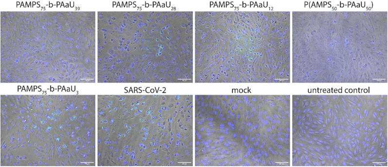
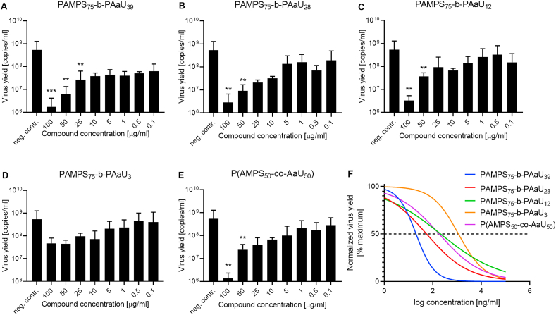
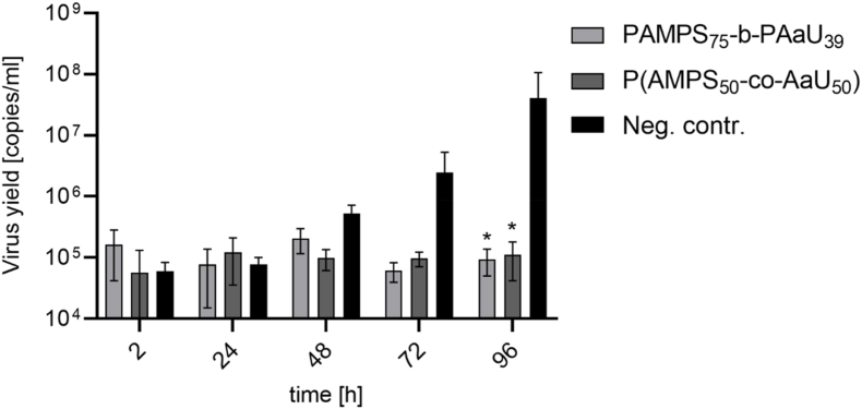
The battle against emerging viral infections has been uneven, as there is currently no broad-spectrum drug available to contain the spread of novel pathogens throughout the population. Consequently, the pandemic outbreak that occurred in early 2020 laid bare the almost empty state of the pandemic box. Therefore, the development of novel treatments with broad specificity has become a paramount concern in this post-pandemic era. Here, we propose copolymers of poly (sodium 2-(acrylamido)-2-methyl-1-propanesulfonate) (PAMPS) and poly (sodium 11-(acrylamido)undecanoate (AaU), both block (PAMPS75-b-PAaUn) and random (P(AMPSm-co-AaUn)) that show efficacy against a broad range of alpha and betacoronaviruses. Owing to their intricate architecture, these polymers exhibit a highly distinctive mode of action, modulating nano-mechanical properties of cells and thereby influencing viral replication. Through the employment of confocal and atomic force microscopy techniques, we discerned perturbations in actin and vimentin filaments, which correlated with modification of cellular elasticity and reduction of glycocalyx layer. Intriguingly, this process was reversible upon polymer removal from the cells. To ascertain the applicability of our findings, we assessed the efficacy and underlying mechanism of the inhibitors using fully differentiated human airway epithelial cultures, wherein near-complete abrogation of viral replication was documented. Given their mode of action, these polymers can be classified as biologically active nanomaterials that exploit a highly conserved molecular target-cellular plasticity-proffering the potential for truly broad-spectrum activity while concurrently for drug resistance development is minimal.

Keywords: Antivirals; Broad-spectrum; Coronaviruses; Nanomaterials; Polymers.


Conflict of interest statement

The authors declare the following financial interests/personal relationships which may be considered as potential competing interests Krzysztof Pyrc reports financial support was provided by 10.13039/501100004281National Science Centre Poland. Maria Nowakowska reports financial support was provided by 10.13039/501100004281National Science Centre Poland.

Figures

Image 1
Graphical abstract
Fig. 1
Fig. 1
Polymers inhibit the cytopathic effect. CPE effect in Vero cells infected with SARS-CoV-2 at 1600 TCID50/ml 48 h post-infection in the presence or absence of 25 μg/ml of polymers. The live cell staining was conducted with Blue/Green Cell Viability Imaging Kit. The blue color denotes the nuclei of all cells, while the green color denotes only the nuclei of cells with compromised plasma membrane integrity. Light microscope images were collected on the EVOS Floid Imaging system using 20× objective. Scale bar 100 μm. (For interpretation of the references to color in this figure legend, the reader is referred to the Web version of this article.)
Fig. 2
Fig. 2
The copolymers are potent SARS-CoV-2 inhibitors. The figure shows RT-qPCR analysis of supernatants collected from cells infected with SARS-CoV-2 at 1600 TCID50/ml 48 h post-infection in the absence (neg.contr.) or presence of compounds in the range of concentrations 0.1–100 μg/ml (A–E) and the dose-response curves of normalized data (F). All experiments were performed in at least two biological repetitions, each in triplicate. The results are presented as average values with standard deviations (error bars). Values statistically significant are indicated by asterisks: **p < 0.05, ***p < 0.005.
Fig. 3
Fig. 3
PAMPS-PAaU copolymers are not toxic to MRC-5 and HCT-8 cells in the low μg/ml concentration range. Results of the XTT assay for PAMPS75-b-PAaU39 and P(AMPS50-co-AaU50) copolymers at concentrations of 4000, 3000, 2000, 1000, 500, and 100, 50, and 10 μg/ml on MRC-5 and HCT-8 cells. All experiments were performed in duplicate. Average values with standard deviation (error bars) are shown.
Fig. 4
Fig. 4
The PAMPS75-b-PAaU39block and P(AMPS50-co-AaU50) random copolymers as broad-spectrum antivirals. The figure shows RT-qPCR analysis of cell culture supernatants collected from cells infected with HCoV-OC43 at 1600 TCID50/ml 72 h post-infection (A, C) and HCoV-229E at 4000 TCID50/ml 72 h post-infection (B, D), neg. contr. denotes cells infected without polymer. All experiments were performed in at least three biological repetitions, each in triplicate. The results are presented as average values with standard deviations (error bars). *Significantly different from the control (p < 0.05).
Fig. 5
Fig. 5
Copolymers block SARS-CoV-2 replication in ex vivo model. The graph presents the number of viral RNA copies collected apically after the indicated time. HAE culture was infected with SARS-CoV-2 at 5000 TCID50/ml until 96 h post-infection, neg. contr. denotes cells infected without polymer. Each experiment was performed in 2 biological repetitions, each in triplicate. The results are presented as average values with standard deviations (error bars). *Significantly different from the control (p < 0.05).
Fig. 6
Fig. 6
PAMPS-b-PAaUs inhibit the SARS-CoV-2 attachment to host cells and the replication cycle. Antiviral activity of copolymers was tested in Vero cells infected with the SARS-CoV-2, as described in the Material and Method section: virus inactivation assay (A), virus attachment assay (B), virus entry assay (C) with the graph showing the number of SARS-CoV-2 virions attached and entered to the cells with line at median with 95% CI (D), and virus replication, assembly and egress assay (E). F-actin – red; SARS-CoV-2 N protein – green; cell nuclei – blue. Scale bar = 25 μm. (For interpretation of the references to color in this figure legend, the reader is referred to the Web version of this article.)
Fig. 7
Fig. 7
The PAMPS75-b-PAaU28quickly penetrates the cells. The figure shows the signal from fluorescein attached to PAMPS75-b-PAaU28 (upper panel) and fluorescein (lower panel) administrated to the cells. The signal distribution was observed for up to 48 min. The results are presented as maximum projections from chosen time points: 2, 16, and 30 min. Scale bar = 20 μm.
Fig. 8
Fig. 8
Changes of the nano-mechanical parameters of Vero cells induced by PAMPS75-b-PAaU39. Box plots depict the quantitative analysis of elastic moduli counted for the entire range of indentation corresponding to the whole cell (A) and a specific range of nanoindentation corresponding to the elasticity of the cortex of the non-treated cells (control) or cells incubated with PAMPS75-b-PAaU39 for 15 min, 1 h, 2 h, 3 h, 4 h and 6 h (B). Box plots depict the quantitative analysis of thickness of the glycocalyx layer for non-treated cells (control) and cells incubated with PAMPS75-b-PAaU39 for 15 min, 1 h, 2 h, 3 h, 4 h, and 6 h (C). The representative AFM maps of height (topography, top row) and elasticity (bottom row) were taken for Vero cells incubated with PAMPS75-b-PAaU39 for 15 min, 1 h, 2 h, 3 h, 4 h, and 6 h. Non-treated cells were used as a reference (control). All images were taken for 0.5% GA fixed cells (D). Statistics: p values were determined by one-way analysis of variance ANOVA followed by Tukey's post-hoc test. *Significantly different from the control (p < 0.05).
Fig. 9
Fig. 9
PAMPS-b-PAaU reversibly affects cell skeleton. Confocal images of Vero cells non-treated (A), treated with 100 μg/ml PAMPS75-b-PAaU39 at 15 min (B), 2 h (C), and 6 h (D) F-actin – red; Vimentin – white; Vinculin – green; cell nuclei – blue. Scale bar = 10 μm. (For interpretation of the references to color in this figure legend, the reader is referred to the Web version of this article.)

References

    1. Zhu N., Zhang D., Wang W., Li X., Yang B., Song J., et al. A novel coronavirus from patients with pneumonia in China. N. Engl. J. Med. 2020 doi: 10.1056/nejmoa2001017. 2019. - DOI - PMC - PubMed
    1. Yang J., Chen X., Deng X., Chen Z., Gong H., Yan H., et al. Disease burden and clinical severity of the first pandemic wave of COVID-19 in Wuhan, China. Nat. Commun. 2020 doi: 10.1038/s41467-020-19238-2. - DOI - PMC - PubMed
    1. Brinkworth J.F., Rusen R.M. SARS-CoV-2 is not special, but the pandemic is: the ecology, evolution, policy, and future of the deadliest pandemic in living memory. Annu. Rev. Anthropol. 2022;51:527–548. doi: 10.1146/annurev-anthro-041420-100047. - DOI
    1. Collaborators E.M. Estimating excess mortality due to the COVID-19 pandemic: a systematic analysis of COVID-19-related mortality, 2020-21. Lancet (London, England) 2022;399:1513–1536. doi: 10.1016/S0140-6736(21)02796-3. - DOI - PMC - PubMed
    1. Fields B.N. Fields virology. Fields Virol. 2013:2664.

LinkOut - more resources