. 2023 Aug 1:14:1223021.
doi: 10.3389/fendo.2023.1223021. eCollection 2023.

Neuropilin 2 in osteoblasts regulates trabecular bone mass in male mice

Affiliations

Neuropilin 2 in osteoblasts regulates trabecular bone mass in male mice

Lieve Verlinden et al. Front Endocrinol (Lausanne). .

Abstract

Introduction: Neuropilin 2 (NRP2) mediates the effects of class 3 semaphorins and vascular endothelial growth factor and is implicated in axonal guidance and angiogenesis. Moreover, NRP2 expression is suggested to be involved in the regulation of bone homeostasis. Indeed, osteoblasts and osteoclasts express NRP2 and male and female global Nrp2 knockout mice have a reduced bone mass accompanied by reduced osteoblast and increased osteoclast counts.

Methods: We first examined the in vitro effect of the calciotropic hormone 1,25-dihydroxyvitamin D3 [1,25(OH)2D3] on Nrp2 transcription in osteoblasts. We next generated mice with a conditional deletion of Nrp2 in the osteoblast cell lineage under control of the paired related homeobox 1 promoter and mice with a conditional Nrp2 knockdown in osteoclasts under control of the Lysozyme promoter. Mice were examined under basal conditions or after treatment with either the bone anabolic vitamin D3 analog WY 1048 or with 1,25(OH)2D3.

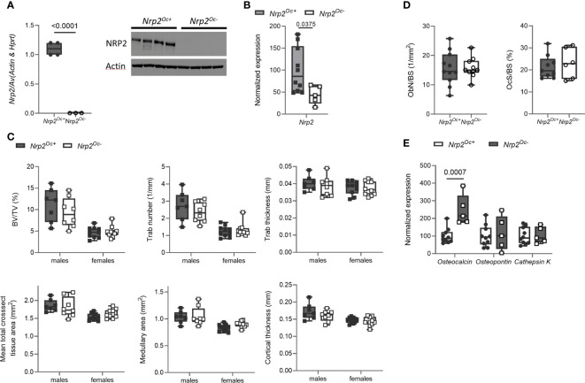
Results and discussion: We show that Nrp2 expression is induced by 1,25(OH)2D3 in osteoblasts and is associated with enrichment of the vitamin D receptor in an intronic region of the Nrp2 gene. In male mice, conditional deletion of Nrp2 in osteoblast precursors and mature osteoblasts recapitulated the bone phenotype of global Nrp2 knockout mice, with a reduced cortical cross-sectional tissue area and lower trabecular bone content. However, female mice with reduced osteoblastic Nrp2 expression display a reduced cross-sectional tissue area but have a normal trabecular bone mass. Treatment with the vitamin D3 analog WY 1048 (0.4 μg/kg/d, 14 days, ip) resulted in a similar increase in bone mass in both genotypes and genders. Deleting Nrp2 from the osteoclast lineage did not result in a bone phenotype, even though in vitro osteoclastogenesis of hematopoietic cells derived from mutant mice was significantly increased. Moreover, treatment with a high dose of 1,25(OH)2D3 (0.5 μg/kg/d, 6 days, ip), to induce osteoclast-mediated bone resorption, resulted in a similar reduction in trabecular and cortical bone mass. In conclusion, osteoblastic Nrp2 expression is suggested to regulate bone homeostasis in a sex-specific manner.

Keywords: bone; neuropilin 2; osteoblast; osteoclast; vitamin D.


Conflict of interest statement

The authors declare that the research was conducted in the absence of any commercial or financial relationships that could be construed as a potential conflict of interest.

Figures

Figure 1
Figure 1
Nrp2 is a direct transcriptional target of 1,25(OH)2D3. (A) Nrp2 transcript levels are induced in MC3T3-E1 and ST2 osteoblast cell lines and in primary calvarial osteoblasts after treatment with 1,25(OH)2D3 (24 h, 10-8 M) (n=2-3). (B) Nrp2 transcript levels in MC3T3-E1 cells stimulated with 1,25(OH)2D3 (10-8 M) and treated with actinomycin D (5 µg/ml, 6 h) or cycloheximide (CHX) (100 ng/ml, 12 h) (n=2). (C) Overlaid triplicate and averaged ChIP-Seq tracks for VDR, RXR, and H4K5 acetylation (H4K5ac) where ethanol vehicle (Veh) are shown in yellow, 1,25(OH)2D3 treatment shown in blue, and overlapping data appear as green. Nrp2 genomic region displayed is chr1:62,726,447-62,867,109 with exons depicted as boxes, introns by arrows. Region of interest is highlighted in the inset below (chr1:62,765,185-62,770,248). Maximum height of tag sequence density for each data track indicated on the Y-axis (normalized to input and 107 tags). (D) ChIP-qPCR results confirmed the 1,25(OH)2D3-inducible association of VDR and RXR in response to treatment at the studied VDR binding site within the Nrp2 gene. ChIP was performed with chromatin samples from MC3T3-E1 cells incubated with 1,25(OH)2D3 (10-7 M) or vehicle for 3 h (n=3). (E) A DNA fragment, containing either the wildtype or a mutated, non-functional VDR binding site, was cloned and inserted into the pGL3-basic vector. Luciferase reporter gene assays were performed in exponentially growing MC3T3-E1 cells to evaluate the functionality of the identified VDR binding sites (10-8 M 1,25(OH)2D3, 24 h). Luciferase activity was normalized to that of empty pGL3-basic reporter vector (n=3). All results are expressed as mean and SEM. Two-tailed Student’s t-tests were performed to determine significant differences between 1,25(OH)2D3-treated and vehicle-treated cells.
Figure 2
Figure 2
Osteoblast-specific deletion of Nrp2 affects bone differently in male and female mice. (A) NRP2 transcript and protein levels were significantly lower in primary calvarial osteoblasts derived from Nrp2Ob- mice compared to osteoblasts from wildtype littermates (n=4). Data are expressed as mean and SEM. Two-tailed Student’s t-tests were performed to detect significant differences (B) Reduced Nrp2 transcript levels in whole bone homogenates from 10-week-old male and female Nrp2Ob- mice (n=5-9). (C, D) µCT analysis and evaluation of trabecular (C) and cortical bone parameters (D) in tibia of 10-week-old male and female Nrp2Ob- mice and their wildtype littermates (n=9-11). (E) Representative 3D models of tibial epiphyses and metaphyses (upper panel) and cross-sections of the tibial mid diaphysis from 10-week-old male Nrp2Ob- mice and their wildtype littermates. Data from panels B-E are expressed as mean and SD. Two-way ANOVA analysis, with genotype and sex as independent variables, followed by Sidak’s multiple comparisons test, was performed to evaluate significant differences between genotypes for male and female mice.
Figure 3
Figure 3
Osteoblast and osteoclast activity in Nrp2Ob- mice. (A) Representative pictures and histomorphometric analysis of osteoblasts (upper panels, n=6-7) and osteoclasts (lower panels, n=5-6) in tibia of 10-week-old male Nrp2Ob- mice and wildtype littermates (scale bars are 500 µm in overview pictures and 100 µm in picture details). (B) Quantification of osteoblast number and osteoclast surface in tibia of 8-week-old female Nrp2Ob- mice and wildtype littermates (n=8-10). (C) Serum P1NP and CTx levels in 10-week-old male Nrp2Ob- mice and wildtype littermates (n=5-7). (D) In vitro evaluation of adhesive and migratory capacity of primary Nrp2-/- and Nrp2+/+ calvarial osteoblasts (n=4-5). All in vivo data are expressed as mean and SD, whereas in vitro data are expressed as mean and SEM. Two-tailed Student’s t-tests were performed to detect significant differences.
Figure 4
Figure 4
Osteoclast-specific deletion of Nrp2 does not affect bone mass. (A) NRP2 transcript and protein levels were significantly lower in primary osteoclast cultures derived from hematopoietic cells that were isolated from Nrp2Oc- or Nrp2Oc+ mice and in vitro stimulated with M-CSF and RANKL (n=3-4). Data are expressed as mean and SEM. Two-tailed Student’s t-tests were performed to detect significant differences. (B) Reduced Nrp2 transcript levels in whole bone homogenates from 10-week-old female Nrp2Oc- mice compared to wildtype littermates (n=5-10). Data are expressed as mean and SS. Two-tailed Student’s t-tests were performed to detect significant differences. (C) µCT analysis and evaluation of trabecular and cortical bone parameters in tibia of 10-week-old male and -female Nrp2Oc- mice and their wildtype littermates (n=7-9). Data are expressed as mean and SD. Two-way ANOVA analysis, with genotype and sex as independent variables, followed by Sidak’s multiple comparisons test, was performed to evaluate significant differences between genotypes for male and female mice. (D) Histomorphometric analysis of osteoblast numbers (left panel) and osteoclast surface (right panel) in tibia of 10-week-old female Nrp2Oc- mice and their wildtype littermates (n=6-11). Data are expressed as mean and SD. Two-tailed Student’s t-tests were performed to detect significant differences. (E) qPCR analysis of the osteoblast markers osteocalcin and osteopontin and the osteoclast marker cathepsin K in whole bone homogenates of 10-week-old female Nrp2Oc- mice and their wildtype littermates. Data are expressed as mean and SD. Two-tailed Student’s t-tests were performed to detect significant differences.
Figure 5
Figure 5
In vitro osteoclastogenesis is enhanced when hematopoietic cells are derived from Nrp2Oc- mice. (A) Hematopoietic cells, derived from Nrp2Oc- mice or their wildtype littermates, were cultured in presence of M-CSF and RANKL. Osteoclasts were quantified on TRAP stainings (n=7-10) and qPCR analysis was performed to evaluate expression of the osteoclast-specific genes cathepsin K, calcitonin receptor and DC-stamp (n=3-4). (B) Hematopoietic cells, derived from Nrp2Oc- mice or their wildtype littermates, were co-cultured with wildtype osteoblasts and stimulated with PGE2 and 1,25(OH)2D3. Osteoclasts were quantified on TRAP stainings (n=7) and qPCR analysis was performed to evaluate expression of the osteoclast-specific genes cathepsin K, calcitonin receptor and DC-stamp (n=7). Data are expressed as mean and SEM. Two-tailed Student’s t-tests were performed to detect significant differences.
Figure 6
Figure 6
Short-term treatment with the vitamin D analog WY 1048 increases bone mass similarly in both genotypes. (A) µCT analysis and evaluation of trabecular bone parameters in femurs of 10-week-old male Nrp2Ob- and Nrp2Ob+ mice, which were treated daily for 2 weeks with 0.4 µg/kg WY 1048 or vehicle (n=7-8). (B) Calcium content in bone, levels in serum, and renal fractional excretion in 10-week-old male Nrp2Ob- and Nrp2Ob+ mice, treated daily with 0.4 µg/kg WY 1048 or vehicle during 2 weeks (n=5-9). All data are expressed as mean and SD. Two-way ANOVA analysis, with genotype and treatment as independent variables, followed by Sidak’s multiple comparisons test was performed to detect significant differences between Nrp2Ob+ and Nrp2Ob- mice and to evaluate the effect of the treatment.
Figure 7
Figure 7
Short-term treatment with 1,25(OH)2D3 does not affect bone differently in Nrp2Oc- mice and their wildtype littermates. (A) µCT analysis and evaluation of trabecular and cortical bone parameters in femurs of 10-week-old male Nrp2Oc- and Nrp2Oc+ mice, which were treated daily for 1 week with 0.5 µg/kg 1,25(OH)2D3 or vehicle (n=7-8). (B) Calcium content in bone, serum, and urine in 10-week-old male Nrp2Oc- and Nrp2Oc+ mice, daily treated with 0.5 µg/kg 1,25(OH)2D3 or vehicle during 1 week (n=5-9). All data are expressed as mean and SD. Two-way ANOVA analysis, with genotype and treatment as independent variables, followed by Sidak’s multiple comparisons test was performed to detect significant differences between Nrp2Oc+ and Nrp2Oc- mice and to evaluate the effect of the treatment.

Similar articles

Cited by

References

    1. Sulpice E, Plouet J, Berge M, Allanic D, Tobelem G, Merkulova-Rainon T. Neuropilin-1 and neuropilin-2 act as coreceptors, potentiating proangiogenic activity. Blood (2008) 111:2036–45. doi: 10.1182/blood-2007-04-084269 - DOI - PubMed
    1. Parker MW, Linkugel AD, Goel HL, Wu T, Mercurio AM, Vander Kooi CW. Structural basis for VEGF-C binding to neuropilin-2 and sequestration by a soluble splice form. Structure (2015) 23:677–87. doi: 10.1016/j.str.2015.01.018 - DOI - PMC - PubMed
    1. Islam R, Mishra J, Bodas S, Bhattacharya S, Batra SK, Dutta S, et al. . Role of Neuropilin-2-mediated signaling axis in cancer progression and therapy resistance. Cancer Metastasis Rev (2022) 41:771–87. doi: 10.1007/s10555-022-10048-0 - DOI - PMC - PubMed
    1. Schellenburg S, Schulz A, Poitz DM, Muders MH. Role of neuropilin-2 in the immune system. Mol Immunol (2017) 90:239–44. doi: 10.1016/j.molimm.2017.08.010 - DOI - PubMed
    1. Zachary I. Neuropilins: role in signalling, angiogenesis and disease. Chem Immunol Allergy (2014) 99:37–70. doi: 10.1159/000354169 - DOI - PubMed

Publication types