. 2023 Aug 4:14:1227766.
doi: 10.3389/fimmu.2023.1227766. eCollection 2023.

Analysis of tumor infiltrating CD4+ and CD8+ CDR3 sequences reveals shared features putatively associated to the anti-tumor immune response

Affiliations

Analysis of tumor infiltrating CD4+ and CD8+ CDR3 sequences reveals shared features putatively associated to the anti-tumor immune response

Andrea Aran et al. Front Immunol. .

Abstract

Introduction: Tumor-infiltrating lymphocytes (TILs) have predictive and prognostic value in breast cancer (BC) and exert a protective function against tumor growth, indicating that it is susceptible to treatment using adoptive cell transfer of TILs or T cell receptor (TCR)-based therapies. TCR can be used to identify naturally tumor-reactive T cells, but little is known about the differences in the TCR repertoires of CD4+ and CD8+ TILs.

Methods: TCR high-throughput sequencing was performed using TILs derived from the initial cultures of 11 BC biopsies and expanded and sorted CD4+ and CD8+ TILs as well as using PBMCs from healthy donors expanded and sorted using the same methodology.

Results: Physicochemical TCR differences between T cell subsets were observed, as CD4+ TILs presented larger N(D)Nnt TRB sequences and with a higher usage of positively charged residues, although only the latest was also observed in peripheral T cells from healthy individuals. Moreover, in CD4+ TILs, a more restricted TCR repertoire with a higher abundance of similar sequences containing certain amino acid motifs was observed.

Discussion: Some differences between CD4+ and CD8+ TCRs were intrinsic to T cell subsets as can also be observed in peripheral T cells from healthy individuals, while other were only found in TILs samples and therefore may be tumor-driven. Notably, the higher similarity among CD4+ TCRs suggests a higher TCR promiscuity in this subset.

Keywords: CD4+ T cells; CD8+ T cells; T cell receptor; breast cancer; tumor-infiltrating lymphocytes.


Conflict of interest statement

The authors declare that the research was conducted in the absence of any commercial or financial relationships that could be construed as a potential conflict of interest.

Figures

Figure 1
Figure 1
CD4+ and CD8+ TILs TCR repertoire analysis. Biopsies were cut in several slices cultured as explants and TILs from the initial cultures were harvested after 7-15 days for both TCR HTS and T cell expansions. After 15 days, expanded CD4+ and CD8+ TILs were purified using a cell sorter and TCR HTS was performed. We analyzed TCR physicochemical properties (CDR3nt and N(D)Nnt length and biochemical properties of the central 5-mer of the CDR3aa), TCR repertoire properties (diversity, convergent recombination, and the presence of public sequences), and the presence of motifs in the CDR3 sequences. Clustered sequences obtained from the motif analysis (GLIPH2 (16, 17)) were compared with the literature (using McPAS-TCR (18)). Created with Biorender.com.
Figure 2
Figure 2
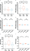
TRB physicochemical properties in TILs and peripheral T cells from healthy donors. CDR3nt length and NDNnt length analysis of TRB CDR3nt sequences in TILs samples (A, B), respectively) and PBMCs from healthy donors (C, D), respectively). TILs-initial and TILs-CD4+ samples presented significantly larger TRB NDNnt sequences compared with TILs-CD8+ samples. This difference was not observed in the PBMCs samples. The charge level of TRB CDR3 sequences in TILs (E) and PBMCs (F) revealed a significantly higher usage of positively charged residues in TILs-CD4+ samples compared to TILs-initial and TILs-CD8+ samples. This difference was also observed in CD4+ samples from PBMCs, both before (P-CD4+ vs. P-CD8+) and after (eP-CD4+ vs. eP-CD8+) expansions. TILs samples derived from BC biopsies and control samples used are summarized in Supplementary Tables 1 and 2 , respectively. Values from each sample are available in Supplementary Data . ns (not significant), p >0.05; *, p ≤ 0.05; **, p ≤ 0.01.
Figure 3
Figure 3
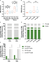
TRB diversity in TILs and peripheral T cells from healthy donors. (A) TRB diversity analysis calculated using all samples did not show differences between groups. (B) The paired analysis of TRB diversity calculated using only the values from slices from which TILs-CD4+ and TILs-CD8+ samples were available revealed a significant higher diversity in the CD8+ group. (C) TRB diversity levels were not significantly different between CD4+ and CD8+ T cells from PBMCs neither before (P-CD4+ vs. P-CD8+) not after (eP-CD4+ vs. eP-CD8+) in vitro expansions. The TRB diversity inversely correlated with the percentage of CD4+ T cells in the TILs-initial samples (D) but no correlation was observed with the percentage of CD8+ T cells (E). TILs samples derived from BC biopsies and control samples used are summarized in Supplementary Tables 1 and 2 , respectively. Values from each sample are available in Supplementary Data . ns (not significant), p >0.05; *, p ≤ 0.05; **, p ≤ 0.01.
Figure 4
Figure 4
TRB CR level and percentage of public sequences in TILs and peripheral T cells from healthy donors. (A) The TRB CR level of TILs samples calculated by biopsy and by section (slices) did not show significant differences between different groups of samples. In the biopsy analysis, a significantly higher CR level was observed bot in the TILs-CD4+ and the TILs-CD8+ samples compared to the TILs-initial samples, indicating that certain convergent sequences in different slices were selected after expansions. (B) No differences were observed in the CR level of T cells from PBMCs, neither before (P-CD4+ vs. P-CD8+) not after (eP-CD4+ vs. eP-CD8+) in vitro expansions. (C) Similar percentages of TRB public sequences were observed in TILs-CD4+ and TILs-CD8+ samples. (D) The percentage of TRB public sequences in PBMCs was higher than in TILs, but no differences were observed between CD4+ and CD8+ T cells, neither before (P-CD4+ vs. P-CD8+) not after (eP-CD4+ vs. eP-CD8+) in vitro expansions. (E) The percentage of TRB public sequences was higher among the convergent compared to the non-convergent group of sequences in both TILs groups of samples. TILs samples derived from BC biopsies and control samples used are summarized in Supplementary Tables 1 and 2 , respectively. Values from each sample are available in Supplementary Data . CR, convergent sequences; nonCR, nonconvergent sequences. ns (not significant), p >0.05; **, p ≤ 0.01; ***, ****, p ≤ 0.0001.
Figure 5
Figure 5
TRB CDR3aa motifs in TILs-CD4+ and TILs-CD8 samples. (A) Number of motifs obtained by GLIPH2 (16, 17) and the number of TRB CDR3 sequences comprising them in the different biopsies analyzed. Normalized fractions of clustered TRB sequences in the TILs-CD4+ (B) and the TILs-CD8+ (C) samples. A higher number of motifs composed by a higher number of sequences was obtained among TILs-CD4+ compared to TILs-CD8+ samples. Only those motifs with a Fisher’s exact test score and expansion score <0.5 were selected. Note that the absence of CD4+ and CD8+ clusters in the BTLQ2 and BTLQ15 biopsies, respectively, was due to the lack of samples in these biopsies as purified populations could not be obtained.
Figure 6
Figure 6
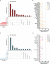
TRB CDR3aa shared motifs and amino acid sequences. UpsetPlots show the number of motifs found in the TILs-CD4+ (A) and the TILs-CD8+ (C) in each biopsy and their exclusivity and/or privacy in the different biopsies analyzed. A higher number of shared motifs was observed among TILs-CD4+ samples. Amino acid sequences of motifs found in the TILs-CD4+ (B) and TILs-CD8+ (D) samples and the number of CDR3aa sequences containing each motif. GLIPH2 (16, 17) was used for clustering and only those motifs with a Fisher’s exact test score and expansion score <0.5 were selected.
Figure 7
Figure 7
TRB CDR3 sequences found in the McPAS-TCR (18) database and annotated sequences using motifs. The number of appearances of sequences described in the literature as tumor-specific, grouped by the cancer type or antigen etiology in TILs-CD4+ (A) and TILs-CD8+ samples (B). Frequency of tumor-specific annotated sequences in the TILs-CD4+ (C) and TILs-CD8+ samples (D). Sequences with an * are those which were described in the literature, while the others are manually annotated. The sequence in bold highlights the motif contained in that cluster and the biopsies in which they were found are noted next to the frequency bars.

Similar articles

Cited by

References

    1. Hanahan D, Weinberg RA. Hallmarks of cancer: the next generation. Cell (2011) 144:646–74. doi: 10.1016/j.cell.2011.02.013 - DOI - PubMed
    1. Couzin-Frankel J. Breakthrough of the year 2013. Cancer immunotherapy. Science (2013) 342:1432–3. doi: 10.1126/science.342.6165.1432 - DOI - PubMed
    1. Cortes J, Rugo HS, Cescon DW, Im S-A, Yusof MM, Gallardo C, et al. . Pembrolizumab plus chemotherapy in advanced triple-negative breast cancer. N Engl J Med (2022) 387:217–26. doi: 10.1056/NEJMoa2202809 - DOI - PubMed
    1. Zacharakis N, Chinnasamy H, Black M, Xu H, Lu Y-C, Zheng Z, et al. . Immune recognition of somatic mutations leading to complete durable regression in metastatic breast cancer. Nat Med (2018) 24:724–30. doi: 10.1038/s41591-018-0040-8 - DOI - PMC - PubMed
    1. Zhang Y, Wang S, Yang B, Lu S, Du Y, Liu H. Adjuvant treatment for triple-negative breast cancer: a retrospective study of immunotherapy with autologous cytokine-induced killer cells in 294 patients. Cancer Biol Med (2019) 16:350–60. doi: 10.20892/j.issn.2095-3941.2018.0378 - DOI - PMC - PubMed

Publication types