. 2023 Mar 3;5(1):zcad013.
doi: 10.1093/narcan/zcad013. eCollection 2023 Mar.

R-loops, type I topoisomerases and cancer

Affiliations

R-loops, type I topoisomerases and cancer

Sourav Saha et al. NAR Cancer. .

Abstract

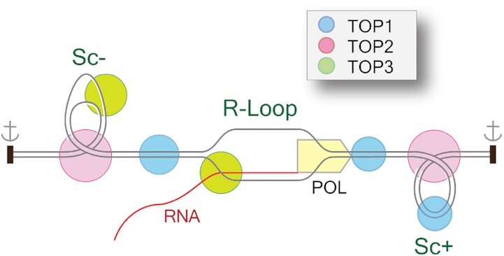
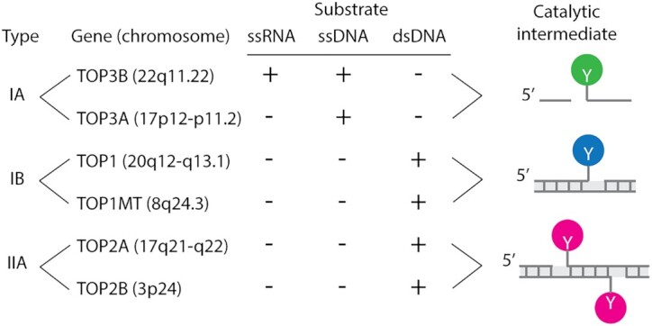
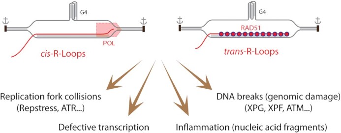
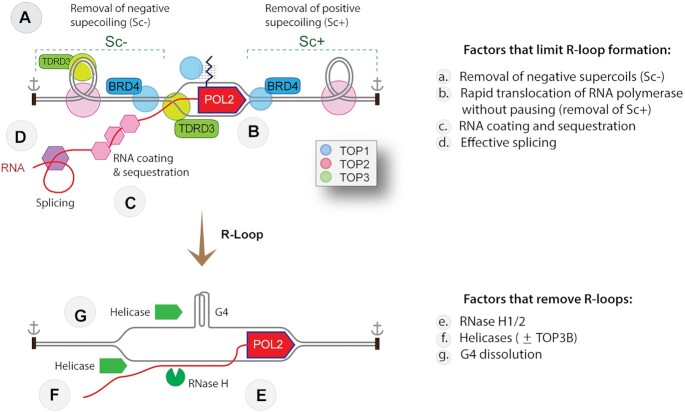
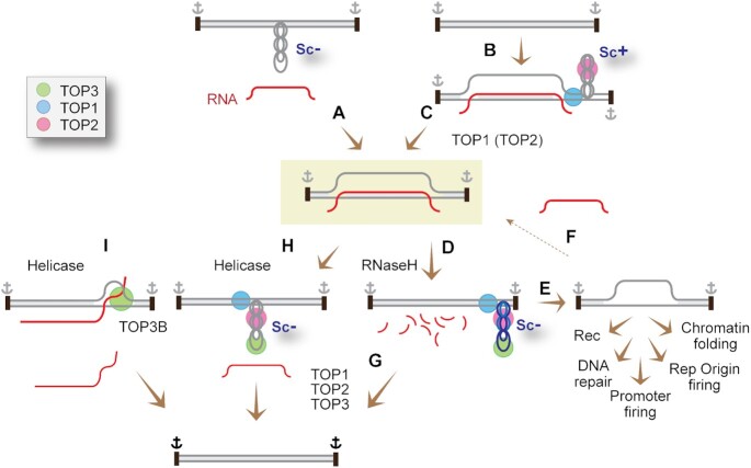
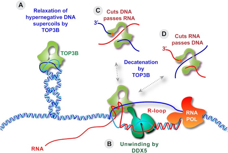
R-loops are abundant and dynamic structures ubiquitously present in human cells both in the nuclear and mitochondrial genomes. They form in cis in the wake of transcription complexes and in trans apart from transcription complexes. In this review, we focus on the relationship between R-loops and topoisomerases, and cancer genomics and therapies. We summarize the topological parameters associated with the formation and resolution of R-loops, which absorb and release high levels of genomic negative supercoiling (Sc-). We review the deleterious consequences of excessive R-loops and rationalize how human type IA (TOP3B) and type IB (TOP1) topoisomerases regulate and resolve R-loops in coordination with helicase and RNase H enzymes. We also review the drugs (topoisomerase inhibitors, splicing inhibitors, G4 stabilizing ligands) and cancer predisposing genes (BRCA1/2, transcription, and splicing genes) known to induce R-loops, and whether stabilizing R-loops and thereby inducing genomic damage can be viewed as a strategy for cancer treatment.


Figures

Graphical Abstract
Graphical Abstract
Topoisomerases (TOP1, TOP2 and TOP3B) can remove DNA supercoiling to prevent R-loop formation while TOP3B coordinates with helicase DDX5 and resolves R-loops by decatenation mechanism.
Figure 1.
Figure 1.
Classification and distinctive features of human topoisomerases. The catalytic intermediates (cleavage complexes) are schematically drawn at right (the Y’s refer to the catalytic tyrosyl residues that form the reversible cleavage complexes). Type IA topoisomerases cleave nucleic acids in single-stranded regions (ssRNA and ssDNA), whereas Type II topoisomerases cleave double-stranded DNA (dsDNA) only. Type IA topoisomerases are conserved in all domains of life and considered the most ancestral topoisomerase enzymes. TOP3B is the only RNA topoisomerase. Type IA enzymes can only relax hypernegative DNA. Type IB topoisomerases are highly efficient ‘swivelases’ (‘DNA untwisting enzymes’) removing both positive (Sc+) and negative DNA supercoiling (Sc−). TOP1 acts on the nuclear genome while TOP1MT acts on the mitochondrial genome (mtDNA). TOP2 enzymes relax both Sc+ and Sc− by forming DNA double-strand breaks (with a 4 bp stagger) in a DNA duplex region allowing the passage of another DNA segment through the cleavage complex. Upon completion of the DNA passage, TOP2 rejoins the DNA. TOP2-mediated strand passage is also essential for DNA decatenation during replication. Shaded rectangles represent canonical helical B-DNA with base pairs as short vertical bars.
Figure 2.
Figure 2.
Deleterious effects of R-loops. Unresolved and excessive cis- and trans-R-loops have been reported to have multiple deleterious effects. Watson-Crick helical double-stranded nucleic acids are shaded in grey. DNA is shown in gray and RNA in red. A guanosine quartet structure is included as stabilizer of R-loops. RAD51 is implicated in the formation of trans-R-loops. Replication fork collisions give rise to replication stress (RepStress) and engagement of the ATR/CHK1 pathways. Stable R-loops also stall transcription complexes. DNA breaks are produced by the nucleotide excision repair nucleases (XPG and XPF), leading to DNA double-strand breaks and activation of ATM. Released nucleic acid fragments can initiate inflammation.
Figure 3.
Figure 3.
Cellular mechanisms controlling R-loops associated with transcription (cis-R-loops). The top scheme represents an elongating RNA POL2 complex with its associated topological features within a chromatin topologically constrained domain whose boundaries are schematized as black rectangles with anchor signs. Duplex canonical DNA is shown as parallel lines (without showing the Watson-Crick helical turns for simplicity). Factors that limit R-loop formation and remove R-loops are listed at right, annotated in the figure, and discussed in the text. Symbols for the human topoisomerases are in the inset.
Figure 4.
Figure 4.
Topological considerations for the formation (A−C) and removal (D−I) of R-loops. The formation of R-loops (A−C) is associated with (and requires) topological adjustments of the DNA within topologically constrained chromatin domains whose boundaries are schematized as black rectangles with anchor signs. (A) R-loops readily form in hypernegatively supercoiled (Sc−) domains (see Figure 2 for co-transcriptional R-loops formed in cis of transcription complexes). In doing so, R-loops constrain the Sc− (one negative superhelical turn (Sc−) per 10.5 bp of helical RNA-DNA hybrid). (B) to form R-loops by RNA strand invasion (R-loops formed in trans) (as in the case of CRISPR-Cas9 and TERRA), the DNA topology needs to be adjusted by removing the Sc+ (C) formed to compensate for hybridization of the RNA-DNA heteroduplex. (D) R-loops can be removed by degradation of the hybridized RNA by RNase H1 (and H2). (E) removal of the RNA releases the corresponding Sc−, promoting chromatin folding (one nucleosome absorbs one Sc−), replication origin and promoter firing, DNA repair and recombinations. (F) Sc- can help capture an RNA to reform R-loops. (G) TOP1 and TOP2 can relax the Sc− released by the digestion of RNA in double-stranded DNA, and TOP3 can relax the hypernegative Sc- in single-stranded segments generated by the hypernegative Sc−. (H) Helicases by dissociating the RNA from the heteroduplex also release the Sc− that was constrained in the R-loop (≈20 Sc− for a 200 bp R-loop). (I) By pushing the RNA−DNA hybrids to the end of the R-loops, the helicases can generate knotted DNA−RNA structures that are resolved (decatenated) by TOP3B in the single-stranded segment of the RNA and/or DNA.
Figure 5.
Figure 5.
Proposed molecular mechanisms by which TOP3B can suppress R-loops (101). (A) By cleaving single-stranded DNA and passing the complementary strand through the gap, TOP3B can reduce hypernegative Sc and prevent the formation or destabilize R-loops. (B) Following the unwinding of the DNA–RNA heteroduplex of the R-loop by DDX5, TOP3B can decatenate the nucleic acids at the junction of the R-loop by cutting a DNA single-strand and passing the RNA through the gap (C), or by cutting the RNA and passing a DNA single-strand through the gap (D).

References

    1. Pommier Y., Nussenzweig A., Takeda S., Austin C. Human topoisomerases and their roles in genome stability and organization. Nat. Rev. Mol. Cell Biol. 2022; 23:407–427. - PMC - PubMed
    1. Pommier Y., Sun Y., Huang S.N., Nitiss J.L. Roles of eukaryotic topoisomerases in transcription, replication and genomic stability. Nat. Rev. Mol. Cell Biol. 2016; 17:703–721. - PMC - PubMed
    1. Champoux J.J. DNA topoisomerases: structure, function, and mechanism. Annu. Rev. Biochem. 2001; 70:369–413. - PubMed
    1. Wang J.C. Cellular roles of DNA topoisomerases: a molecular perspective. Nat. Rev. Mol. Cell Biol. 2002; 3:430–440. - PubMed
    1. Vos S.M., Tretter E.M., Schmidt B.H., Berger J.M. All tangled up: how cells direct, manage and exploit topoisomerase function. Nat. Rev. Mol. Cell Biol. 2011; 12:827–841. - PMC - PubMed