

Case Reports
doi: 10.1016/j.radcr.2023.07.070.
eCollection 2023 Oct.
Myocardial metastasis within lipomatous hypertrophy of the interatrial septum
Affiliations
- PMID: 37601124
- PMCID: PMC10432261
- DOI: 10.1016/j.radcr.2023.07.070
Item in Clipboard
Case Reports
Myocardial metastasis within lipomatous hypertrophy of the interatrial septum
Radiol Case Rep.
.
Abstract
A 74-year-old lady with lipomatous hypertrophy of the interatrial septum presented with symptomatic anemia. Imaging revealed a new diagnosis of metastatic cancer of presumed lung origin, with a new soft tissue myocardial lesion adjacent to the right atrium within the region of lipomatous hypertrophy. This was favored to represent a myocardial metastasis within concurrent lipomatous hypertrophy of the interatrial septum.
Keywords: Cardiac mass; Computed tomography; Lipomatous hypertrophy of interatrial septum; Lung cancer; Myocardial metastasis.
© 2023 The Authors. Published by Elsevier Inc. on behalf of University of Washington.
Figures
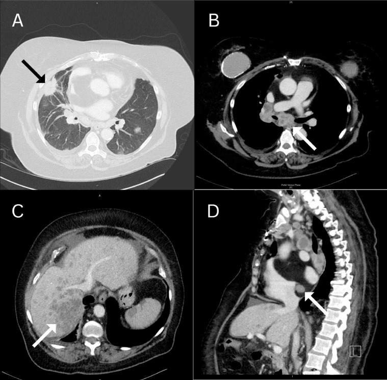
Computed tomography scans demonstrate the presence of a spiculated right middle lobe pulmonary parenchymal mass (black arrow, A) suspicious of a primary lung malignancy. There are enlarged mediastinal necrotic lymph nodes (white arrow, B) as well as multiple hypodense hepatic lesions (white arrow, C), favored to represent nodal and hepatic metastases. There is a myocardial soft tissue lesion seen adjacent to the right atrium within fatty tissue (white arrow, D).
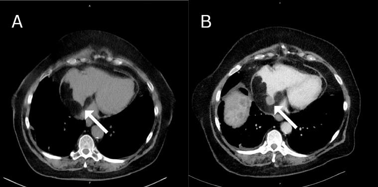
Axial noncontrast chest CT from 1 year prior demonstrates lipomatous hypertrophy of the interatrial septum (white arrow, A). Side-by-side comparison to the axial contrast-enhanced chest CT 1 year after demonstrates interval development of the new myocardial soft tissue lesion within the pre-existing LHIS (white arrow, B).
Similar articles
-
Lipomatous hypertrophy of the interatrial septum: the typical echographic aspect is worth being known.Acta Anaesthesiol Belg. 2011;62(3):157-9. Acta Anaesthesiol Belg. 2011. PMID: 22145258
-
A rare case of cardiac tumor of the interventricular septum complicated with atrioventricular block.J Cardiol Cases. 2022 Sep 29;26(6):419-422. doi: 10.1016/j.jccase.2022.09.002. eCollection 2022 Dec. J Cardiol Cases. 2022. PMID: 36506495 Free PMC article.
-
A rare case of regressively changed lipomatous hypertrophy of the interatrial septum presenting with anemia and recurrent fever.Cardiovasc Pathol. 2016 Mar-Apr;25(2):161-4. doi: 10.1016/j.carpath.2015.09.002. Epub 2015 Sep 24. Cardiovasc Pathol. 2016. PMID: 26453091
-
Lipomatous hypertrophy of the interatrial septum: an overview.Arch Pathol Lab Med. 2006 Mar;130(3):397-9. doi: 10.5858/2006-130-397-LHOTIS. Arch Pathol Lab Med. 2006. PMID: 16519573 Review.
-
[Lipomatous hypertrophy of the interatrial septum: its assessment with TEE, CT and MRI].G Ital Cardiol. 1998 Nov;28(11):1273-7. G Ital Cardiol. 1998. PMID: 9866805 Review. Italian.
References
-
- Prior J. Lipomatous hypertrophy of cardiac interatrial septum: a lesion resembling hibernoma, lipoblastomatosis and infiltrating lipoma. Arch Pathol. 1964;78:11–15. - PubMed
-
- Maleszewski J, Bols M, Bois J, Young P, Stulak J, Klarich K. Neoplasia and the heart: pathological review of effects with clinical and radiological correlation. J Am Coll Cardiol. 2018;72:202–227. - PubMed
-
- Patsia L, Lartsuliani K, Intskirveli N, Ratiani L. Lipomatous hypertrophy of the interatrial septum- a benign heart anomaly causing unexpected problem in electrophysiology (case report) Georgian Med News. 2021;318:72–74. - PubMed
Publication types
LinkOut - more resources
Full Text Sources
