Review
. 2023 Aug 8:31:53-62.
doi: 10.1016/j.bioactmat.2023.07.026. eCollection 2024 Jan.

Manganese molybdate nanodots with dual amplification of STING activation for "cycle" treatment of metalloimmunotherapy

Affiliations
Review

Manganese molybdate nanodots with dual amplification of STING activation for "cycle" treatment of metalloimmunotherapy

Huali Lei et al. Bioact Mater. .

Abstract

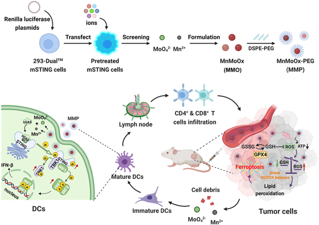
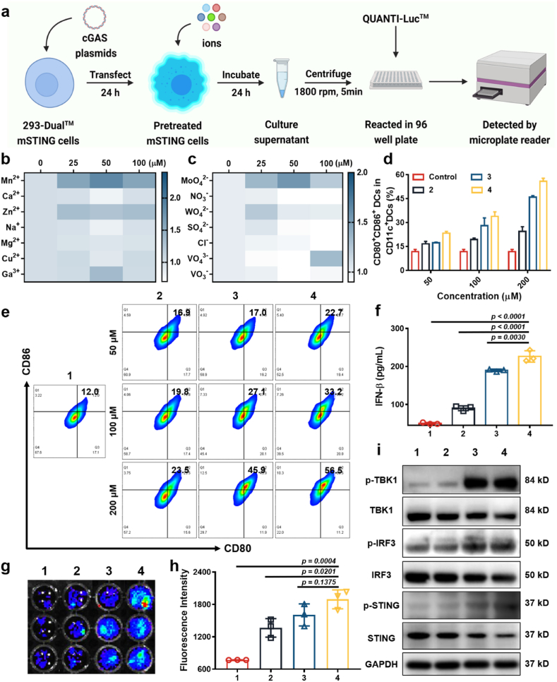
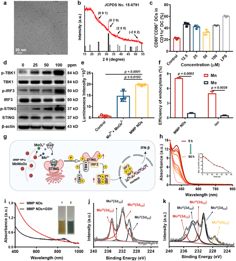
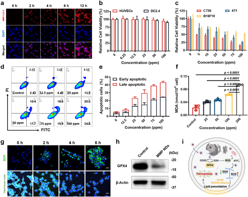
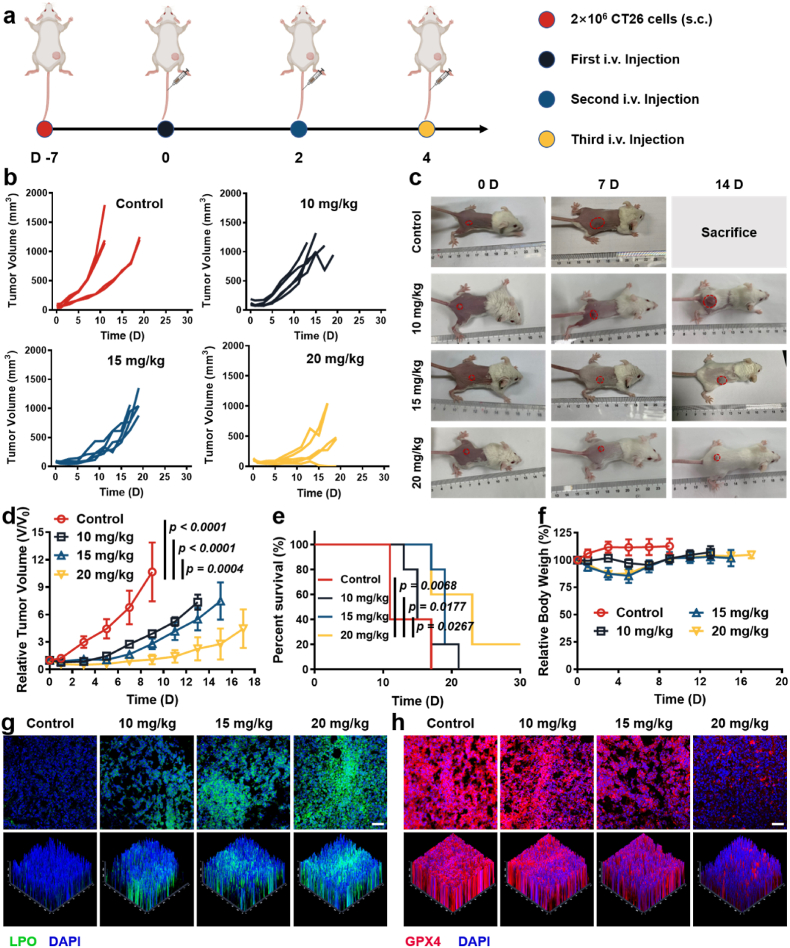
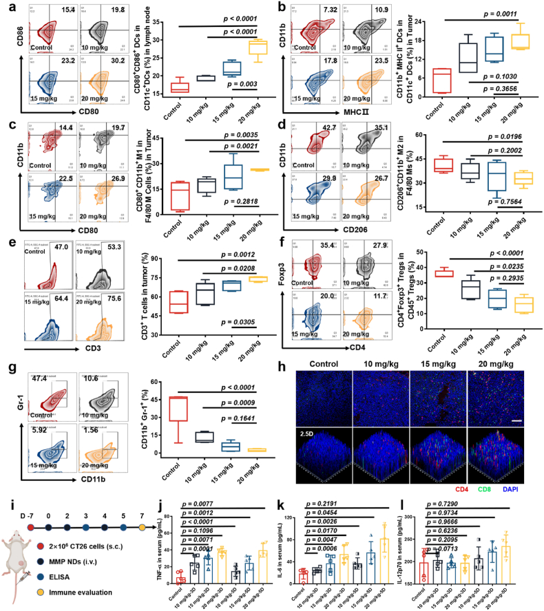
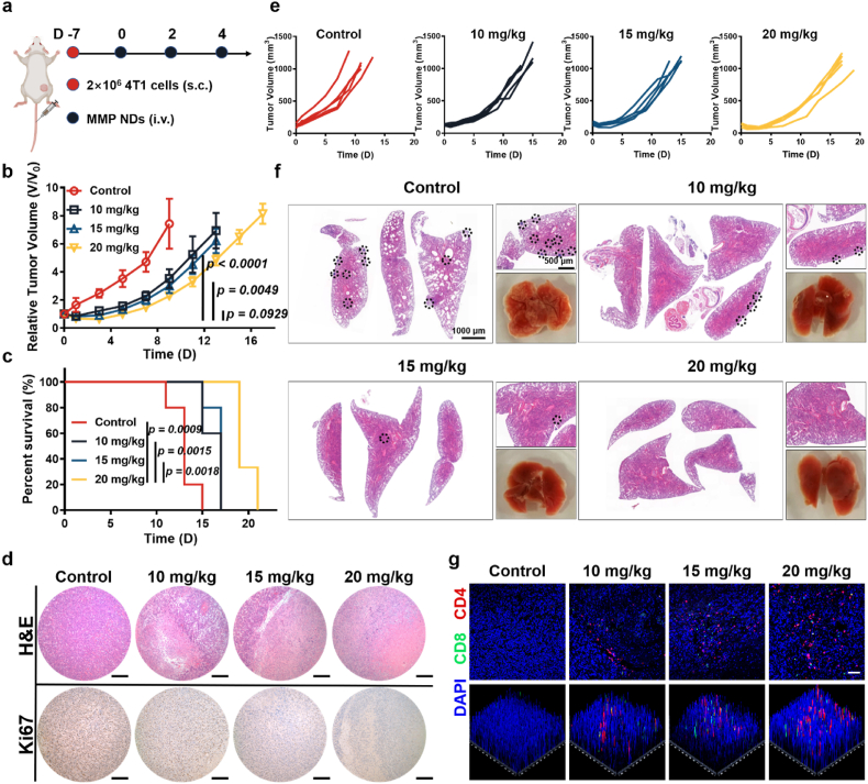
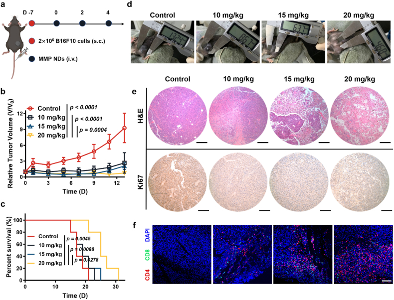
Certain types of cationic metal ions, such as Mn2+ are able to activate immune functions via the stimulator of interferon genes (STING) pathway, showing potential applications in eliciting antitumor immunity. How anionic ions interact with immune cells remains largely unknown. Herein, selecting from a range of cationic and anionic ions, we were excited to discover that MoO42- could act as a cGAS-STING agonist and further confirmed the capability of Mn2+ to activate the cGAS-STING pathway. Inspired by such findings, we synthesized manganese molybdate nanoparticles with polyethylene glycol modification (MMP NDs) for cancer metalloimmunotherapy. Meanwhile, MMP NDs could consume glutathione (GSH) over-expressed in tumors and induce ferroptosis owing to high-valence Mo and Mn to elicit tumor-specific immune responses, which was further amplified by MMP-triggered the cGAS-STING activation. In turn, activated CD8+ T cells to secrete high levels of interferon γ (IFN-γ) and reduced GPX4 expression in tumor cells to trigger ferroptosis-specific lipid peroxidation, which constituted a "cycle" of therapy. As a result, the metalloimmunotherapy with systemic administration of MMP NDs offered a remarkable tumor inhibition effect for a variety of tumor models. Our work for the first time discovered the ability of anionic metal ions to activate the immune system and rationally designed bimetallic oxide nanostructures as a multifunctional therapeutic nanoplatform for tumor immunotherapy.

Keywords: DCs maturation; Ferroptosis; Manganese molybdate nanoparticles; Metalloimmunotherapy; cGAS-STING pathway.


Conflict of interest statement

The authors declare that they have no known competing financial interests or personal relationships that could have appeared to influence the work reported in this paper.

Figures

Image 1
Graphical abstract
Scheme 1
Scheme 1
MMP NDs with dual cGAS-STING activation for cancer metalloimmunotherapy. Metal cations and anions with the strongest cGAS-STING activation were screened using 293-Dual™ mSTING cells and then employed to construct bimetallic oxide MMO NDs, then MMP NDs were obtained by DSPE-PEG5k modification. MMP NDs containing Mn2+ and MoO42− showed effective cGAS-STING activation. After systemic administration, MMP NDs could consume the highly expressed GSH in tumor cells, break the redox balance, inhibit the activity of the GPX4 enzyme, and subsequently lead tumor cells ferroptosis. Tumor debris in the presence of MMP NDs would then trigger tumor-specific cell immunity.
Fig. 1
Fig. 1
Screening for ions with the ability to activate the cGAS-STING pathway. (a) Scheme of ion screening with 293-Dual™ mSTING cells. (b&c) 293-Dual™ mSTING cells containing cGAS were treated with different concentrations of metal cations (b) or anions (c). (d) The percentages of mature DCs in (e). (e) The detection of BMDCs maturation after incubation with different concentrations of Mn2+, MoO42− and the mixtures of Mn2+ and MoO42− for 16 h. (f) Quantitative analysis of IFN-β secreted by DCs. (g) Qualification of bioluminescence intensities of mSTING cells after incubation with Mn2+, MoO42− and mixtures of Mn2+ and MoO42− for 24 h. (h) Fluorescence intensity of mSTING cells in (g). (i) HeLa cells were incubated with Mn2+, MoO42− and mixtures of Mn2+ and MoO42− for 16 h, following with WB detection of marker proteins in the cGAS-STING pathway, including TBK1/p-TBK1, STING/p-STING, and IRF-3/p-IRF-3. (1: control, 2: MoO42−, 3: Mn2+, and 4: Mn2++MoO42−).
Fig. 2
Fig. 2
Characterization and properties of MMP NDs. (a) TEM image of MMO NDs. (b) XRD patterns of MMO NDs. (c) The detection of BMDCs maturation (CD80+CD86+ cells among CD11c+ DCs) after incubation with MMP NDs for 16 h. (d) HeLa cells were incubated with different concentrations of MMP NDs for 16 h, followed by WB for marker proteins in the STING–IFN–β pathway including TBK1/p-TBK1, STING/p-STING, and IRF-3/p-IRF-3. (e) Quantification of the bioluminescence intensities of mSTING cells after incubation with MMP NDs and the mixtures of Mn2+ and MoO42− for 24 h. (f) The cellular uptake of Mn2+ or MMP NDs by HeLa cells was detected by ICP-MS. (g) Illustration showing that MMP NDs after endocytosis may be gradually decomposed into Mn2+ and MoO42−, which would activate the cGAS-STING pathway. NDs. (h) Time-dependent GSH (1 mM) consumption by MMP NDs. (i) UV absorption and optical photos of MMP NDs before and after reaction with GSH, 1) MMP NDs, 2) MMP NDs + GSH. (j&k) XPS of Mo in MMP NDs before (j) and after (k) reaction with GSH.
Fig. 3
Fig. 3
MMP NDs induced ferroptosis. (a) Endocytosis of MMP-Cy5.5 NDs (scale bar: 20 μm). (b&c) Relative viabilities of (b) normal cells (HUVECs and DC2.4) and (c) tumor cells (CT26, 4T1 and B16F10) after incubation with different concentrations of MMP NDs for 12 h. (d) Apoptosis detection of CT26 cells after incubation with MMP NDs. (e) Statistical data of apoptosis in (d). (f) MDA content in MMP NDs treated CT26 cells. (g) MMP NDs treated CT26 cells were stained with DCFH-DA and ThiolTracker violet probe (scale bar: 20 μm). (h) The expression of GPX4 in MMP NDs treated CT26 cells was evaluated by WB. (i) Scheme of ferroptosis of tumor cells triggered by MMP NDs.
Fig. 4
Fig. 4
Systemic administration of MMP NDs to treat CT26 tumors. (a) Scheme of systemic administration of MMP NDs in CT26 tumor bearing mice. (b) Tumor sizes of mice after various treatments, including 1) control, 2) MMP NDs (i.v. 10 mg/kg), 3) MMP NDs (i.v. 15 mg/kg) and 4) MMP NDs (i.v. 20 mg/kg). (c) Optical photos of mice after various treatments in (b). (d) Relative CT26 tumor volume in (b). (e) Percent survival of mice in (b). (f) Relative body weights of the mice with different treatments in (b). (g&h) Fluorescence images of tumor slices after staining with LPO (g) and GPX4 (h) (scale bar: 50 μm).
Fig. 5
Fig. 5
Evaluations of the immune responses triggered by MMP NDs in mice. (ag) Flow cytometric plots for the analysis of DC maturation in the TDLNs (a), DC maturation in tumors (b), CD11b+ CD80+ M1 macrophages in tumors (c), M2 cells in tumors (d), CD3+ T cells (e), Tregs in tumors (f) and CD11b+Gr-1+ MDSCs in tumors (g). (h) Immunofluorescence staining images showing CD4+CD8+ T cells in CT26 tumors (scale bar: 100 μm). (i) Scheme of immune evaluation after various treatments in CT26 tumor bearing mice. (jl) TNF-α (j), IL-6 (k) and IL-12p70 (l) level in sera from mice post MMP NDs treatment on the day 3 and 5.
Fig. 6
Fig. 6
Systemic administration of MMP NDs to treat 4T1 tumors. (a) Scheme of systemic administration of MMP NDs in 4T1 tumor bearing mice. (b) Relative 4T1 tumor volume in (e). (c) Percent survival of mice in (e). (d) Images of 4T1 tumor slices after staining with H&E and Ki67 (scale bar: 200 μm). (e) Tumor sizes of mice with 4T1 tumors after various treatments, including 1) control, 2) MMP NDs (i.v. 10 mg/kg), 3) MMP NDs (i.v. 15 mg/kg) and 4) MMP NDs (i.v. 20 mg/kg). (f) H&E staining images of lungs collected from mice after various treatments in (e). (g) CD4+CD8+ T cells in CD3+ T cells in 4T1 tumors.
Fig. 7
Fig. 7
Systemic administration of MMP NDs to treat B16F10 tumors. (a) Scheme of systemic administration of MMP NDs in B16F10 tumor bearing mice. (b) Relative B16F10 tumor volume in (d). (c) Percent survival of mice in (d). (d) Optical photos of mice bearing B16F10 tumors after various treatments including 1) control, 2) MMP NDs (i.v. 10 mg/kg), 3) MMP NDs (i.v. 15 mg/kg) and 4) MMP NDs (i.v. 20 mg/kg). (e) Images of B16F10 tumor slices after staining with H&E and Ki67 (scale bar: 200 μm). (f) CD4+CD8+ T cells in CD3+ T cells in B16F10 tumors (scale bar: 100 μm).

References

    1. Kuai R., Ochyl L.J., Bahjat K.S., Schwendeman A., Moon J.J. Designer vaccine nanodiscs for personalized cancer immunotherapy. Nat. Mater. 2017;16:489–496. - PMC - PubMed
    1. Liang J.L., Luo G.F., Chen W.H., Zhang X.Z. Recent advances in engineered materials for immunotherapy-involved combination cancer therapy. Adv. Mater. 2021;33 202007630. - PubMed
    1. Gravitz L. Cancer immunotherapy. Nature. 2013;504 S1–S1. - PubMed
    1. Ledford H. Immunotherapy’s cancer remit widens. Nature. 2013;497 544–544. - PubMed
    1. Yang N.L., Gong F., Cheng L., Lei H.L., Li W., Sun Z.B., Ni C.F., Wang Z.H., Liu Z. Biodegradable magnesium alloy with eddy thermal effect for effective and accurate magnetic hyperthermia ablation of tumors. Natl. Sci. Rev. 2021;8:nwaa122. - PMC - PubMed