Review
. 2023 Jul 31:13:1226289.
doi: 10.3389/fonc.2023.1226289. eCollection 2023.

Targeting MCL-1 protein to treat cancer: opportunities and challenges

Affiliations
Review

Targeting MCL-1 protein to treat cancer: opportunities and challenges

Shady I Tantawy et al. Front Oncol. .

Abstract

Evading apoptosis has been linked to tumor development and chemoresistance. One mechanism for this evasion is the overexpression of prosurvival B-cell lymphoma-2 (BCL-2) family proteins, which gives cancer cells a survival advantage. Mcl-1, a member of the BCL-2 family, is among the most frequently amplified genes in cancer. Targeting myeloid cell leukemia-1 (MCL-1) protein is a successful strategy to induce apoptosis and overcome tumor resistance to chemotherapy and targeted therapy. Various strategies to inhibit the antiapoptotic activity of MCL-1 protein, including transcription, translation, and the degradation of MCL-1 protein, have been tested. Neutralizing MCL-1's function by targeting its interactions with other proteins via BCL-2 interacting mediator (BIM)S2A has been shown to be an equally effective approach. Encouraged by the design of venetoclax and its efficacy in chronic lymphocytic leukemia, scientists have developed other BCL-2 homology (BH3) mimetics-particularly MCL-1 inhibitors (MCL-1i)-that are currently in clinical trials for various cancers. While extensive reviews of MCL-1i are available, critical analyses focusing on the challenges of MCL-1i and their optimization are lacking. In this review, we discuss the current knowledge regarding clinically relevant MCL-1i and focus on predictive biomarkers of response, mechanisms of resistance, major issues associated with use of MCL-1i, and the future use of and maximization of the benefits from these agents.

Keywords: BCL-2 family; BCL-2 family proteins; MCL-1 inhibitors; MCL-1 protein; apoptosis; cancer therapy.


Conflict of interest statement

Author VG has received research grants from Pharmacyclics, Acerta, Gilead, Sunesis, ClearCreek Bio, Infinity, and Loxo Oncology. The remaining authors declare that the research was conducted in the absence of any commercial or financial relationships that could be construed as a potential conflict of interest.

Figures

Figure 1
Figure 1
Structure of myeloid cell leukemia-1 (MCL-1) protein. MCL-1 protein consists of 350 amino acids, highlighting several important post-translational modification sites. Two caspase cleavage sites (aspartic acid [Asp] 127 and 157), 5 lysine [Lys] residues (5, 40, 136, 194, and 196) for ubiquitination, and 6 phosphorylation sites (serine [Ser] 64, threonine [Thr] 92, Ser 121, 155, and 159, and Thr 163) are indicated. The N-terminus of the protein is largely unstructured and includes 4 proline, glutamate, serine, and threonine (PEST) regions (2 major and 2 minor, labeled as “PEST” and “pest,” respectively). Four BCL-2 homology (BH) domains (BH1-BH4) are located near the C-terminus, which also contains the transmembrane (TM) domain necessary for mitochondrial localization.
Figure 2
Figure 2
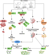
Targeting myeloid cell leukemia-1 (MCL-1) protein. MCL-1 protein can be targeted either directly, by disrupting MCL-1 interaction with proapoptotic proteins (#3), or indirectly by targeting MCL-1 protein at stages of transcription(#1), translation(#2) or degradation(#4). Cyclin-dependent kinase (CDK)7/9 inhibitors or microRNA can block the transcription of MCL-1 messenger RNA (mRNA). SF3B1 inhibitors can generate a proapoptotic MCL-1S isoform by switching on alternative splicing. Once transcribed, MCL-1 mRNA moves to the cytoplasm for translation into MCL-1 protein (depicted in green). This translation process can be prevented with phosphatidylinositol-3 kinase (PI3K)/mammalian target of rapamycin (mTOR) inhibitors or epidermal growth factor receptor (EGFR)/vascular endothelial growth factor receptor (VEGFR) inhibitors. After translation, MCL-1 protein binds and deactivates the BAK/BAX complex (depicted in red/pink) in the outer mitochondrial membrane. This binding can be disrupted with direct MCL-1 inhibitors), leading to BAX/BAK dissociation and apoptosis. Additionally, the proteasomal degradation of MCL-1 can be enhanced using glycogen synthase kinase 3β (GSK3β) stimulants, extracellular signal-regulated kinase (ERK) inhibitors, the proteolysis-targeting chimera (PROTAC), or deubiquitinating enzymes (DUB) inhibitors (e.g., WP1130), thereby increasing MCL-1 polyubiquitination and subsequent degradation. CREB, cAMP response element-binding protein; GMCSF, Granulocyte-macrophage colony-stimulating factor; HIF-1α, Hypoxia-inducible factor 1-alpha; IL, interleukin; Ser, serine; STAT, signal transducer and activator of transcription; Thr, threonine.
Figure 3
Figure 3
Mechanisms of myeloid cell leukemia-1 (MCL-1) protein upregulation by MCL-1 inhibitors. MCL-1i bind to the BCL-2 homology 3 (BH3) domain of MCL-1 and directly induce a conformation change/a state of MCL-1 that favors deubiquitination by deubiquitinating enzymes (DUBs [e.g., USP9x] rather than ubiquitination. This deubiquitination is further enhanced by Noxa disruption and rapid degradation, leading to enhanced DUBs activity on the MCL-1 protein. Additionally, the binding of MCL-1i transiently decreases the expression of the E3 ligase Mule and increases MEK/ERK-mediated threonine (Thr)163 phosphorylation of MCL-1, thus further contributing to the observed protein stability. Despite this upregulation of MCL-1 protein, MCL-1i disrupted the MCL-1–BAK/BAX interaction to induce apoptosis. The exact mechanism for MCL-1 upregulation was not studied using S63415, however, it is predicted to be the same owing to similar structure and function. Figure is adapted from Tantawy, et al, Clinical Cancer Research).
Figure 4
Figure 4
The rationale for combination therapy using MCL-1 inhibitors. Myeloid cell leukemia-1 (MCL-1), B-cell lymphoma (BCL)-2 extra large (BCL-xL), and BCL-2 prosurvival proteins cooperate to bind and inactivate BAX, BAK complex, and other proapoptotic BCL-2 homology (BH)3-only proteins (Noxa, BIM, PUMA, BID, and BAD). When one of these prosurvival proteins loses its function (e.g., from MCL-1 inhibition by MCL-1i), the other proteins (BCL-2 and BCL-xL) may at least partially compensate for this loss. Thus, combination therapy using MCL-1i with a BCL-2 inhibitor (e.g., venetoclax) or BCL-2/BCL-xL inhibitor (e.g., navitoclax) appears promising and has been shown to be effective. Another way to enhance the activity of MCL-1i is to increase the expression of BH3-only proteins. Trametinib can upregulate BIM, which can then bind to MCL-1, thus increasing dependency on MCL-1 protein and subsequently increasing sensitivity to MCL-1i. BIM can also bind to BCL-2 or BCL-xL, indirectly inactivating them. Carfilzomib can upregulate Noxa, which is a BH3-only sensitizer protein that binds to MCL-1, leading to more BH3-only activator proteins (BIM, PUMA, and BID) becoming available to bind to BAX/BAK and activate downstream apoptosis. Chemotherapy induces DNA damage with subsequent activation of TP53 and induction of BAX and Noxa. In addition, various chemotherapeutic agents can downregulate MCL-1. MCL-1i can synergize with chemotherapy by inducing more DNA damage, possibly affecting DNA repair, and neutralizing MCL-1. Effect of the clinically relevant MCL-1i (S63845, AMG-176 and AZD5991) on DNA repair mechanisms needs to be further explored.
Figure 5
Figure 5
Effect of myeloid cell leukemia-1 (MCL-1) deletion or MCL-1 inhibitors on the heart. (A) Mcl-1 knockout in a mouse model led to dilated cardiomyopathy with thin ventricle walls and atrial thrombus, features of impaired cardiac hemodynamics. Ultrastructure changes in the mitochondria, myocyte degeneration, fibrosis, inflammation, and necrosis were observed. Mitochondrial dysfunction and impaired autophagy may have contributed to the observed toxicity. (B) Treating hiPSC-CMs with S68345 induced dynamin-related protein 1 (DRP-1–dependent mitochondrial dysfunction, cytoskeleton disruption, and impaired/irregular beating of the cardiomyocytes. The potential role of Noxa and Mule downregulation on cardiotoxicity needs to be further explored. ATP, adenosine triphosphate; OCR, oxygen consumption rate; OMM, outer mitochondrial membrane.

Similar articles

Cited by

References

    1. Cory S, Adams JM. The Bcl2 family: regulators of the cellular life-or-death switch. Nat Rev Cancer (2002) 2(9):647–56. doi: 10.1038/nrc883 - DOI - PubMed
    1. Kerr JF, Wyllie AH, Currie AR. Apoptosis: a basic biological phenomenon with wideranging implications in tissue kinetics. Br J Cancer (1972) 26(4):239–57. doi: 10.1038/bjc.1972.33 - DOI - PMC - PubMed
    1. Beroukhim R, Mermel CH, Porter D, Wei G, Raychaudhuri S, Donovan J, et al. . The landscape of somatic copy-number alteration across human cancers. Nature (2010) 463(7283):899–905. doi: 10.1038/nature08822 - DOI - PMC - PubMed
    1. Campbell KJ, Dhayade S, Ferrari N, Sims AH, Johnson E, Mason SM, et al. . MCL-1 is a prognostic indicator and drug target in breast cancer. Cell Death Dis (2018) 9(2):1–14. doi: 10.1038/s41419-017-0035-2 - DOI - PMC - PubMed
    1. Kotschy A, Szlavik Z, Murray J, Davidson J, Maragno AL, Le Toumelin-Braizat G, et al. . The MCL1 inhibitor S63845 is tolerable and effective in diverse cancer models. Nature (2016) 538(7626):477–82. doi: 10.1038/nature19830 - DOI - PubMed

LinkOut - more resources