Case Reports
. 2023 Aug 10:2023:1439919.
doi: 10.1155/2023/1439919. eCollection 2023.

"Extreme Nephroptosis": A Kidney in the Inguinal Hernia

Affiliations
Case Reports

"Extreme Nephroptosis": A Kidney in the Inguinal Hernia

Dmytro Shchukin et al. Case Rep Med. .

Abstract

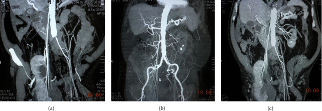
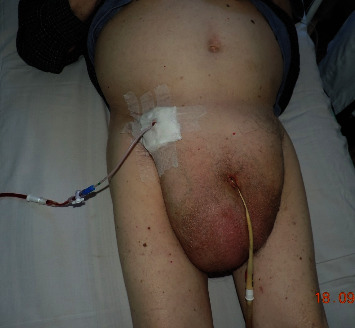
We present an extremely rare case of renal ptosis from the normal orthotopic position into the cavity of inguinal hernia in a 93-year-old male patient. The following clinical case was accompanied by renal insufficiency, which was associated with the obstruction of the right ureter in the hernial sac and the stenosis of the left renal artery. The differential diagnosis between nephroptosis and dystopic kidney was based on MDCT scan images, which demonstrated the length of the right renal artery to be more than 20 cm. The patient underwent percutaneous nephrostomy through the right inguinal area and was successfully followed up for two years. We also analyzed six similar clinical cases described in the literature. This disease has, thus far, been observed exclusively in elderly men with long-standing and large inguinal hernias. The most frequent complications in these patients include ureteral strangulation in the area of the hernial gate and renal failure.


Conflict of interest statement

The authors declare that they have no conflicts of interest.

Figures

Figure 1
Figure 1
Multidetector-computed tomography (MDCT) demonstrates the location of the right kidney in the right-sided inguinal hernia. (a) Frontal reconstruction. (b) Sagittal reconstruction.
Figure 2
Figure 2
Multidetector-computed tomography. Frontal views present a severely elongated right renal artery and stenosis of the left renal artery (a–c).
Figure 3
Figure 3
MDCT. Axial (a) and sagittal (b) views of an enlarged prostate gland and the right kidney located in the right half of the scrotum.
Figure 4
Figure 4
Percutaneous nephrostomy via the right inguinal area.

References

    1. Srirangam S. J., Pollard A. J., Adeyoju A. A. B., O’Reilly P. H. Nephroptosis: seriously misunderstood? BJU International . 2009;103(3):296–300. doi: 10.1111/j.1464-410x.2008.08082.x. - DOI - PubMed
    1. Perone R. W., Del Prado D., Katz D. S. Pelvic kidney presenting as inguinal hernia and acute renal failure. American Journal of Roentgenology . 2007;188(3):p. W305. doi: 10.2214/ajr.06.0502. - DOI - PubMed
    1. Wendler J. J., Baumunk D., Liehr U.-B., Schostak M. Kidney dislocation in a monstrous inguinal intestinal hernia with ureteropelvic junction obstruction and acute on chronic renal failure. Urologia Internationalis . 2013;91(3):370–372. doi: 10.1159/000347042. - DOI - PubMed
    1. Farrell M. R., Coogan C., Hibbeln J., Millikan K., Benson J. Inguinal hernia containing a kidney with a duplicated system: an exceptionally rare case. International Journal of Urology . 2014;21(3):338–340. doi: 10.1111/iju.12257. - DOI - PubMed
    1. Osmani H. T., Boulton R., Wyatt H., Saunders S. M. An unusual inguinoscrotal hernia with renal involvement. BMJ Case Reports . 2015;2015 doi: 10.1136/bcr-2015-211519.bcr2015211519 - DOI - PMC - PubMed

Publication types

LinkOut - more resources