. 2023 Jul 6;3(8):100356.
doi: 10.1016/j.xgen.2023.100356. eCollection 2023 Aug 9.

Schizophrenia-associated somatic copy-number variants from 12,834 cases reveal recurrent NRXN1 and ABCB11 disruptions

Collaborators, Affiliations

Schizophrenia-associated somatic copy-number variants from 12,834 cases reveal recurrent NRXN1 and ABCB11 disruptions

Eduardo A Maury et al. Cell Genom. .

Abstract

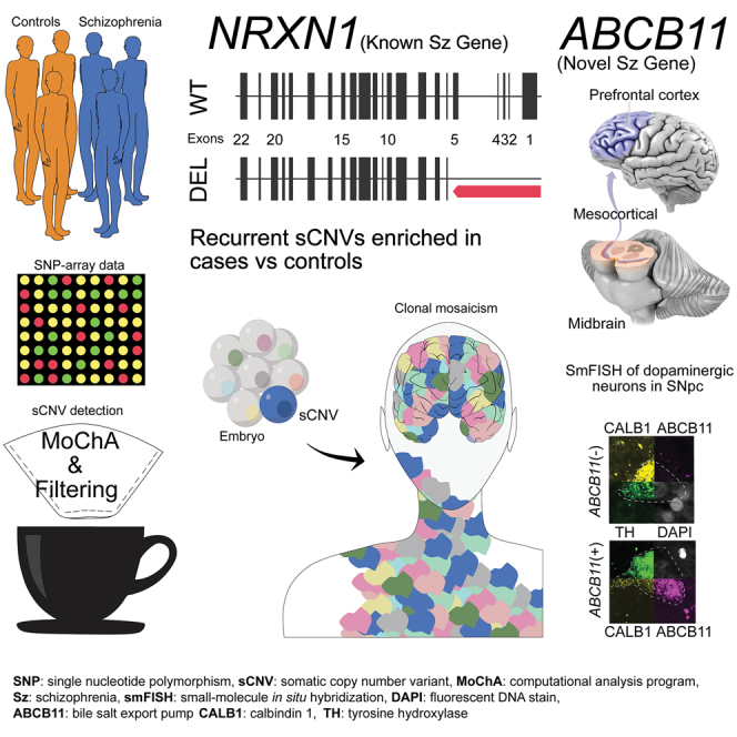
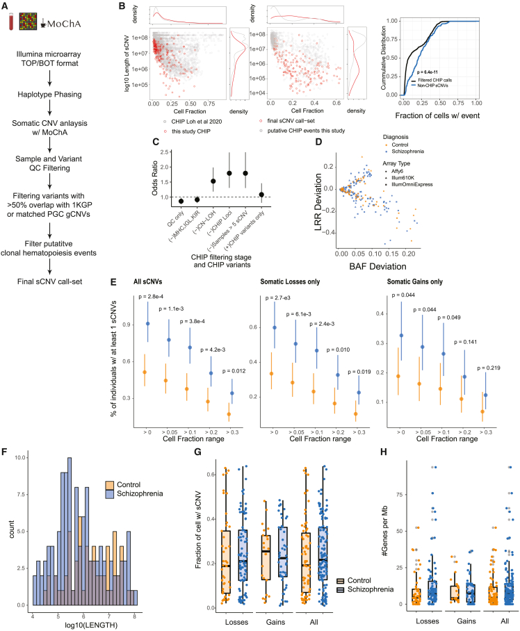
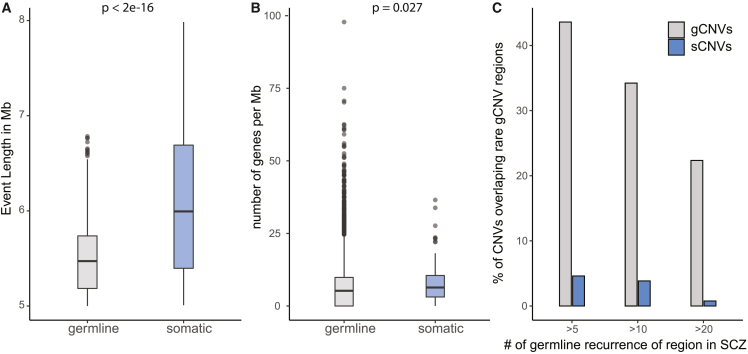
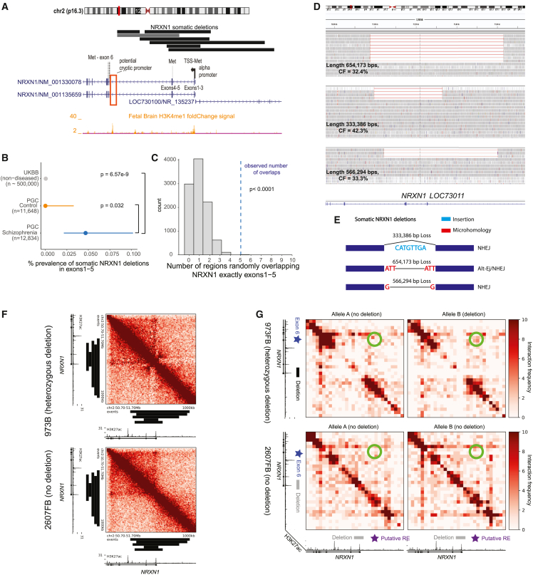
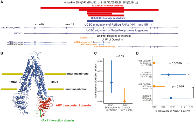
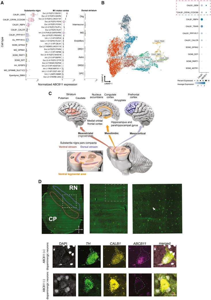
While germline copy-number variants (CNVs) contribute to schizophrenia (SCZ) risk, the contribution of somatic CNVs (sCNVs)-present in some but not all cells-remains unknown. We identified sCNVs using blood-derived genotype arrays from 12,834 SCZ cases and 11,648 controls, filtering sCNVs at loci recurrently mutated in clonal blood disorders. Likely early-developmental sCNVs were more common in cases (0.91%) than controls (0.51%, p = 2.68e-4), with recurrent somatic deletions of exons 1-5 of the NRXN1 gene in five SCZ cases. Hi-C maps revealed ectopic, allele-specific loops forming between a potential cryptic promoter and non-coding cis-regulatory elements upon 5' deletions in NRXN1. We also observed recurrent intragenic deletions of ABCB11, encoding a transporter implicated in anti-psychotic response, in five treatment-resistant SCZ cases and showed that ABCB11 is specifically enriched in neurons forming mesocortical and mesolimbic dopaminergic projections. Our results indicate potential roles of sCNVs in SCZ risk.

Keywords: ABCB11; NRXN1; genomics; mosaicism; schizophrenia; somatic; structural variants; treatment resistance.


Conflict of interest statement

The authors declare no competing interests.

Figures

None
Graphical abstract
Figure 1
Figure 1
Somatic CNV burden in SCZ (A) Schematic of sCNV calling and filtering. (B) Left: scatterplot and marginal distributions of length and CF of sCNVs identified as CHIP vs. non-CHIP. Middle: distribution of canonical CHIP events in sCNVs identified as CHIP in our call set compared with CHIP events identified in the UK Biobank. Right: cumulative distributions of CF of CHIP vs. non-CHIP events; p value from Kolmologorov-Smirnov test. (C) Odds ratio plots comparing sCNV burden across different CHIP filtering stages. Odds ratios and 95% CI were derived from Fisher’s exact test. CHIP variants were defined as those overlapping canonical CHIP events. (D) Trident plot of final call set. Each point represents an event, with colors and shapes indicating subject’s diagnoses and array type. (E) Percentage of individuals with ≥1 sCNV in cases and controls across different minimum CF thresholds. Dots represent mean fraction and lines represent 95% CI from the binomial distribution using Wilson’s score interval with Newcombe modification; p values calculated with two-sided Fisher’s exact test. (F) Histogram of sCNV size (log10 scale) in cases and controls. (G) Boxplots of sCNV CFs in cases vs. controls. (H) Boxplots of the number genes per megabase of sCNVs in cases and controls.
Figure 2
Figure 2
Somatic CNVs differ in size, gene content, and location from gCNVs in SCZ (A) Boxplot of event length in SCZ in somatic and germline state. (B) Plot of number of genes affected per megabase; p values for (A) and (B) were calculated using mixed-effect model log-normal and negative binomial regression, respectively, with batch as a random effect. (C) Bar plots showing percentage of CNVs in each category that overlapped recurrent germline rare CNV regions in SCZ across three different minimum recurrence thresholds.
Figure 3
Figure 3
Somatic deletions of NRXN1 exons 1–5 (A) Adapted GenomeBrowser view of seven somatic deletions of NRXN1. The alpha promoter and in-frame ATG/methionine sites on exons are annotated for NRXN1. Histone marks were obtained from Roadmap epigenomics tracks. Potential cryptic promoter/enhancer is marked by a red box. Gray horizontal bar indicates CNV previously called germline that was found to be somatic. (B) Prevalence of somatic deletions of NRXN1 exons 1–5 in SCZ, controls, and UK Biobank; p values were estimated using two-sided Fisher’s exact test, and 95% CIs were obtained using the Wilson’s score interval with Newcombe modification. (C) Histogram of the distribution of number of overlaps of NRXN1 exons 1-5 from randomly shuffling the discovered NRXN1 sCNVs across the NRXN1 locus. The blue dashed line is the observed number of overlaps, which is equal to six. (D) IGV plots of the deletions of three SCZ subjects with somatic deletions in NRXN1 exons 1–5 from WGS. For clarity, not all the reads are shown. (E) Breakpoint analysis schematic showing observed insertions and microhomology at breakpoints of NRXN1 sCNVs along with event length. NHEJ, non-homologous end-joining repair; Alt-EJ, alternative end joining. (F) Unphased Hi-C heatmap for hiPSC-derived neurons with and without 5′ (exon 1and 2) deletions. Black bars indicate regions of somatic NRXN1 deletions. (G) Phased Hi-C heatmaps for hiPSC-derived neurons. Green circles indicate areas of higher signal with 5′ deletion of NRXN1 in the affected allele. Black bar indicates germline NRXN1 deletion of exons 1and 2. RE, regulatory element.
Figure 4
Figure 4
Somatic CNVs in treatment-resistant SCZ subjects overlap the ABCB11 gene (A) Adapted GenomeBrowser view of five somatic deletions and one somatic duplication of ABCB11. Protein domains of interest overlapped by the sCNVs have orange font. (B) PyMOL schematic of the ABCB11 protein shows HAX1 protein interaction region and the ABC transporter 1 domain, which are affected by somatic deletions of ABCB11. The protein is on an “inner-open” conformation, not bound to ATP. (C) Prevalence of intragenic sCNV in ABCB11 in SCZ and controls. (D) Prevalence of intragenic sCNV in ABCB11 in CLOZUK cohort samples. For (C) and (D), p values were estimated using two-sided Fisher’s exact test, and 95% CIs were obtained using the Wilson’s score interval with Newcombe modification.
Figure 5
Figure 5
Expression of ABCB11 in human brain DA neurons (A) Boxplot of log-normalized ABCB11 expression across three brain regions. Each point indicates an individual sample. Cell type annotations obtained from Kamath et al. (for SN and dorsal striatum samples) and Bakken et al. (for M1 motor cortex samples). Ex/Exc, excitatory neurons; Inh, inhibitory interneurons; Olig, oligodendrocytes; MG, microglia/macrophages; Endofibro, endothelial cells/pericytes; DRD1, direct spiny projection neurons; DRD2, indirect spiny projection neurons; Astro, astrocytes; OPC, oligodendrocyte precursor cells. (B) Left: uniform manifold approximation projection (UMAP) of low-dimensional embedding of 15,684 DA neurons from eight neurotypical donors. Points are colored by clusters obtained from Kamath et al. Right: dot plot of normalized ABCB11 expression across 10 DA subtypes. (C) Schematic of major DA projections from dorsal and ventral streams of SN pars compacta to cortical areas associated with SCZ. (D) Top row: tiled image of a postmortem midbrain tissue section with increasing magnification. Right: white dashed box corresponds to approximate location of middle image and similarly, for the middle image, white boxed arrow with the right image. Red outline indicates approximate ventral tier and blue is approximate dorsal tier. Bottom row: representative image of smFISH of human DA neurons. Scale bar, 15 μm. Colors are DAPI (gray), TH (green), CALB1 (yellow), and ABCB11 (magenta). Outline indicates approximate boundary of DA neuron as identifiable by TH. RN, red nucleus; CP, cerebral peduncles; cartesian arrow labels are D, dorsal, V, ventral; M, medial; L, lateral.

References

    1. Kirov G., Pocklington A.J., Holmans P., Ivanov D., Ikeda M., Ruderfer D., Moran J., Chambert K., Toncheva D., Georgieva L., et al. De novo CNV analysis implicates specific abnormalities of postsynaptic signalling complexes in the pathogenesis of schizophrenia. Mol. Psychiatry. 2012;17:142–153. doi: 10.1038/mp.2011.154. - DOI - PMC - PubMed
    1. Kirov G., Rujescu D., Ingason A., Collier D.A., O’Donovan M.C., Owen M.J. Neurexin 1 (NRXN1) Deletions in Schizophrenia. Schizophr. Bull. 2009;35:851–854. doi: 10.1093/schbul/sbp079. - DOI - PMC - PubMed
    1. Sherman M.A., Rodin R.E., Genovese G., Dias C., Barton A.R., Mukamel R.E., Berger B., Park P.J., Walsh C.A., Loh P.R. Large mosaic copy number variations confer autism risk. Nat. Neurosci. 2021;24:197–203. doi: 10.1038/s41593-020-00766-5. - DOI - PMC - PubMed
    1. Maury E.A., Walsh C.A. Somatic copy number variants in neuropsychiatric disorders. Curr. Opin. Genet. Dev. 2021;68:9–17. doi: 10.1016/j.gde.2020.12.013. - DOI - PMC - PubMed
    1. Ruderfer D.M., Chambert K., Moran J., Talkowski M., Chen E.S., Gigek C., Gusella J.F., Blackwood D.H., Corvin A., Gurling H.M., et al. Mosaic copy number variation in schizophrenia. Eur. J. Hum. Genet. 2013;21:1007–1011. doi: 10.1038/ejhg.2012.287. - DOI - PMC - PubMed

LinkOut - more resources