A case of branch retinal artery occlusion postcataract surgery in an antiphospholipid syndrome patient
- PMID: 37602169
- PMCID: PMC10433039
- DOI: 10.4103/ojo.ojo_302_22
A case of branch retinal artery occlusion postcataract surgery in an antiphospholipid syndrome patient
Abstract
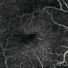
A 46-year-old female with preoperative vision 6/18 N18 (LogMar 0.5) in re and posterior subcapsular cataract underwent an uneventful phacoemulsification surgery under a peribulbar block. On the postoperative day 2, she complained of no visual gain in the operated eye. The reported vision was counting fingers close to the face. Through multimodal imaging (MMI), a diagnosis of branched retinal artery occlusion (BRAO) was made. A detailed consultation and history taking with the patient revealed a concealed history of four miscarriages in the past. A detailed systemic blood workup revealed antiphospholipid antibody (APLA) positive. BRAO postuneventful cataract surgery is a devasting outcome for the surgeon and patient undergoing surgery. The report focuses on the importance of taking detailed past medical history and usage of MMI early to rule out and diagnose unexpected scenarios. We suggest BRAO in our patient was a result of emboli formation, which is a common element in APLA-positive patients.
Keywords: Antiphospholipid syndrome; branched retinal artery occlusion; cataract surgery.
Copyright: © 2023 Oman Ophthalmic Society.
Conflict of interest statement
There are no conflicts of interest.
Figures


Similar articles
-
A novel PAX3 mutation in a Korean patient with Waardenburg syndrome type 1 and unilateral branch retinal vein and artery occlusion: a case report.BMC Ophthalmol. 2018 Oct 11;18(1):266. doi: 10.1186/s12886-018-0933-9. BMC Ophthalmol. 2018. PMID: 30314436 Free PMC article.
-
Branch Retinal Artery Occlusion as a Presentation of Seronegative Antiphospholipid Syndrome in Pregnancy.Cureus. 2024 Nov 22;16(11):e74198. doi: 10.7759/cureus.74198. eCollection 2024 Nov. Cureus. 2024. PMID: 39712706 Free PMC article.
-
Central retinal arterial occlusion (CRAO) after phacoemulsification-a rare complication.Nepal J Ophthalmol. 2013 Jul-Dec;5(2):281-3. doi: 10.3126/nepjoph.v5i2.8746. Nepal J Ophthalmol. 2013. PMID: 24172572
-
Transient branch retinal artery occlusion in a 15-year-old girl and review of the literature.Biomed Pap Med Fac Univ Palacky Olomouc Czech Repub. 2015 Sep;159(3):508-11. doi: 10.5507/bp.2015.031. Epub 2015 Jul 3. Biomed Pap Med Fac Univ Palacky Olomouc Czech Repub. 2015. PMID: 26160228 Review.
-
Complications of Intra-Arterial tPA for Iatrogenic Branch Retinal Artery Occlusion: A Case Report through Multimodal Imaging and Literature Review.Medicina (Kaunas). 2021 Sep 13;57(9):963. doi: 10.3390/medicina57090963. Medicina (Kaunas). 2021. PMID: 34577886 Free PMC article. Review.
Cited by
-
Eyes are a window to the body: A journey from subconjunctival hemorrhage to SLE and inferior vena cava stenting.Indian J Ophthalmol. 2024 Oct 1;72(10):1390. doi: 10.4103/IJO.IJO_486_24. Epub 2024 Sep 27. Indian J Ophthalmol. 2024. PMID: 39331426 Free PMC article. No abstract available.
References
-
- Chan E, Mahroo OA, Spalton DJ. Complications of cataract surgery. Clin Exp Optom. 2010;93:379–89. - PubMed
-
- Swamy BN, Merani R, Hunyor A. Central retinal artery occlusion after phacoemulsification. Retin Cases Brief Rep. 2010;4:281–3. - PubMed
-
- Hanley ME, Hendriksen S, Cooper JS. StatPearls [Internet] Treasure Island (FL): StatPearls Publishing; 2022. Hyperbaric Treatment Of Central Retinal Artery Occlusion. [Updated 2022 Aug 10] - PubMed
-
- Azmon B, Alster Y, Lazar M, Geyer O. Effectiveness of sub-Tenon's versus peribulbar anesthesia in extracapsular cataract surgery. J Cataract Refract Surg. 1999;25:1646–50. - PubMed
Publication types
LinkOut - more resources
Full Text Sources
Miscellaneous
