. 2023 Oct 1;211(7):1123-1133.
doi: 10.4049/jimmunol.2300373.

cGAS-STING Pathway Activation during Trypanosoma cruzi Infection Leads to Tissue-Dependent Parasite Control

Affiliations

cGAS-STING Pathway Activation during Trypanosoma cruzi Infection Leads to Tissue-Dependent Parasite Control

Natasha Perumal et al. J Immunol. .

Abstract

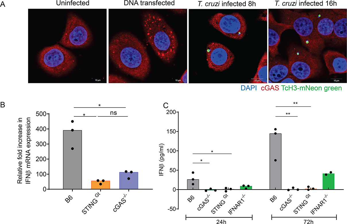
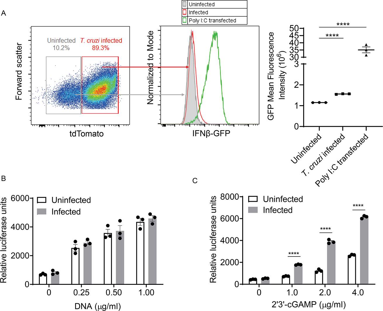
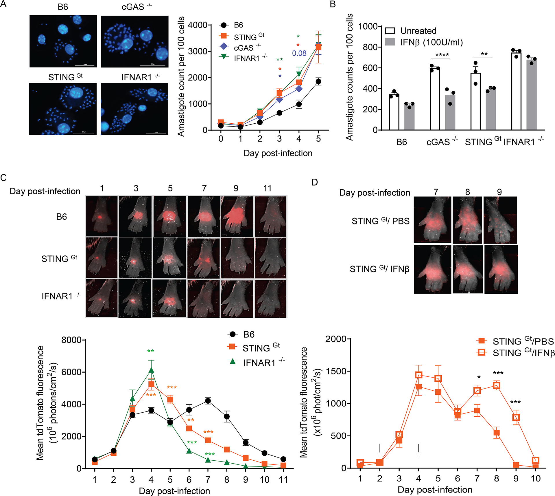
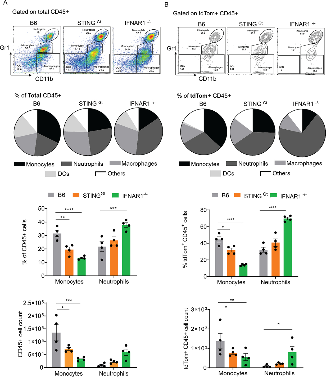
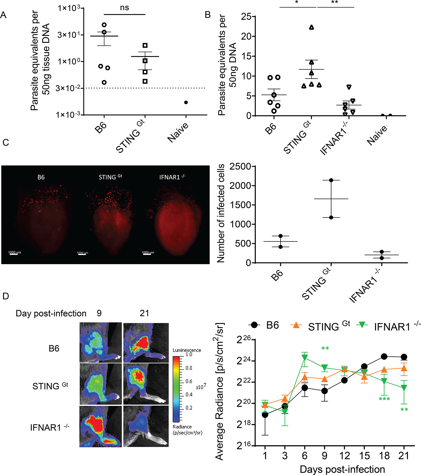
Host cell invasion by Trypanosoma cruzi is a markedly silent process, with limited host transcriptional changes indicative of innate immune recognition, except for a modest type I IFN (IFN-I) response. In this study, we show that T. cruzi-induced IFN-β production was nearly abolished in primary murine cGAS-/- or stimulator of IFN genes (STING)-deficient (STINGGt) macrophages and fibroblasts. T. cruzi infection did not impact the ability of IFN-regulatory factor reporter macrophages to respond to classical cGAS-STING agonists, indicating that the limited IFN-β induction is not due to active parasite suppression. cGAS-/-, STINGGt, and IFN-α/β receptor-/- (IFNAR-/-) macrophages infected with T. cruzi yielded significantly higher numbers of amastigotes compared with wild-type macrophages; however, the impact of the STING pathway during infection in vivo is more complex. Despite an initial increase in parasite growth, STINGGt and IFNAR-/- mice ultimately had lower parasite burden in footpads as compared with wild-type mice, demonstrating a role for IFN-I expression in potentiating parasite growth at the infection site. STING pathway activation had little impact on parasite levels in the skeletal muscle; however, in the heart, cGAS-/- and STINGGt mice, but not IFNAR-/- mice, accumulated higher acute parasite loads, suggesting a protective role of STING sensing of T. cruzi in this organ that was independent of IFN-I. Together, these results demonstrate that host cGAS-STING senses T. cruzi infection, enhancing parasite growth at the site of entry, and contributes to acute-phase parasite restriction in the heart, a major site of tissue damage in chronic T. cruzi infection.


Figures

Figure 1:
Figure 1:
T. cruzi activates the cGAS-STING pathway in primary cells to induce a modest IFN-I response. A) Microscopic detection of cGAS activation in uninfected, DNA-transfected, and infected HeLa cells 8h post-transfection, and 8h and 16h post-infection. Cells were transfected with DNA (2μg/ml). Cells were infected with transgenic T. cruzi overexpressing a histone 3 (TcCLB.509471.59)-mNeon green fusion protein which localizes in the parasite nucleus. B) The IFNβ transcript levels of bone marrow-derived macrophages (BMDMs) from C57BL/6 (B6), STINGGt, and cGAS−/− mice infected with T. cruzi and measured by qRT-PCR at 24h post-infection. Values reported are normalized to GAPDH transcript levels and expressed as a fold change compared to uninfected controls. Data are representative of 2 independent experiments of n=3 infection replicates. C) The levels of IFNβ in supernatants from infected primary mouse fibroblasts were quantified using ELISA after 24h and 72h. Data are representative of 2 independent experiments of n=2–3 infection replicates.
Figure 2:
Figure 2:
Modest IFNβ levels are induced in all infected cells and is not a consequence of active parasite suppression of the pathway. A) IFNβ-GFP reporter THF cells were infected with tdTomato-expressing CL T. cruzi or transfected with Poly I:C for 16hrs under IFNAR blocking conditions. The level of reporter expression in tdTomato -ve (uninfected) or tdTomato +ve (infected) cell subsets within a representative infected sample is displayed alongside levels of reporter activation following poly I:C transfection as measured by flow cytometry. The mean florescence intensities are displayed (right). B-C) IRF-reporter macrophage activation by transfected DNA (B) or IFNβ (C) in previously infected (for 16h) or uninfected cells to stimulation. All data are representative of 2 or more independent experiments of n=3 infection replicates.
Figure 3:
Figure 3:
Activation of the cGAS-STING pathway restricts intracellular parasite growth in vitro but promotes parasite expansion at the infection site. A) In vitro parasite growth in primary bone marrow-derived macrophages from B6, cGAS−/−, STING Gt and IFNAR1−/− mice with representative images at 3days post-infection (left) and amastigote counts (right). n=3 replicate infections. B) Amastigote counts at 3 days post-infection with or without treatment of infected BMDM with IFNβ (100U/ml) added after 3hr of infection with parasites. n=3 replicate infections. C) Differential parasite growth kinetics of STING Gt and IFNAR1−/− compared to B6 mice infected with tdTomato-expressing parasites in each footpad and quantification of fluorescence intensity to assess parasite levels daily post-infection. Infected footpads were imaged, and fluorescence intensity was quantified. Representative images of infected feet at different time points post-infection (top) and quantification of mean fluorescence intensity (bottom). n=5–6 mice (10–12 footpads). D) Parasite growth kinetics of STINGGt mice following footpad injections with PBS or recombinant mouse IFNβ at 2- and 4-days post-infection (represented by black arrowheads). Values reported are from n=5 mice (10 footpads). All data are representative of 2 or more independent experiments.
Figure 4:
Figure 4:
STING activation at the infection site results in a differential infiltration of monocyte and neutrophils. Cells recruited to the ears of mice infected at the site with tdTomato expressing parasites were isolated at 5 days post-infection and immunophenotyped using the gating strategy described in Supplemental 3A. Differences in the proportion of A) total or B) Infected (tdTom+) CD45+ leukocyte subsets in B6, STINGGt and IFNAR1−/− are shown. Representative flow plots (top) denote populations of monocytes (CD45+ CD11bHi Gr-1Int), neutrophils (CD45+ CD11bHi Gr-1Hi), macrophages (CD45+ CD11bHi Gr-1lo) and DCs (CD45+ CD11blo Gr-1lo). Pie charts (middle) show average (of n=4 mice) proportions of each population in the different mouse groups and bar graphs (bottom) show significant differences in the proportion and absolute count of monocyte and neutrophil subsets. Data are representative of 2 or more independent experiments.
Figure 5:
Figure 5:
Specific depletion or augmentation of cellular recruitment had limited impact on parasite growth at the infection site. A) Specific depletion of monocytes in B6 mice by intraperitoneal injection with either isotype control or anti-CCR2 (MC-21) antibody as demonstrated by representative flow plots of the cellular infiltrate at the infection site at 6 days post-infection. B) Parasite growth kinetics at the site of infection in WT mice with and without monocyte depletion, with black arrowheads representing the timing of treatment. n=4–5 mice/group. C) Specific depletion neutrophils in STING GT mice by intraperitoneal administration of either PBS or anti- Ly6G (mAb 1A8) at 2- and 4-days post-infection, demonstrated by peripheral blood staining at 5 days post-infection. D) Parasite growth kinetics following neutrophil depletion after infection, with black arrowheads representing the timing of treatment. n=4–5 mice. Data are representative of 2 independent experiments.
Figure 6:
Figure 6:
STING activation does not impact the initial establishment of infection nor long-term parasite persistence but constrains acute parasite load in the heart independently of IFN-I A) The overall control of infection as determined by quantification of parasite loads in the skeletal muscle after >300days post footpad infection in B6 and STINGGt mice. n>4 mice per group. The dotted line represents the threshold of detection for the assay. B) T. cruzi DNA in the hearts of B6, STINGGt and IFNAR1−/− mice infected intraperitoneally with 200,000 Colombiana strain parasites as determined by quantitative real-time PCR at 14dpi. n= 6 mice per group. C) Light-sheet microscopic detection of tdTomato- expressing parasites in clarified hearts of B6, STING Gt and IFNAR1−/− mice 14 days post-infection with 200,000 Colombiana strain parasites. Representative images (left) with quantification (right) of n=2 mice per group. D) Parasite growth kinetics in the calf muscle of B6, STING Gt and IFNAR1−/− following direct infection of the gastrocnemius with parasites expressing luciferase determined by bioluminescence intensity. Representative images (left) and luminescent intensity with time (right). n> 4 mice per infection. All data are representative of 2 independent experiments.

Similar articles

Cited by

References

    1. Jain A, and Pasare C. 2017. Innate Control of Adaptive Immunity: Beyond the Three-Signal Paradigm. J Immunol 198: 3791–3800. - PMC - PubMed
    1. Sullivan BM, Teijaro JR, de la Torre JC, and Oldstone MB. 2015. Early virus-host interactions dictate the course of a persistent infection. PLoS Pathog 11: e1004588. - PMC - PubMed
    1. Frolov I, Akhrymuk M, Akhrymuk I, Atasheva S, and Frolova EI. 2012. Early events in alphavirus replication determine the outcome of infection. J Virol 86: 5055–5066. - PMC - PubMed
    1. Subbian S, Bandyopadhyay N, Tsenova L, O’Brien P, Khetani V, Kushner NL, Peixoto B, Soteropoulos P, Bader JS, Karakousis PC, Fallows D, and Kaplan G. 2013. Early innate immunity determines outcome of Mycobacterium tuberculosis pulmonary infection in rabbits. Cell Commun Signal 11: 60. - PMC - PubMed
    1. Pontiroli F, Dussurget O, Zanoni I, Urbano M, Beretta O, Granucci F, Ricciardi-Castagnoli P, Cossart P, and Foti M. 2012. The timing of IFNbeta production affects early innate responses to Listeria monocytogenes and determines the overall outcome of lethal infection. PLoS One 7: e43455. - PMC - PubMed

Publication types