. 2023 Dec;270(12):5849-5865.
doi: 10.1007/s00415-023-11862-4. Epub 2023 Aug 21.

Analysis of muscle magnetic resonance imaging of a large cohort of patient with VCP-mediated disease reveals characteristic features useful for diagnosis

Diana Esteller  1 Marianela Schiava  2 José Verdú-Díaz  2 Rocío-Nur Villar-Quiles  3 Boris Dibowski  4 Nadia Venturelli  4 Pascal Laforet  5 Jorge Alonso-Pérez  6   7 Montse Olive  7   8 Cristina Domínguez-González  8   9 Carmen Paradas  10   11 Beatriz Vélez  10   11 Anna Kostera-Pruszczyk  12   13 Biruta Kierdaszuk  12   13 Carmelo Rodolico  14 Kristl Claeys  15 Endre Pál  16 Edoardo Malfatti  17 Sarah Souvannanorath  17 Alicia Alonso-Jiménez  18 Willem de Ridder  18 Eline De Smet  18 George Papadimas  19 Constantinos Papadopoulos  19 Sofia Xirou  19 Sushan Luo  20 Nuria Muelas  8   21   22   23 Juan J Vilchez  8   21   22   23 Alba Ramos-Fransi  24 Mauro Monforte  25 Giorgio Tasca  2 Bjarne Udd  26   27 Johanna Palmio  26   27 Srtuhi Sri  28 Sabine Krause  29 Benedikt Schoser  29 Roberto Fernández-Torrón  11   30 Adolfo López de Munain  11   30 Elena Pegoraro  31 Maria Elena Farrugia  32 Mathias Vorgerd  33 Georgious Manousakis  34 Jean Baptiste Chanson  35 Aleksandra Nadaj-Pakleza  35 Hakan Cetin  36 Umesh Badrising  37 Jodi Warman-Chardon  38 Jorge Bevilacqua  39 Nicholas Earle  39 Mario Campero  39 Jorge Díaz  39 Chiseko Ikenaga  40 Thomas E Lloyd  40 Ichizo Nishino  41 Yukako Nishimori  41 Yoshihiko Saito  41 Yasushi Oya  42 Yoshiaki Takahashi  43 Atsuko Nishikawa  44 Ryo Sasaki  45 Chiara Marini-Bettolo  2 Michela Guglieri  2 Volker Straub  2 Tanya Stojkovic  3 Robert Y Carlier #  4 Jordi Díaz-Manera #  46   47   48
Affiliations

Analysis of muscle magnetic resonance imaging of a large cohort of patient with VCP-mediated disease reveals characteristic features useful for diagnosis

Diana Esteller et al. J Neurol. 2023 Dec.

Erratum in

  • Correction to: Analysis of muscle magnetic resonance imaging of a large cohort of patient with VCP‑mediated disease reveals characteristic features useful for diagnosis.
    Esteller D, Schiava M, Verdú-Díaz J, Villar-Quiles RN, Dibowski B, Venturelli N, Laforet P, Alonso-Pérez J, Olive M, Domínguez-González C, Paradas C, Vélez B, Kostera-Pruszczyk A, Kierdaszuk B, Rodolico C, Claeys K, Pál E, Malfatti E, Souvannanorath S, Alonso-Jiménez A, de Ridder W, De Smet E, Papadimas G, Papadopoulos C, Xirou S, Luo S, Muelas N, Vilchez JJ, Ramos-Fransi A, Monforte M, Tasca G, Udd B, Palmio J, Sri S, Krause S, Schoser B, Fernández-Torrón R, López de Munain A, Pegoraro E, Farrugia ME, Vorgerd M, Manousakis G, Chanson JB, Nadaj-Pakleza A, Cetin H, Badrising U, Warman-Chardon J, Bevilacqua J, Earle N, Campero M, Díaz J, Ikenaga C, Lloyd TE, Nishino I, Nishimori Y, Saito Y, Oya Y, Takahashi Y, Nishikawa A, Sasaki R, Marini-Bettolo C, Guglieri M, Straub V, Stojkovic T, Carlier RY, Díaz-Manera J. Esteller D, et al. J Neurol. 2024 Apr;271(4):2147-2148. doi: 10.1007/s00415-023-12178-z. J Neurol. 2024. PMID: 38349561 Free PMC article. No abstract available.

Abstract

Background: The diagnosis of patients with mutations in the VCP gene can be complicated due to their broad phenotypic spectrum including myopathy, motor neuron disease and peripheral neuropathy. Muscle MRI guides the diagnosis in neuromuscular diseases (NMDs); however, comprehensive muscle MRI features for VCP patients have not been reported so far.

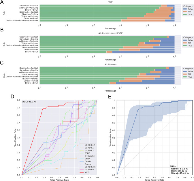
Methods: We collected muscle MRIs of 80 of the 255 patients who participated in the "VCP International Study" and reviewed the T1-weighted (T1w) and short tau inversion recovery (STIR) sequences. We identified a series of potential diagnostic MRI based characteristics useful for the diagnosis of VCP disease and validated them in 1089 MRIs from patients with other genetically confirmed NMDs.

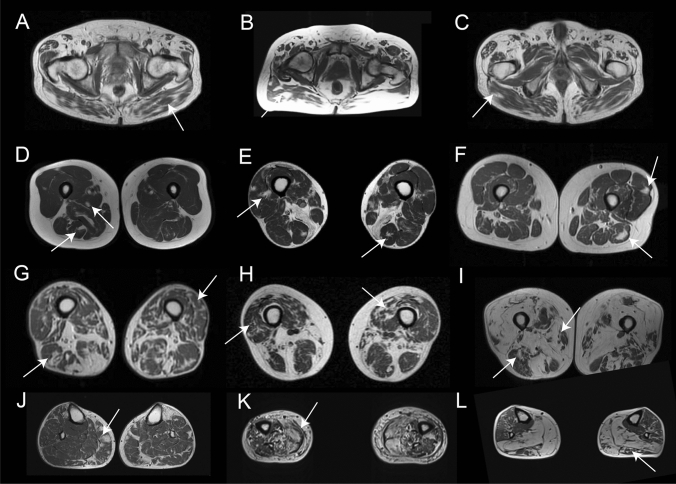
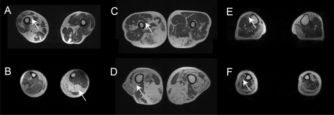
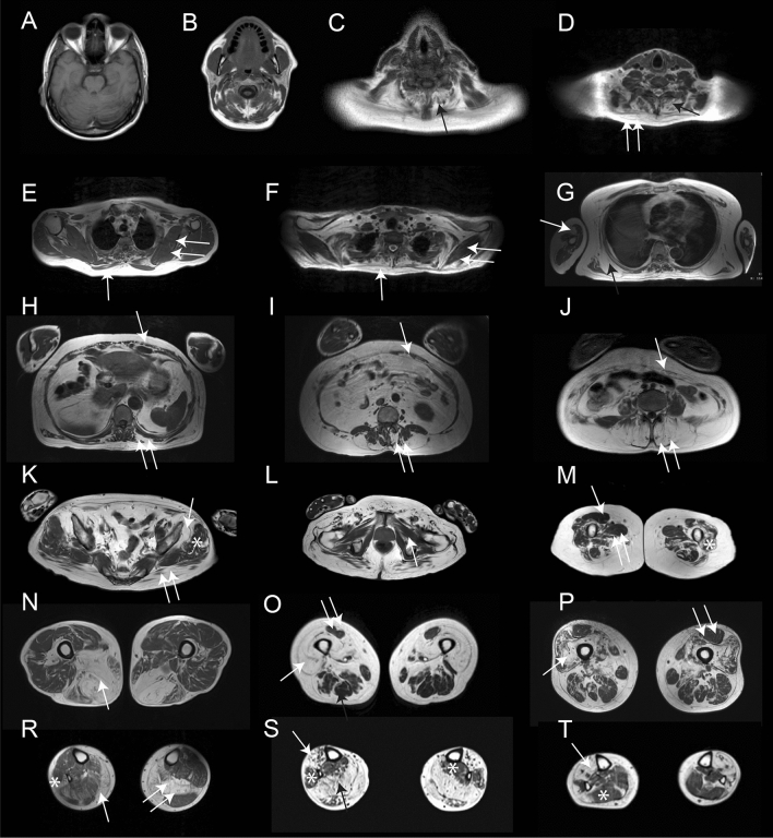
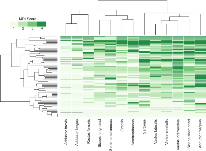
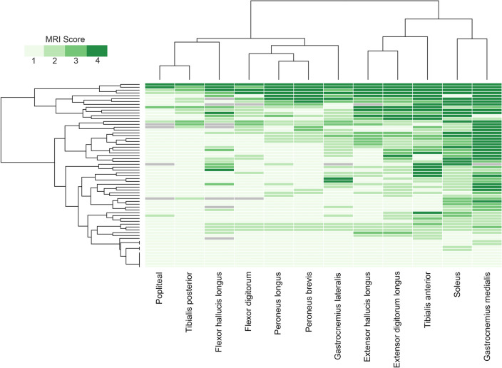
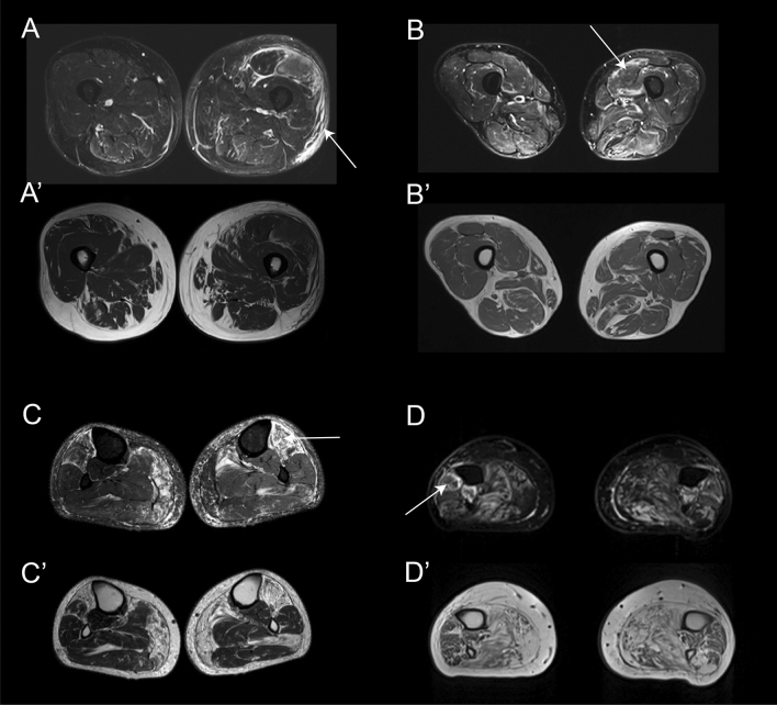
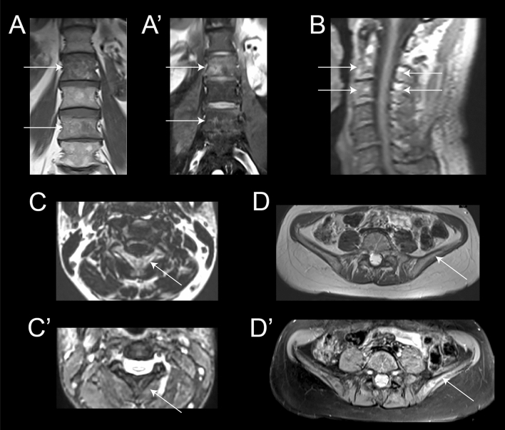
Results: Fat replacement of at least one muscle was identified in all symptomatic patients. The most common finding was the existence of patchy areas of fat replacement. Although there was a wide variability of muscles affected, we observed a common pattern characterized by the involvement of periscapular, paraspinal, gluteal and quadriceps muscles. STIR signal was enhanced in 67% of the patients, either in the muscle itself or in the surrounding fascia. We identified 10 diagnostic characteristics based on the pattern identified that allowed us to distinguish VCP disease from other neuromuscular diseases with high accuracy.

Conclusions: Patients with mutations in the VCP gene had common features on muscle MRI that are helpful for diagnosis purposes, including the presence of patchy fat replacement and a prominent involvement of the periscapular, paraspinal, abdominal and thigh muscles.

Keywords: Multisystemic proteinopathy; Muscle MRI; VCP myopathy; Valosin.


Conflict of interest statement

The authors declare no competing interests related to the content of this work.

Figures

Fig. 1
Fig. 1
Distribution of fat replacement in the skeletal muscles of patients with mutations in the VCP gene. The figure shows examples of the distribution of fat replacement in patients with mutations in the VCP gene at different stages of disease progression. A, B and C show examples of fat replacement in the gluteus maximus. D, E and F show patients at early stages of disease progression where fat can be seen as spots of fat in the muscles as highlighted by the arrows. G, H and I show images of fat replacement in the thigh of patients at middle (G and H) and advanced (I) stages of disease progression. J, K and L show examples of the fat replacement in the muscles of the legs in patients in early (J) and middle (K and L) stages of disease progression
Fig. 2
Fig. 2
Asymmetric and distal to proximal gradient of fat replacement. A and B show MRI of two patients with asymmetric fat replacement in the vastus intermedius (arrow in A) and the soleus (arrow in B) muscles. C and D show a distal to proximal gradient of fat replacement in the vastus intermedius muscle (arrow). E and F show a distal to proximal gradient of fat replacement in the tibialis anterior muscle (arrow)
Fig. 3
Fig. 3
Fat replacement pattern in patients with mutations in the VCP gene. The figure shows the pattern of fat replacement observed in different patients with mutations in the VCP gene. A and B show normal cranial muscles without fat replacement. C shows fat replacement of the paraspinal cervical muscles (black arrow). D shows fat replacement of the paraspinal cervical muscles (black arrow) and trapezius (double arrow). E and F show a fat spot in the trapezius muscle (arrow) while scapular muscles are spared (double arrow). G shows fat replacement of the serratus anterior (black arrow) and the biceps brachii (double arrow). H, I and J show different examples of fat replacement in the trunk muscles, including abdominal (arrow) and paraspinal (double arrow) muscles. K shows involvement of the gluteus maximus (double arrow) and gluteus minimus (arrow) muscles, while gluteus medius (asterisk) is spared. L shows no involvement of the pelvic floor muscles (arrow). M shows characteristic involvement of upper thigh characterized by sparing of rectus femoris (arrow) and adductor longus (double arrow), with involvement of the vasti muscles (asterisk). N, O and P show different combinations of muscle fatty replacement observed in the thigh, N shows predominant posterior thigh involvement (arrow), while O and P show predominant anterior involvement (arrow) with sparing of rectus femoris (double arrow). R, S and T show different combinations of muscle fatty replacement observed in the leg, R shows fat replacement of gastrocnemius medialis (arrow) associated with asymmetric involvement of soleus (double arrow) and peroneus (asterisk). S shows involvement of the anterior (arrow) and posterior compartment (black arrow) and sparing of peroneus and tibialis posterior muscles (asterisk). T shows predominant anterior involvement (arrow) with sparing of soleus (asterisk)
Fig. 4
Fig. 4
Heatmap showing muscle fatty replacement of muscles of the thigh. Heatmap showing fat replacement of the muscles of the thigh. Patients and muscles are ordered according to hierarchical clustering. The score of a muscle in a patient is indicated by the color of the square. As shown in the figure, adductor magnus, biceps short head, vasti muscles and the sartorius were the most frequently affected muscles, while adductor longus, adductor brevis and rectus femoris were commonly spared or less affected
Fig. 5
Fig. 5
Heatmap showing muscle fatty replacement of muscles of the leg. Heatmap showing fat replacement of the muscles of the leg. Patients and muscles are ordered according to hierarchical clustering. The score of a muscle in a patient is indicated by the color of the square. As shown in the figure, gastrocnemius medialis and soleus were the muscles more commonly affected followed by the distal anterior compartment muscles: tibialis anterior, extensor digitorum longus and extensor hallucis longus, while tibialis posterior and popliteal muscles are commonly spared
Fig. 6
Fig. 6
Examples of STIR enhancement in patients with mutations in the VCP gene. The figure shows different examples of STIR enhancement in patients with mutations in the VCP gene. A Enhancement of the subcutaneous tissue (arrow), perifascicular (double arrow) and vastus lateralis muscle (asterisk). A’ T1w image of the same slice showing. No fat replacement in the area enhanced in STIR. B Enhancement of the periphery of the vastus lateralis muscle in STIR while B’ shows no fat replacement in that muscle. C Enhacement of the whole volume of tibialis anterior which is already replaced by fat in T1 as shown in C’. D Enhancement of periphery of peroneus muscle (arrow) which is partially replaced by fat as shown in D’
Fig. 7
Fig. 7
Validation of diagnostic rules identified in VCP myopathy. Figure shows the metrics obtained to validate the rules proposed for VCP myopathy. A Results of the analysis of each rule applied to the cohort of VCP patients. B Results of the analysis of each rule applied to a cohort of 978 MRIs of 10 different neuromuscular diseases. C Results of the analysis of each rule applied to a cohort of VCP patients and 978 MRIs of 10 different neuromuscular diseases. Green, orange and blue in A, B and C corresponds to percentage of patients meeting the rule, percentage of patients with missing data and percentage of patients not meeting the rules, respectively. D Comparison of ROC curves obtained after applying the rules generated to MRIs of patients with different neuromuscular diseases. E ROC curve obtained after applying the rules generated to the cohort of patients with VCP myopathy. The blue area shows an interval of confidence including of missing variables where the upper limit shows the ROC curve if all missing variables were meeting the rules and the lower limit shows the ROC curve if all missing variables were not meeting the rules. AUC: Area under the curve
Fig. 8
Fig. 8
Examples of MRI finding in patients with Paget disease of the bone included in this study. Coronal views T1 (A) and T2 with fat saturation (A’)-weighted images of the lumbar spine showing heterogeneity of the signal of bone marrow at L2 and L4 levels compared to L1 and L3 (arrows). Coarsed trabeculae are more visible on T1 and edema of the vertebral body on T2 fat sat. Sagital (B) and axial (C and C’) T1-weighted and axial T2 with fat saturation views centered on the upper cervical spine. Signal heterogeneity, cortical thickening and bone enlargement of the entirety of C2 and C3 vertebrae is detectable (arrows). Axial T1 (D) and T2 (D’) with fat saturation-weighted images centered on the iliac crest. Bone changes are very subtle on T1 but bone edema and heterogeneity is more visible on T2 on the left side (arrow) especially in comparison with the right side

Similar articles

Cited by

  • Muscle Biopsy Findings in Valosin-Containing Protein Multisystem Proteinopathy.
    Schiava M, Parkhurst Y, Henderson M, Polvikoski T, Valtcheva MV, Nishino I, Inoue M, Nishimori Y, Saito Y, Stojkovic T, Villar-Quiles RN, Romero NB, Evangelista T, Malfatti E, Souvannanorath S, Pegoraro E, Riguzzi P, Monforte M, Bortolani S, Torchia E, Sabatelli M, Tasca G, Straub V, Marini-Bettolo C, Guglieri M, Cetin H, Gelpi E, Klotz S, De Bleecker JL, Alonso-Jimenez A, Baets J, De Ridder W, De Jonghe P, Claeys KG, Thal DR, Bevilacqua JA, Luo S, Zhu W, Lin J, Papadimas G, Papadopoulos C, Zamba-Papanicolaou E, Xirou S, Pal E, Rodolico C, Kostera-Pruszczyk A, Kierdaszuk B, Kaminska A, Muelas N, Vilchez JJ, Domínguez-González C, Hernandez-Lain A, Alonso-Perez J, Nedkova-Hristova V, Aledo C, Oldfors A, Badrising UA, Kushlaf H, Lloyd TE, Ikenaga C, Alfano LN, Quinn CC, Walk D, Vorgerd M, Weihl C, Olivé M, Diaz-Manera J. Schiava M, et al. Neurol Genet. 2025 Jul 16;11(4):e200265. doi: 10.1212/NXG.0000000000200265. eCollection 2025 Aug. Neurol Genet. 2025. PMID: 40678441 Free PMC article.
  • 2024 VCP International Conference: Exploring multi-disciplinary approaches from basic science of valosin containing protein, an AAA+ ATPase protein, to the therapeutic advancement for VCP-associated multisystem proteinopathy.
    Peck A, Dadi A, Yavarow Z, Alfano LN, Anderson D, Arkin MR, Chou TF, D'Ambrosio ES, Diaz-Manera J, Dudley JP, Elder AG, Ghoshal N, Hart CE, Hart MM, Huryn DM, Johnson AE, Jones KB, Kimonis V, Kiskinis E, Lee EB, Lloyd TE, Mapstone M, Martin A, Meyer H, Mozaffar T, Onyike CU, Pfeffer G, Pindon A, Raman M, Richard I, Rubinsztein DC, Schiava M, Schütz AK, Shen PS, Southworth DR, Staffaroni AM, Taralio-Gravovac M, Weihl CC, Yao Q, Ye Y, Peck N. Peck A, et al. Neurobiol Dis. 2025 Apr;207:106861. doi: 10.1016/j.nbd.2025.106861. Epub 2025 Mar 2. Neurobiol Dis. 2025. PMID: 40037468 Review.
  • Cross-sectional study of patients with VCP multisystem proteinopathy 1 using dual-energy x-ray absorptiometry.
    Columbres RCA, Luu V, Nguyen M, Kimonis V. Columbres RCA, et al. Muscle Nerve. 2024 Jun;69(6):699-707. doi: 10.1002/mus.28095. Epub 2024 Mar 29. Muscle Nerve. 2024. PMID: 38551101 Free PMC article.
  • French National Protocol for diagnosis and care of facioscapulohumeral muscular dystrophy (FSHD).
    Attarian S, Beloribi-Djefaflia S, Bernard R, Nguyen K, Cances C, Gavazza C, Echaniz-Laguna A, Espil C, Evangelista T, Feasson L, Audic F, Zagorda B, Milhe De Bovis V, Stojkovic T, Sole G, Salort-Campana E, Sacconi S. Attarian S, et al. J Neurol. 2024 Sep;271(9):5778-5803. doi: 10.1007/s00415-024-12538-3. Epub 2024 Jul 2. J Neurol. 2024. PMID: 38955828 Review.
  • Validity and Reliability of Clinical and Patient-Reported Outcomes in Multisystem Proteinopathy 1.
    Alfano LN, Iammarino MA, Reash NF, Lowes LP, Pietruszewski L, Adderley K, Humphrey L, Knight AB, Steiner CL, Smith MA, Sahenk Z, Connolly AM, Almomen M, D'Ambrosio ES, Peck N, Peck A. Alfano LN, et al. Ann Clin Transl Neurol. 2025 Jul;12(7):1324-1333. doi: 10.1002/acn3.70064. Epub 2025 Apr 28. Ann Clin Transl Neurol. 2025. PMID: 40294152 Free PMC article.

References

    1. Johnson JO, Mandrioli J, Benatar M, et al. Exome sequencing reveals VCP mutations as a cause of familial ALS. Neuron. 2010;68:857–864. doi: 10.1016/j.neuron.2010.11.036. - DOI - PMC - PubMed
    1. Sun X, Qiu H. Valosin-containing protein, a calcium-associated ATPase protein, in endoplasmic reticulum and mitochondrial function and its implications for diseases. Int J Mol Sci. 2020;21:3842. doi: 10.3390/ijms21113842. - DOI - PMC - PubMed
    1. Yeo BK, Hong CJ, Chung KM, et al. Valosin-containing protein is a key mediator between autophagic cell death and apoptosis in adult hippocampal neural stem cells following insulin withdrawal. Mol Brain. 2016;9:31. doi: 10.1186/s13041-016-0212-8. - DOI - PMC - PubMed
    1. Kimonis VE, Kovach MJ, Waggoner B, et al. Clinical and molecular studies in a unique family with autosomal dominant limb-girdle muscular dystrophy and Paget disease of bone. Genet Med. 2000;2:232–241. doi: 10.1097/00125817-200007000-00006. - DOI - PMC - PubMed
    1. Kovach MJ, Waggoner B, Leal SM, et al. Clinical delineation and localization to chromosome 9p13.3-p12 of a unique dominant disorder in four families: hereditary inclusion body myopathy. Paget disease of bone, and frontotemporal dementia. Mol Genet Metab. 2001;74:458–475. doi: 10.1006/mgme.2001.3256. - DOI - PMC - PubMed

MeSH terms

Substances