. 2023 Aug 21;15(1):34.
doi: 10.1186/s13089-023-00334-5.

Feasibility of chest ultrasound up to 42 m underwater

Affiliations

Feasibility of chest ultrasound up to 42 m underwater

Matteo Paganini et al. Ultrasound J. .

Abstract

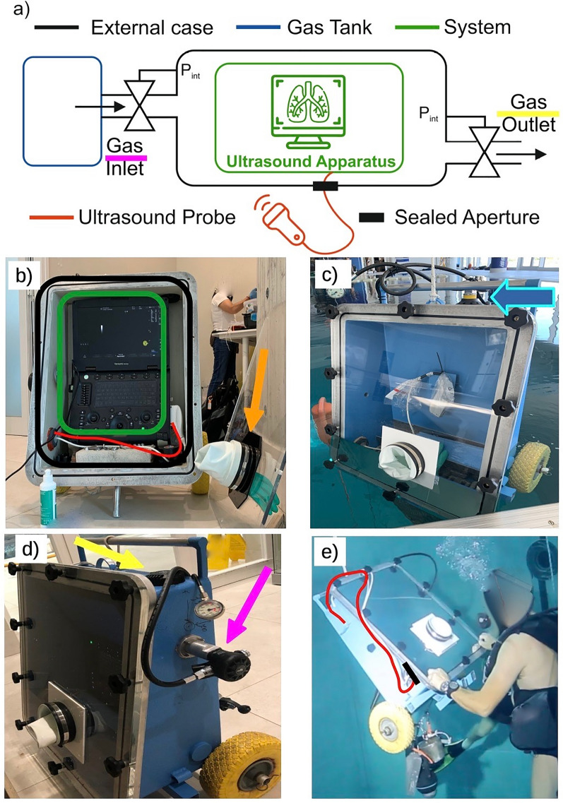
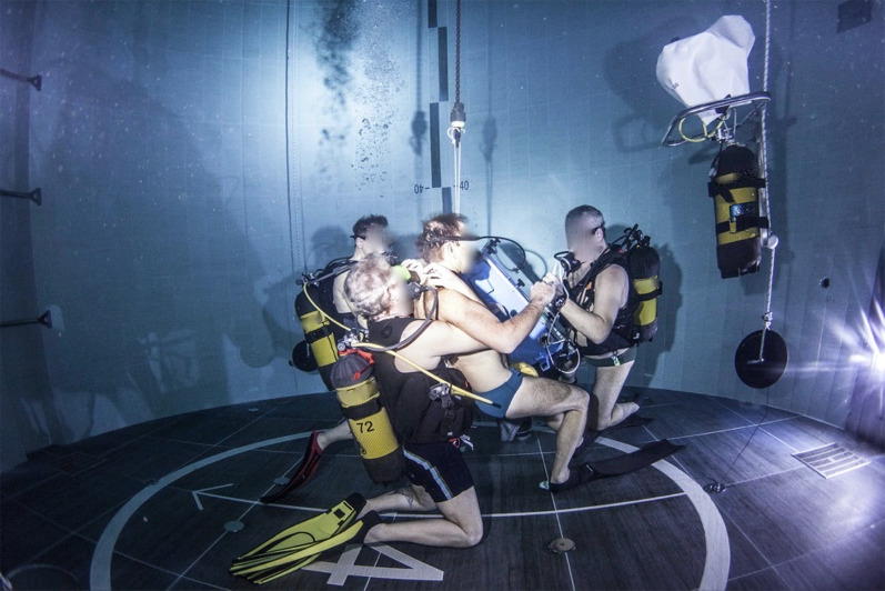
After recent advancements, ultrasound has extended its applications from bedside clinical practice to wilderness medicine. Performing ultrasound scans in extreme environments can allow direct visualization of unique pathophysiological adaptations but can be technically challenging. This paper summarizes how a portable ultrasound apparatus was marinized to let scientific divers and sonographers perform ultrasound scans of the lungs underwater up to - 42 m. A metallic case protected the ultrasound apparatus inside; a frontal transparent panel with a glove allowed visualization and operation of the ultrasound by the diving sonographer. The inner pressure was equalized with environmental pressure through a compressed air tank connected with circuits similar to those used in SCUBA diving. Finally, the ultrasound probe exited the metallic case through a sealed aperture. No technical issues were reported after the first testing step and the real experiments.

Keywords: Chest ultrasound; Diving medicine; Environmental physiology; Extreme environments; Lung ultrasound; Physiology; Ultrasound; Underwater medicine.


Conflict of interest statement

The authors declare that they have no competing interests.

Figures

Fig. 1
Fig. 1
The insulating device used to allow underwater chest ultrasound. a Schematic representation of the setup. b The opened case (black), with the ultrasound apparatus inside (still charging; green) and the ultrasound probe connected (red); the orange arrow indicates the frontal panel and the glove used to operate the ultrasound. c First test of the empty case; the blue arrow indicates the air tank. d Sides of the insulating device with the wheels used to move it, the inlet circuit (purple arrow), and the outlet valve (yellow arrow). e One of the authors (EG) testing the device underwater; on the right side, the ultrasound probe (red line) sticks out of the metallic case through a dedicated sealed aperture (black line)
Fig. 2
Fig. 2
Chest ultrasound performed by the authors at − 42 m underwater

References

    1. Díaz-Gómez JL, Mayo PH, Koenig SJ. Point-of-care ultrasonography. N Engl J Med. 2021;385:1593–1602. doi: 10.1056/NEJMra1916062. - DOI - PubMed
    1. Whitson MR, Mayo PH. Ultrasonography in the emergency department. Crit Care. 2016;20:227. doi: 10.1186/s13054-016-1399-x. - DOI - PMC - PubMed
    1. Feletti F, Mucci V, Aliverti A. Chest ultrasonography in modern day extreme settings: from military setting and natural disasters to space flights and extreme sports. Can Respir J. 2018;2018:1–9. doi: 10.1155/2018/8739704. - DOI - PMC - PubMed
    1. Fagenholz PJ, Murray AF, Noble VE, et al. Ultrasound for high altitude research. Ultrasound Med Biol. 2012;38:1–12. doi: 10.1016/j.ultrasmedbio.2011.10.004. - DOI - PubMed
    1. Fagenholz PJ, Gutman JA, Murray AF, et al. Chest ultrasonography for the diagnosis and monitoring of high-altitude pulmonary edema. Chest. 2007;131:1013–1018. doi: 10.1378/chest.06-1864. - DOI - PubMed

LinkOut - more resources